Copyright
©The Author(s) 2015.
World J Stem Cells. Jun 26, 2015; 7(5): 793-805
Published online Jun 26, 2015. doi: 10.4252/wjsc.v7.i5.793
Published online Jun 26, 2015. doi: 10.4252/wjsc.v7.i5.793
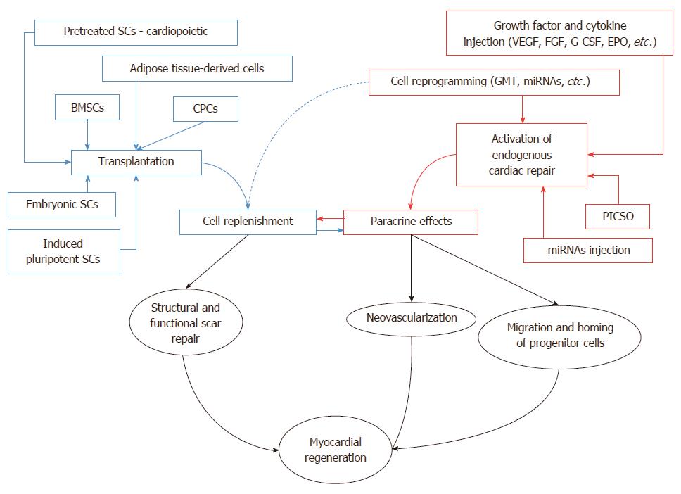
Figure 1 Transplantation of stem cells vs initiation of endogenous myocardial regeneration without cell transplantation.
Depicted are two basic approaches to myocardial regeneration: (1) via transplantation of different types of stem cells into the damaged myocardium (on the left, in blue); and (2) by activation of endogenous pathways of cardiac repair, without cell transplantation (on the right, in red). Both sets of techniques are thought to ultimately exert their effects on the paracrine route and by replenishing lost cardiomyocytes. SCs: Stem cells; BMSCs: Bone marrow-derived stem cells; CPCs: Cardiac progenitor cells; VEGF: Vascular endothelial growth factor; FGF: Fibroblast growth factor; G-CSF: Granulocyte colony stimulating factor; EPO: Erythropoietin; GMT: Gata4, Mef2c, Tbx5; miRNAs: microRNAs; PICSO: Pressure-controlled intermittent coronary sinus occlusion.
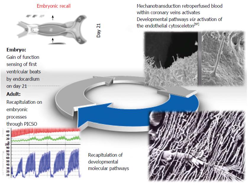
Figure 2 Hypothesis of embryonic recall.
In embryos, the first heart beat is sensed by mechanotransduction of the endocardium, leading to a burst of developmental signals thriving cardiac morphogenesis. In analogy, elevated pressures in cardiac veins act via stretch and shear stress on perivascular cells, thus reiterating the same mechanotransductory signals in the adult failing heart. Pressure elevation in cardiac veins have to be pressure controlled not to increase arterial resistance to nutritive flow (PICSO).
- Citation: Milasinovic D, Mohl W. Contemporary perspective on endogenous myocardial regeneration. World J Stem Cells 2015; 7(5): 793-805
- URL: https://www.wjgnet.com/1948-0210/full/v7/i5/793.htm
- DOI: https://dx.doi.org/10.4252/wjsc.v7.i5.793


