Copyright
©The Author(s) 2015.
World J Stem Cells. May 26, 2015; 7(4): 728-744
Published online May 26, 2015. doi: 10.4252/wjsc.v7.i4.728
Published online May 26, 2015. doi: 10.4252/wjsc.v7.i4.728
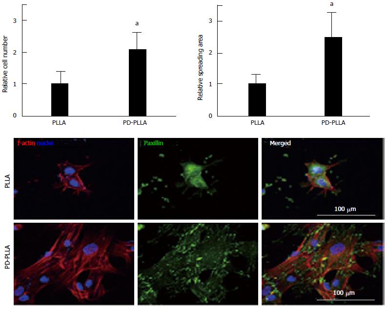
Figure 1 Quantitative analysis of initial cell adhesion from human mesenchymal stem cells cultured on the fibers.
Relative adherent cell numbers and spreading area of human mesenchymal stem cells (hMSCs) cultured on PLLA and PD-PLLA fibers were analyzed after 12 h of culture. aP < 0.05, PD-PLLA vs PLLA group. Adherent morphology of hMSCs on PLLA and PD-PLLA fibers was observed by confocal microscopy. Scale bars represent 100 μm. Reproduced with permission from Rim et al[64]. PLLA: Poly(l-lactide); PD-PLLA: Poly(l-lactide) (PLLA) fibers coated with polydopamine.
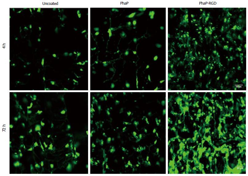
Figure 2 Confocal microscopic imaging of human bone marrow mesenchymal stem cells grown in uncoated, PhaP and PhaP-RGD coated 3-hydroxybutyrate-cohydroxyhexanoate scaffolds after 4 or 72 h of incubation, respectively.
Phalloidin-fluorescein isothiocyanate was used to F-actin of cells grown in the scaffolds (Green). Reproduced with permission from You et al[61]. PhaP-RGD: PhaP binding protein fused with arginyl-glycyl-aspartic acid.
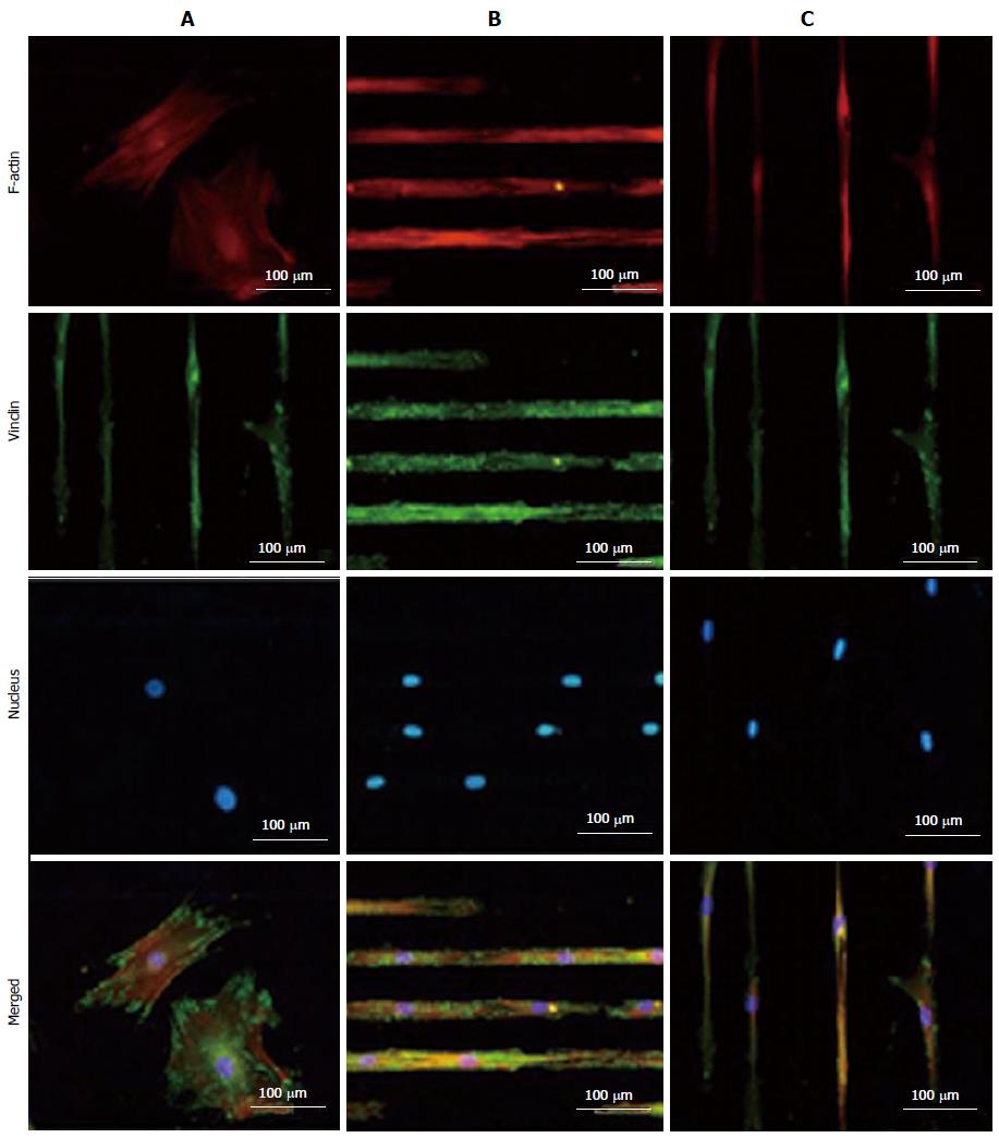
Figure 3 TRITC-Phalloidin labeled F-actin (red), AlexaFluor 488 labeled vinculin (green), diamidino-2-phenylindole nuclear staining (blue) and overlaid fluorescent image of immuno-stained cellular components (merged) for the unpatterned (A) and patterned human mesenchymal stem cells (B and C).
Samples were cultured in Dulbecco's Modified Eagle's Medium supplemented with 10% fetal bovine serum and 1% antibiotic/antimycotic solution for 4 d before they were fixed and stained. All images were taken with a 20 × objective lens. (Scale bar = 100 μm) (For interpretation of the references to colour in this figure legend, the reader is referred to the web version of this article.). Reproduced with permission from Tay et al[86].
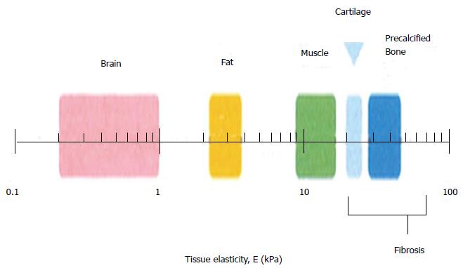
Figure 4 Schematic depicting the normal variation in elasticity of the indicated tissue[92].
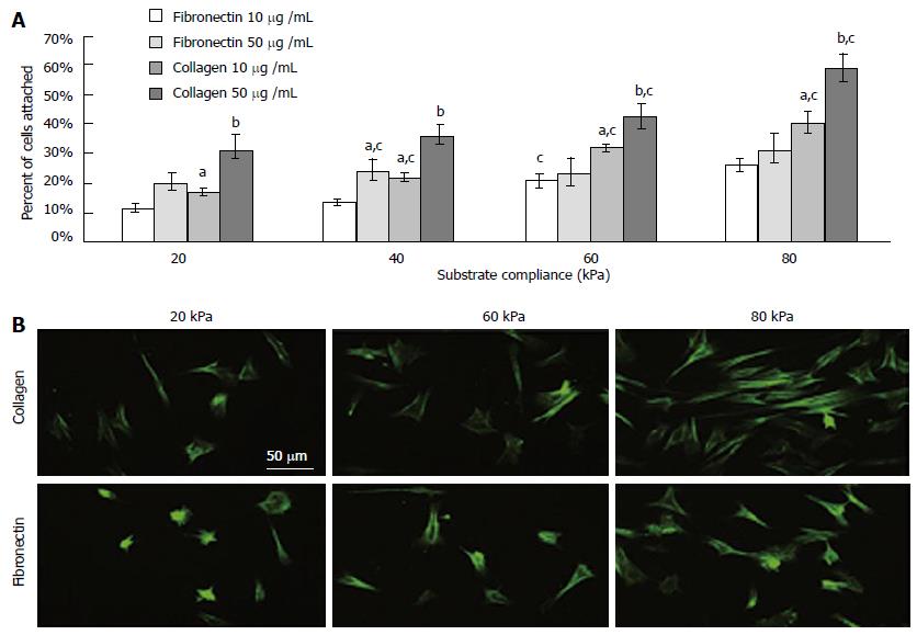
Figure 5 (A) As the rigidity increased, so did cell attachment.
Collagen elicited a greater degree of cell attachment, and increased bulk density of the ligand further enhanced this (B) Morphology on functionalized substrates at 8000 cells/cm2 for 24 h. astatistical difference compared to fibronectin at 10 mg/mL, bdifference compared to collagen at 10 mg/mL, cdifference compared to preceding substrate compliance. Cells were stained with FITC-phalloidin. (Scale bar represents 50 μm). Reproduced with permission from Sharma et al[109].
- Citation: Ghasemi-Mobarakeh L, Prabhakaran MP, Tian L, Shamirzaei-Jeshvaghani E, Dehghani L, Ramakrishna S. Structural properties of scaffolds: Crucial parameters towards stem cells differentiation. World J Stem Cells 2015; 7(4): 728-744
- URL: https://www.wjgnet.com/1948-0210/full/v7/i4/728.htm
- DOI: https://dx.doi.org/10.4252/wjsc.v7.i4.728


