Copyright
©The Author(s) 2015.
World J Stem Cells. May 26, 2015; 7(4): 691-699
Published online May 26, 2015. doi: 10.4252/wjsc.v7.i4.691
Published online May 26, 2015. doi: 10.4252/wjsc.v7.i4.691
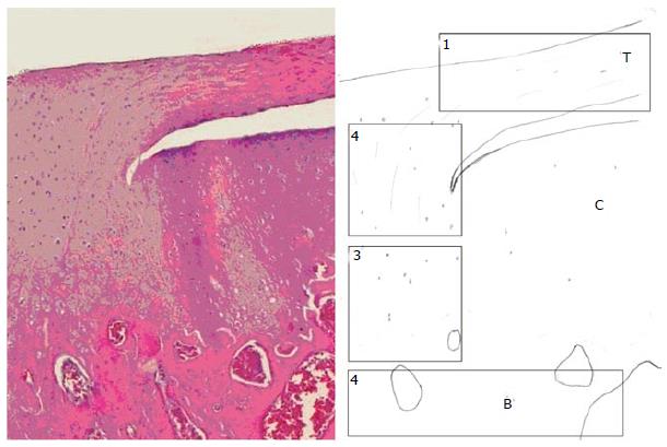
Figure 1 The normal enthesis (longitudinal image and diagram of the bone-tendon junction of the supraspinatus tendon of a rat; hematoxilin-eosine, × 10): the supraspinatus tendon (T) approaches the humeral bone (B) immediately adyacent to the normal carlilage (C).
The normal tendon (zone 1) gradually transforms into a fibrocartilaginous tissue with large mononucleated cells (zone 2). As the fibers progress into the bone the extracellular matrix is progresivelyy calcified (zone 3) until it turns into normal bone (zone 4). Further explanation of the biochemical environment of these zones is shown in Table 1.
- Citation: Mora MV, Ibán MAR, Heredia JD, Laakso RB, Cuéllar R, Arranz MG. Stem cell therapy in the management of shoulder rotator cuff disorders. World J Stem Cells 2015; 7(4): 691-699
- URL: https://www.wjgnet.com/1948-0210/full/v7/i4/691.htm
- DOI: https://dx.doi.org/10.4252/wjsc.v7.i4.691


