Copyright
©2014 Baishideng Publishing Group Inc.
World J Stem Cells. Nov 26, 2014; 6(5): 571-578
Published online Nov 26, 2014. doi: 10.4252/wjsc.v6.i5.571
Published online Nov 26, 2014. doi: 10.4252/wjsc.v6.i5.571
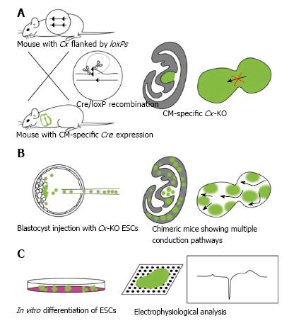
Figure 1 Cre/loxP-mediated tissue-specific knockout mouse models and analysis of embryonic stem cell differentiation.
Mutant cells and regions are shown in green. Mouse and heart drawings, respectively, constitute the middle and right pictures in (A) and (B). A: In the Cre/loxP model shown here, the connexin (Cx) gene, which when lost causes lethality, is deleted specifically in the CM. This results in relatively consistent delay or block in conduction[13,49]; B: Chimeric mice containing embryonic stem cell (ESCs) lacking the Cx43 gene. The example shown here reveals multiple conduction pathways in the heart[52]; C: ESCs can be differentiated in vitro. In this example, the induced CMs are subjected to planar multielectrode array analyses (middle); a typical extracellular recording data is shown in the right graph[53,67]. KO: Knockout
- Citation: Nishii K, Shibata Y, Kobayashi Y. Connexin mutant embryonic stem cells and human diseases. World J Stem Cells 2014; 6(5): 571-578
- URL: https://www.wjgnet.com/1948-0210/full/v6/i5/571.htm
- DOI: https://dx.doi.org/10.4252/wjsc.v6.i5.571


