Copyright
©2014 Baishideng Publishing Group Inc.
World J Stem Cells. Sep 26, 2014; 6(4): 391-403
Published online Sep 26, 2014. doi: 10.4252/wjsc.v6.i4.391
Published online Sep 26, 2014. doi: 10.4252/wjsc.v6.i4.391
Figure 1 Anatomy of the eye.
The cornea (A) comprises the colourless front portion of the eye immediately anterior to the iris and pupil (B). The limbus, located at the corneoscleral junction (B) is the transitional zone where the corneal and conjuctival epithelia merge, is shown in section using Haematoxalin and Eosin stain (C) and is considered a reservoir of stem cells which migrate centripetally to form the 5-7 cell layer corneal epithelium (DAPI fluorescence to highlight cell nuclei in corneal section, D).
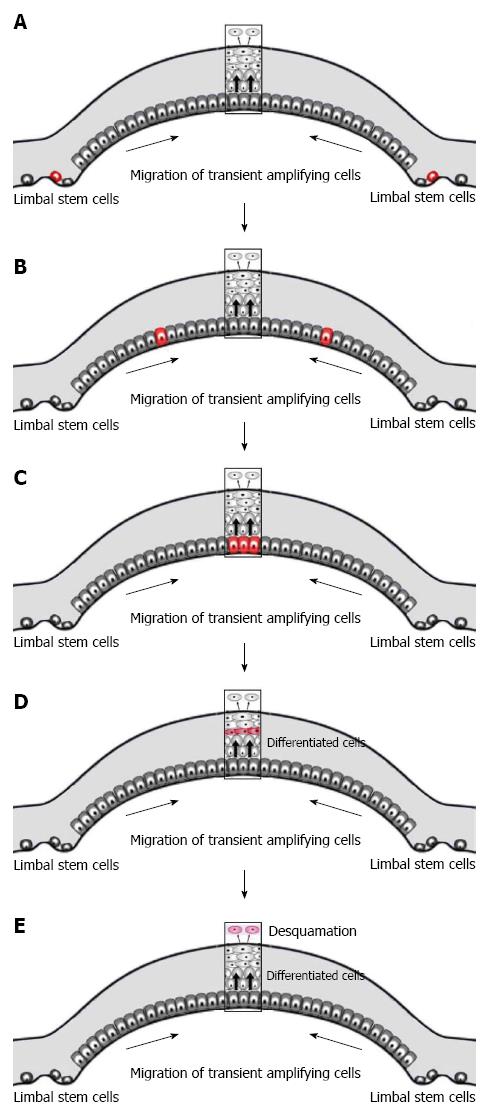
Figure 2 The X, Y, Z hypothesis of corneal maintenance.
Limbal stem cells at the peripheral cornea divide and give rise to transient amplifying cells (TACs) (A). These TACs migrate centripetally through the basal epithelium (B) and undergo a limited number of divisions on the central cornea (C). The differentiated daughter cells move anteriorly to replenish the upper layers of the cornea (D) where they are eventually shed from the corneal surface (E). Hence the sum of X (proliferation and anterior migration) and Y (centripetal migration) must equal Z (desquamation of superficial cells) for corneal maintenance. Red cells: Continuum of transient amplifying migrating and/or differentiated cells.
- Citation: Yoon JJ, Ismail S, Sherwin T. Limbal stem cells: Central concepts of corneal epithelial homeostasis. World J Stem Cells 2014; 6(4): 391-403
- URL: https://www.wjgnet.com/1948-0210/full/v6/i4/391.htm
- DOI: https://dx.doi.org/10.4252/wjsc.v6.i4.391


