Copyright
©The Author(s) 2025.
World J Stem Cells. Jan 26, 2025; 17(1): 101485
Published online Jan 26, 2025. doi: 10.4252/wjsc.v17.i1.101485
Published online Jan 26, 2025. doi: 10.4252/wjsc.v17.i1.101485
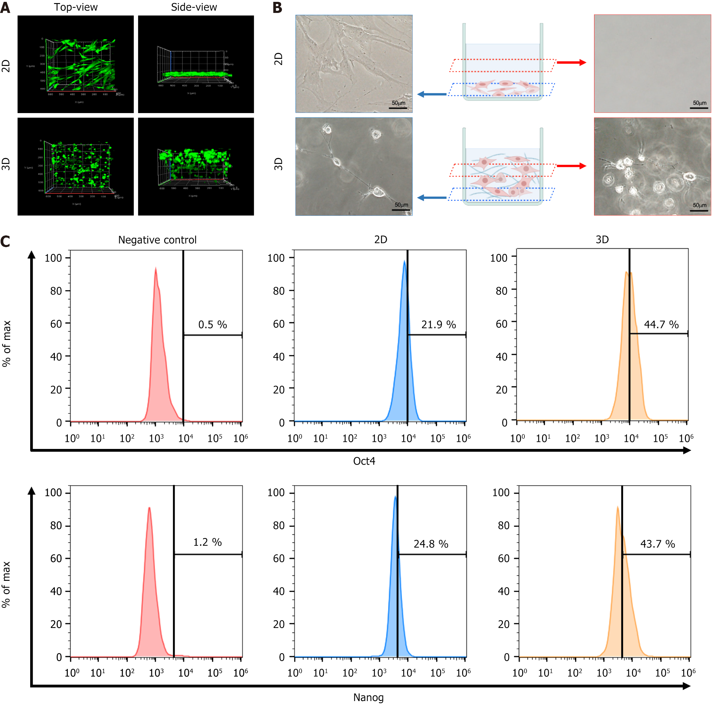
Figure 1 Functional polymer-based three-dimensional culture enhances stemness.
A: Difference between distribution of mesenchymal stromal cells in the two-dimensional (2D) and 3D culture systems; B: Cell morphology of mesenchymal stromal cells cultured in 2D and 3D. Scale bars represent 50 μm; C: Flow cytometry analysis of stemness markers octamer-binding transcription factor 4 and Nanog homeobox in 2D and 3D. Representative histogram with geometric mean fluorescence intensity were shown. 2D: Two-dimensional; 3D: Three-dimensional; Oct4: Octamer-binding transcription factor 4; Nanog: Nanog homeobox.
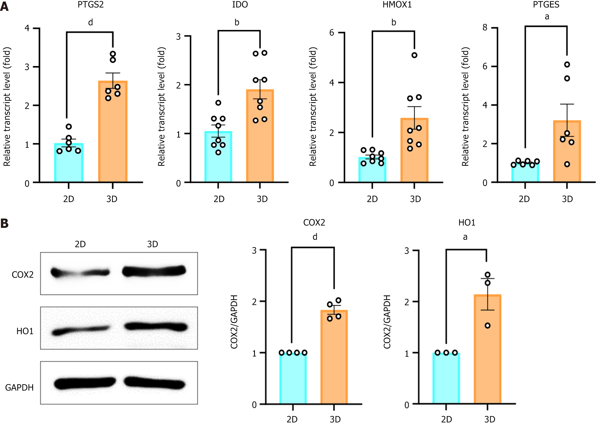
Figure 2 Three-dimensional-cultured mesenchymal stromal cells increase the release of immunomodulation related cytokines.
A: Relative mRNA level of prostaglandin-endoperoxide synthase 2, indoleamine-2,3-dioxygenase, heme oxygenase 1, and prostaglandin E synthase after 24 hours incubation. Expression levels were normalized to two-dimensional (2D); B: Immunoblot analysis measured the protein expression of cyclooxygenase-2 and heme-oxygenase 1 after 24 hours incubation in 2D and 3D (left) and graph showing quantification of the expression level of each protein compared to the expression of glyceraldehyde-3-phosphate dehydrogenase (right). n = 3-4 independent experiments, unpaired t-test, aP < 0.05, bP < 0.01, dP < 0.0001. 2D: Two-dimensional; 3D: Three-dimensional; PTGS2: Prostaglandin-endoperoxide synthase 2; IDO: Indoleamine-2,3-dioxygenase; HMOX1: Heme oxygenase 1; PTGES: Prostaglandin E synthase; COX2: Cyclooxygenase-2; HO1: Heme-oxygenase 1; GAPDH: Glyceraldehyde-3-phosphate dehydrogenase.
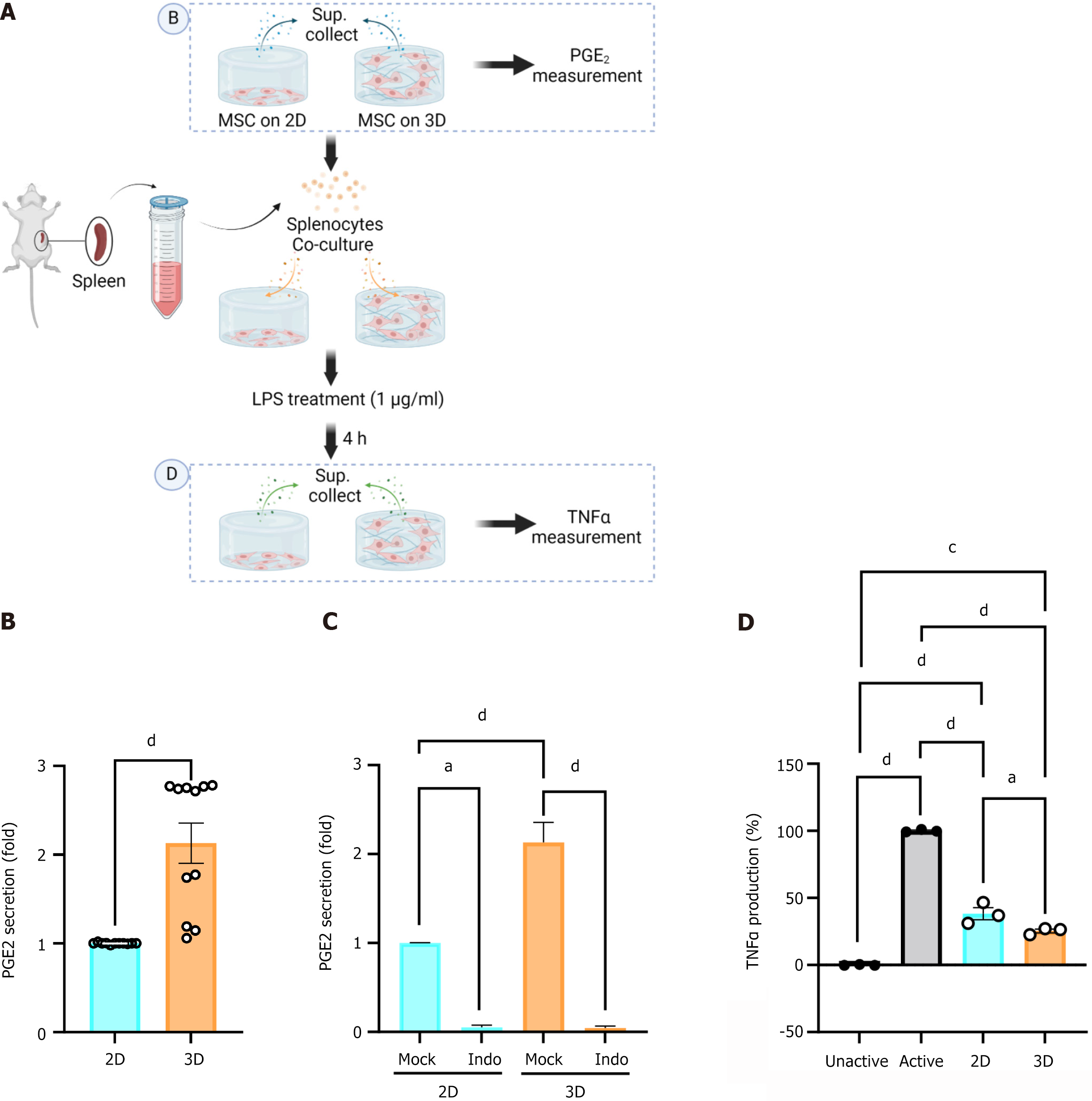
Figure 3 Three-dimensional-cultured mesenchymal stromal cells elevate the secretion of prostaglandin E2 and suppress tumor necrosis factor-alpha from lipopolysaccharide-stimulated splenocytes.
A: Schematic of the method for collection of supernatant from mesenchymal stromal cells (MSCs) or co-cultured MSCs and lipopolysaccharide-stimulated murine splenocytes; B: Secreted prostaglandin E2 (PGE2) in a MSC culture medium was determined by enzyme linked immunosorbent assay (ELISA) (n = 4 independent experiments, unpaired t-test, dP < 0.0001); C: MSCs co-cultured with splenocytes were treated with 10 mmol/L indomethacin (marked as Indo) and secreted PGE2 was assessed using ELISA (n = 3 independent experiment, one-way ANOVA, aP < 0.05, dP < 0.0001); D: Secreted tumor necrosis factor alpha from MSCs co-cultured with murine splenocytes was assessed by ELISA (n = 3 independent experiment, one-way ANOVA, aP < 0.05, cP < 0.001, dP < 0.0001). 2D: Two-dimensional; 3D: Three-dimensional; PGE2: Prostaglandin E2; LPS: Lipopolysaccharide; TNFα: Tumor necrosis factor alpha.
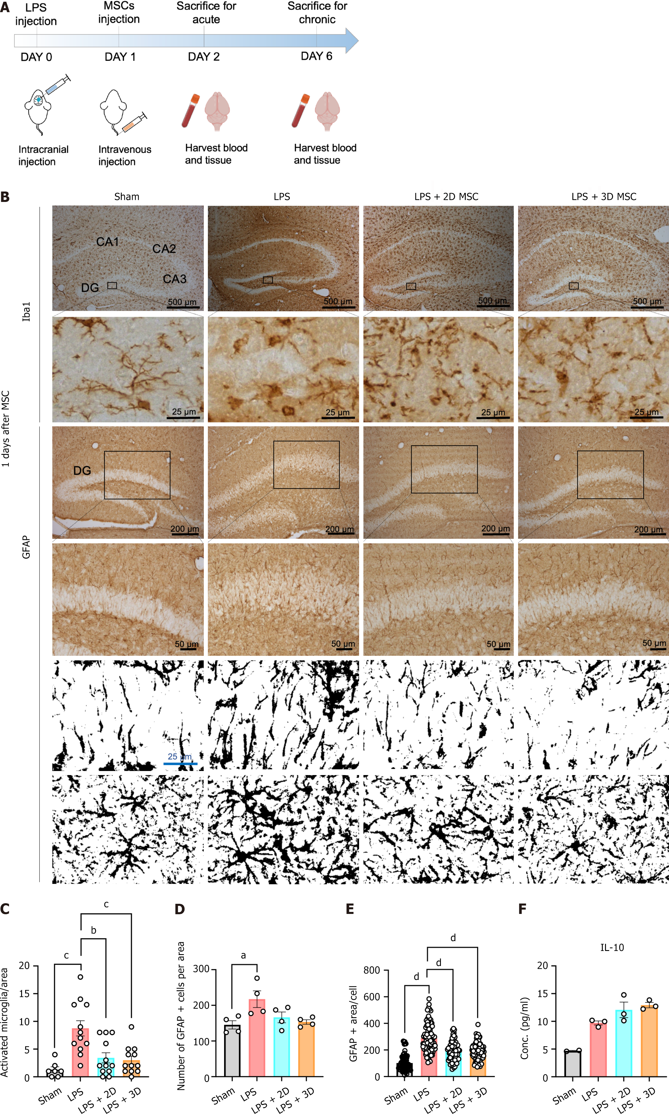
Figure 4 Application of both two-dimensional and three-dimensional-cultured mesenchymal stromal cells reduces the inflammatory response in murine acute lipopolysaccharide-induced neuroinflammation model.
A: Representation of the experimental scheme; B: Immunohistochemistry staining of ionized calcium-binding adaptor molecule 1, microglia marker, and glial fibrillary acidic protein (GFAP), an astrocyte marker, in the hippocampal region after induction of acute neuroinflammation with lipopolysaccharide. Enlarged inset windows represent a small representative portion of top layer in each staining. The black/white images of GFAP were contrasted by flipping the stained and unstained sections to black and white. Scale bars represent 25 μm, 50 μm, 200 μm, 500 μm; C: Quantify the area of activated microglia in acute model (n = 3-4, Kruskal-Wallis One-way ANOVA with Dunnett’s multiple comparisons, bP < 0.01, cP < 0.001); D: Quantify the number of positive GFAP cells per area in acute model (n = 4, Kruskal-Wallis One-way ANOVA with Dunnett’s multiple comparisons, aP < 0.05); E: Quantify the area of positive GFAP per cell in acute model (n = 3-4, Kruskal-Wallis One-way ANOVA with Dunnett’s multiple comparisons, dP < 0.0001); F: Measurement of plasma interleukin-10, an anti-inflammatory cytokine, in the blood of acute inflammation model by enzyme linked immunosorbent assay (n = 3 independent experiment, Kruskal-Wallis One-way ANOVA with Dunnett’s multiple comparisons). LPS: Lipopolysaccharide; MSC: Mesenchymal stromal cell; Iba1: Ionized calcium-binding adaptor molecule 1; GFAP: Glial fibrillary acidic protein; 2D: Two-dimensional; 3D: Three-dimensional; IL: Interleukin.
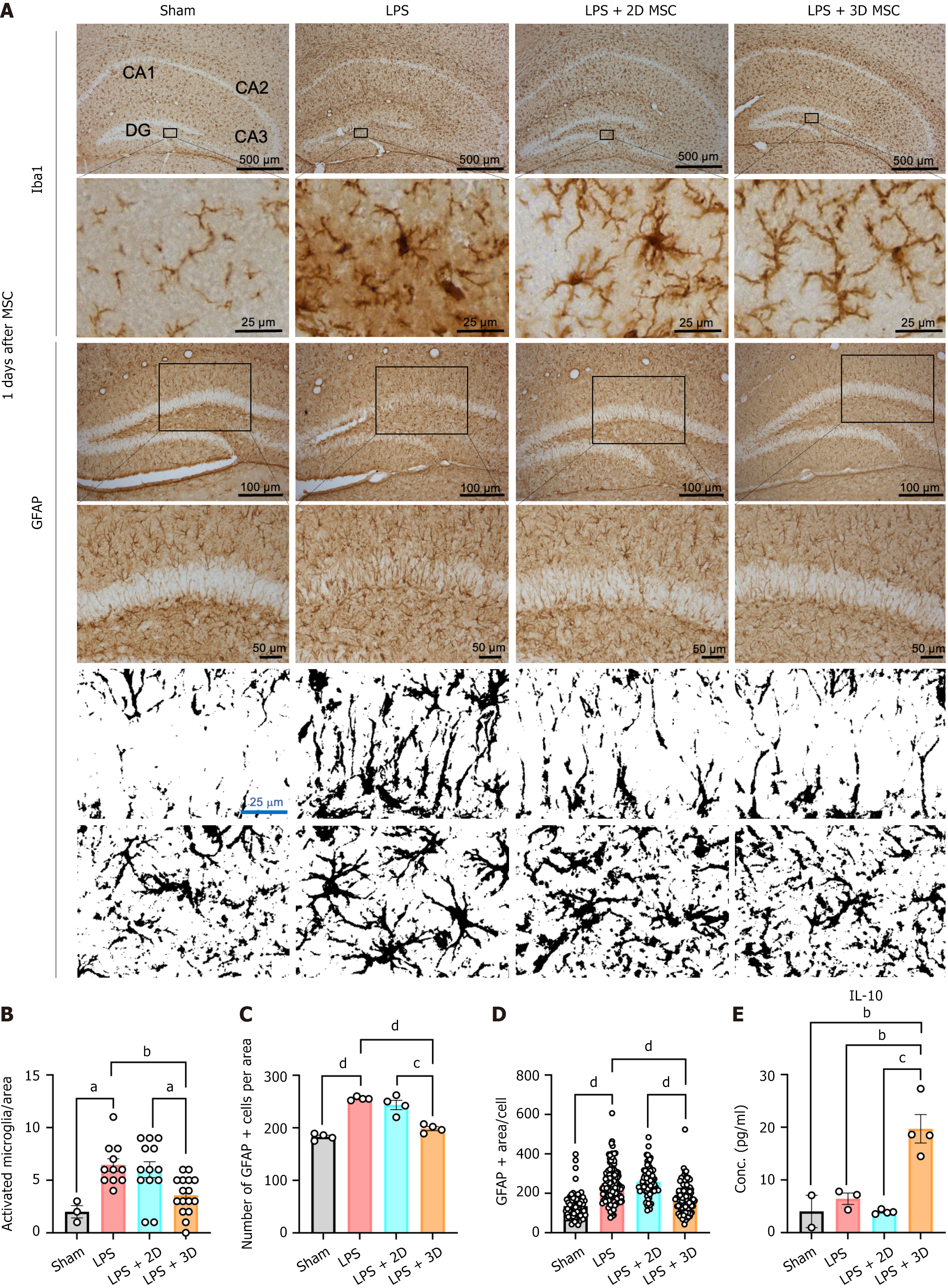
Figure 5 Application of three-dimensional-cultured mesenchymal stromal cells reduces the inflammatory response in murine chronic lipopolysaccharide-induced neuroinflammation model.
A: Immunostaining of ionized calcium-binding adaptor molecule 1 (microglial marker), and GFAP (astrocyte marker), in the hippocampal region after induction of chronic neuroinflammation with lipopolysaccharide. Enlarged inset windows represent a small representative portion of top layer in each staining. The black/white images of GFAP were contrasted by flipping the stained and unstained sections to black and white; B: Quantification of the area of activated microglia in a chronic neuroinflammation model. Cell counts were obtained from 3-4 images per mouse (n = 3-4, Kruskal-Wallis One-way ANOVA with Dunnett’s multiple comparisons, aP < 0.05, bP < 0.01); C: Quantification of the number of positive GFAP cells per area (n = 4, Kruskal-Wallis One-way ANOVA with Dunnett’s multiple comparisons, cP < 0.001, dP < 0.0001); D: Quantification of the area of positive GFAP per cell (n = 3-4, Kruskal-Wallis One-way ANOVA with Dunnett’s multiple comparisons, dP < 0.0001); E: Measurement of plasma interleukin-10, an anti-inflammatory cytokine, in the blood of chronic inflammation model by enzyme linked immunosorbent assay (n = 4, Kruskal-Wallis One-way ANOVA with Dunnett’s multiple comparisions, bP < 0.01, cP < 0.001). LPS: Lipopolysaccharide; MSC: Mesenchymal stromal cell; Iba1: Ionized calcium-binding adaptor molecule 1; GFAP: Glial fibrillary acidic protein; 2D: Two-dimensional; 3D: Three-dimensional; IL: Interleukin.
- Citation: Kim OH, Kang H, Chang ES, Lim Y, Seo YJ, Lee HJ. Extended protective effects of three dimensional cultured human mesenchymal stromal cells in a neuroinflammation model. World J Stem Cells 2025; 17(1): 101485
- URL: https://www.wjgnet.com/1948-0210/full/v17/i1/101485.htm
- DOI: https://dx.doi.org/10.4252/wjsc.v17.i1.101485


