Copyright
©The Author(s) 2024.
World J Stem Cells. Aug 26, 2024; 16(8): 811-823
Published online Aug 26, 2024. doi: 10.4252/wjsc.v16.i8.811
Published online Aug 26, 2024. doi: 10.4252/wjsc.v16.i8.811
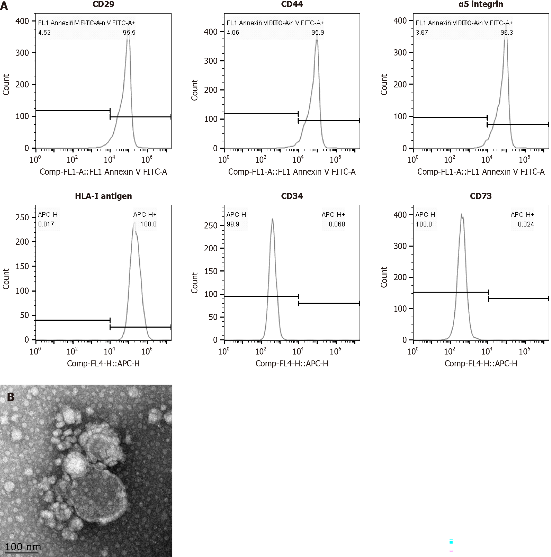
Figure 1 Detection of mesenchymal stromal cell-derived microvesicle markers by flow cytometry and transmission and scanning electron microscopy.
A: The expression of surface molecules (CD29, CD44, α5 integrins and the HLA-I antigen) was positive, whereas the expression of CD34 and CD73 was negative; B: Transmission and scanning electron microscopy were performed on purified mesenchymal stromal cell-derived microvesicles to reveal their spheroid morphologies and confirm their sizes.
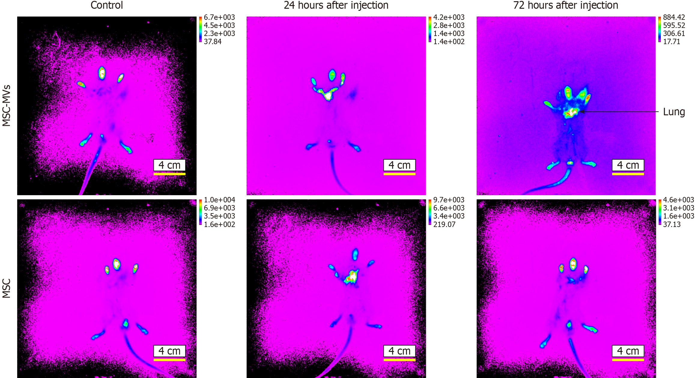
Figure 2 Microvesicles derived from mesenchymal stromal cells homing detection by the near-infrared dye NIR815.
At 24 hours after injection with mesenchymal stromal cell-derived microvesicles (MSC-MVs), the fluorescence appears mainly yellow in lung, and at 72 hours after injection, the fluorescence appears mainly yellow and red with significantly increased fluorescence intensity.
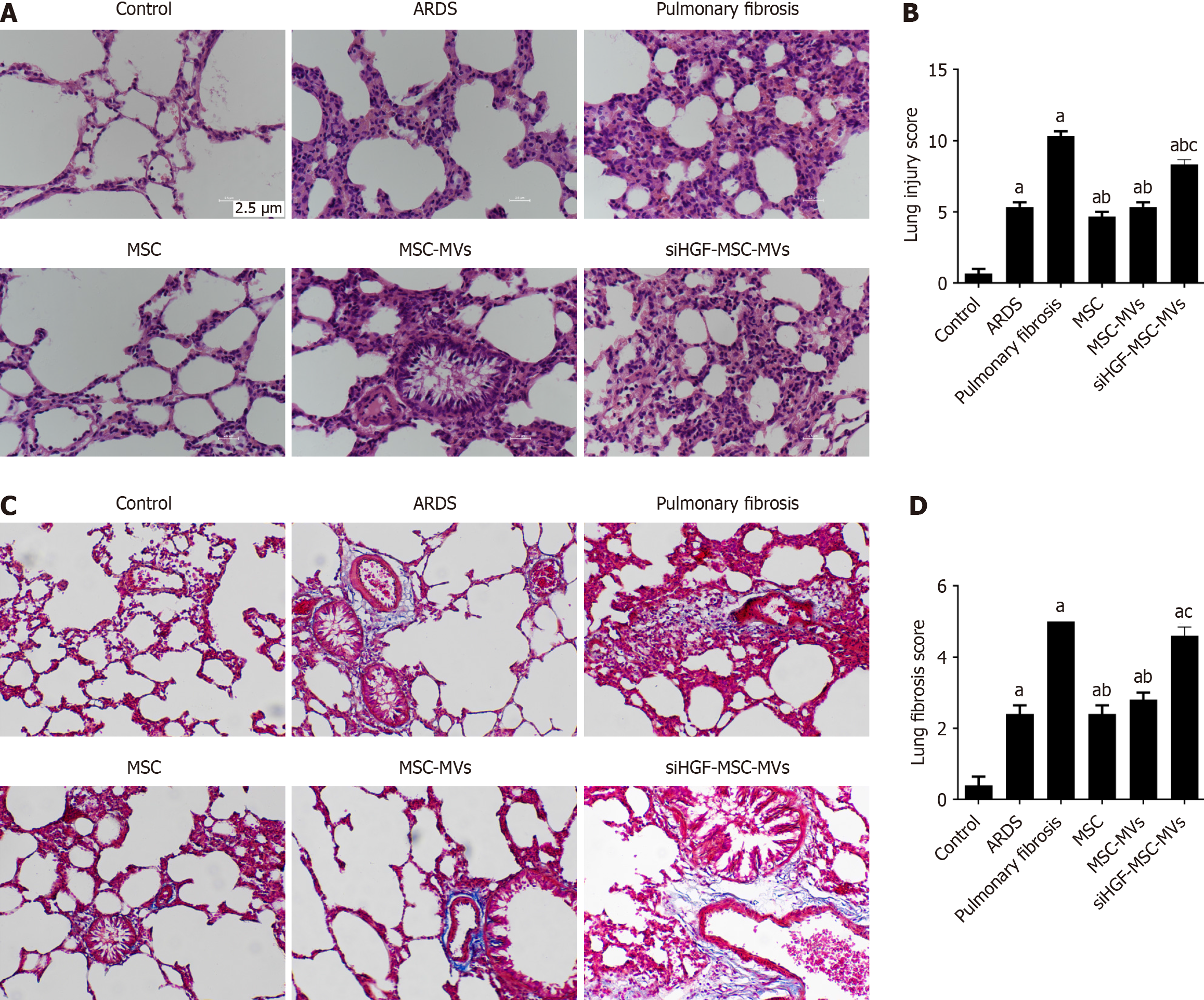
Figure 3 Immunohistochemical detection of the effects of mesenchymal stromal cell-derived microvesicles on pulmonary and fibrosis-related indicators in acute respiratory distress syndrome pulmonary fibrosis mouse models.
A: Hematoxylin and eosin staining of lung tissues from each group; B: Lung injury scores of pathological sections from each group; C: Masson staining of lung tissues from each group; D: Lung fibrosis scores of pathological sections from each group. aP < 0.05, vs the control group; bP < 0.05, vs the pulmonary fibrosis group; cP < 0.05, vs the mesenchymal stromal cell-derived microvesicles (MSC-MVs) group (n = 6). Control, acute respiratory distress syndrome (ARDS), pulmonary fibrosis, MSC, MSC-MVs and low hepatocyte growth factor (HGF)-MSC-MVs represent the control group, ARDS group, pulmonary fibrosis group, MSC group, MSC-MVs group and low HGF-MSC-MVs group, respectively.
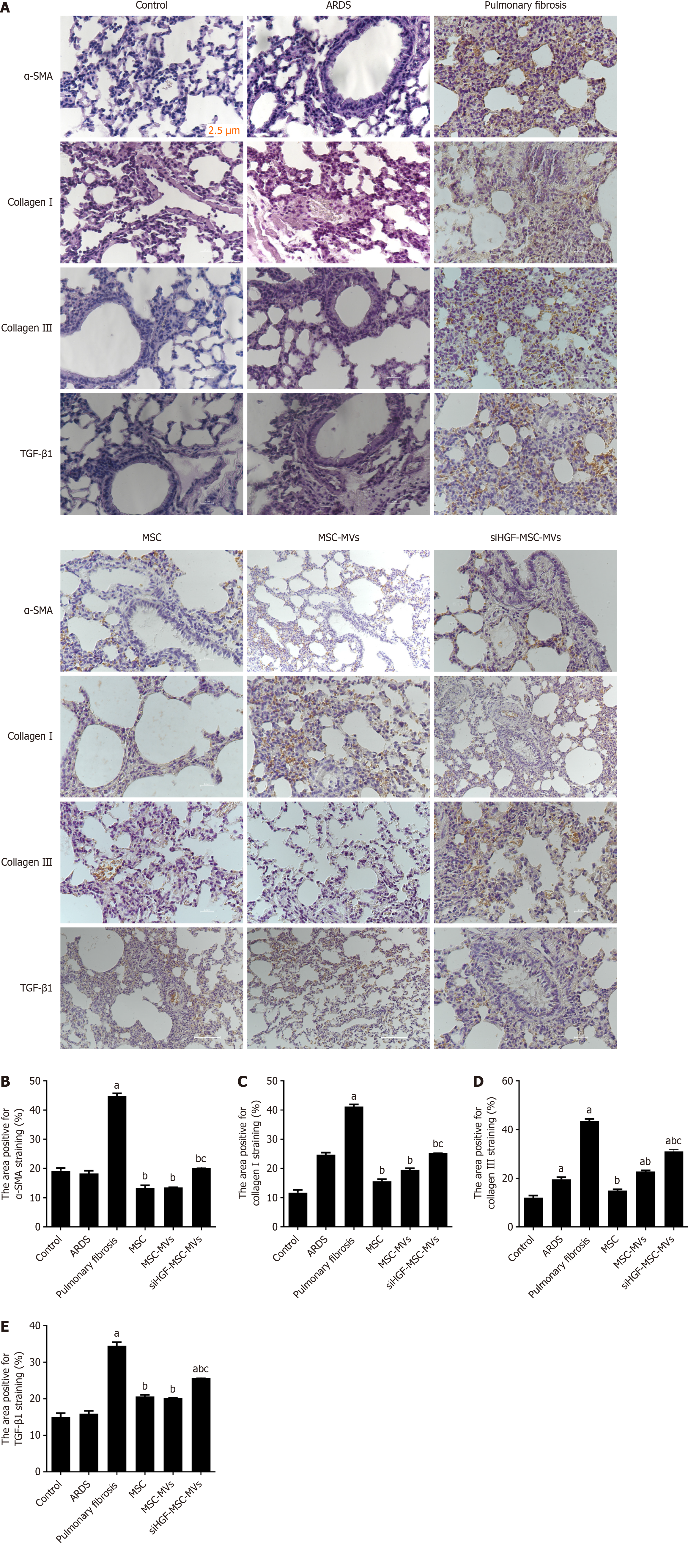
Figure 4 Effects of mesenchymal stromal cell-derived microvesicles on acute respiratory distress syndrome-related pulmonary fibrosis and lung injury.
A: Immunohistochemical images of pathological sections of lung tissues from each group; B: α-smooth muscle actin (α-SMA) protein expression; C: Type I collagen protein expression; D: Type III collagen protein expression; E: Transforming growth factor-β1 (TGF-β1) protein expression. aP < 0.05, vs the control group; bP < 0.05 vs the pulmonary fibrosis group; cP < 0.05 vs the mesenchymal stromal cell-derived microvesicles (MSC-MVs) group (n = 6). Control, acute respiratory distress syndrome (ARDS), pulmonary fibrosis, MSC, MSC-MVs and low hepatocyte growth factor (HGF)-MSC-MVs represent the control group, ARDS group, pulmonary fibrosis group, MSC group, MSC-MVs group and low HGF-MSC-MVs group, respectively.
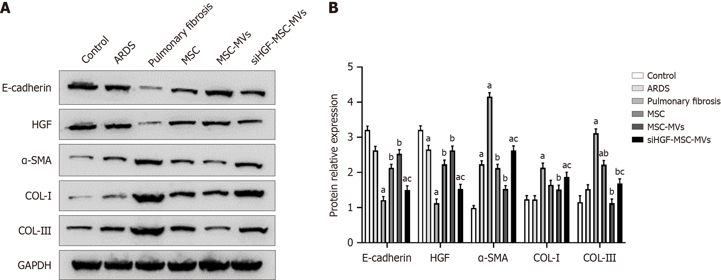
Figure 5 Western blot analysis of the effects of mesenchymal stromal cell-derived microvesicles on pulmonary fibrosis-related proteins in acute respiratory distress syndrome pulmonary fibrosis model mice.
A: Western blot bands of fibrosis-related proteins in the lung tissues of each group; B: Relative expression of fibrosis-related proteins. aP < 0.05, vs the control group; bP < 0.05, vs the pulmonary fibrosis group; cP < 0.05, vs the mesenchymal stromal cell-derived microvesicles (MSC-MVs) group (n = 6). Control, acute respiratory distress syndrome (ARDS), pulmonary fibrosis, MSC, MSC-MVs and low hepatocyte growth factor (HGF)-MSC-MVs represent the control group, ARDS group, pulmonary fibrosis group, MSC group, MSC-MVs group and low HGF-MSC-MVs group, respectively. α-SMA: α-smooth muscle actin; COL: Collagen.
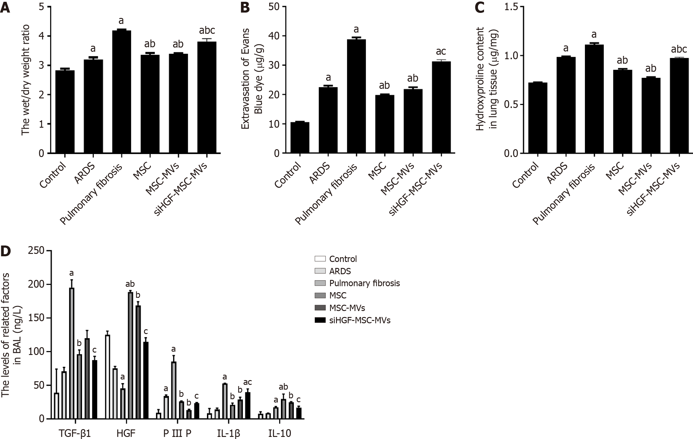
Figure 6 Effects of mesenchymal stromal cell-derived microvesicles on pulmonary vascular endothelial permeability and related proteins in acute respiratory distress syndrome pulmonary fibrosis mouse models.
A: Lung wet/dry weight ratios for each group; B: Evans blue detection of vascular endothelial permeability in each group; C: Hydroxyproline levels in each group; D: Expression of inflammatory factors in each group detected by ELISA. aP < 0.05, vs the control group; bP < 0.05, vs the pulmonary fibrosis group; cP < 0.05, vs the mesenchymal stromal cell-derived microvesicles (MSC-MVs) group (n = 6). Control, acute respiratory distress syndrome (ARDS), pulmonary fibrosis, MSC, MSC-MVs and low hepatocyte growth factor (HGF)-MSC-MVs represent the control group, ARDS group, pulmonary fibrosis group, MSC group, MSC-MVs group and low HGF-MSC-MVs group, respectively. IL: Interleukin; PIIIP: Type III procollagen N-terminal aminopeptide; TGF-β1: Transforming growth factor-β1.
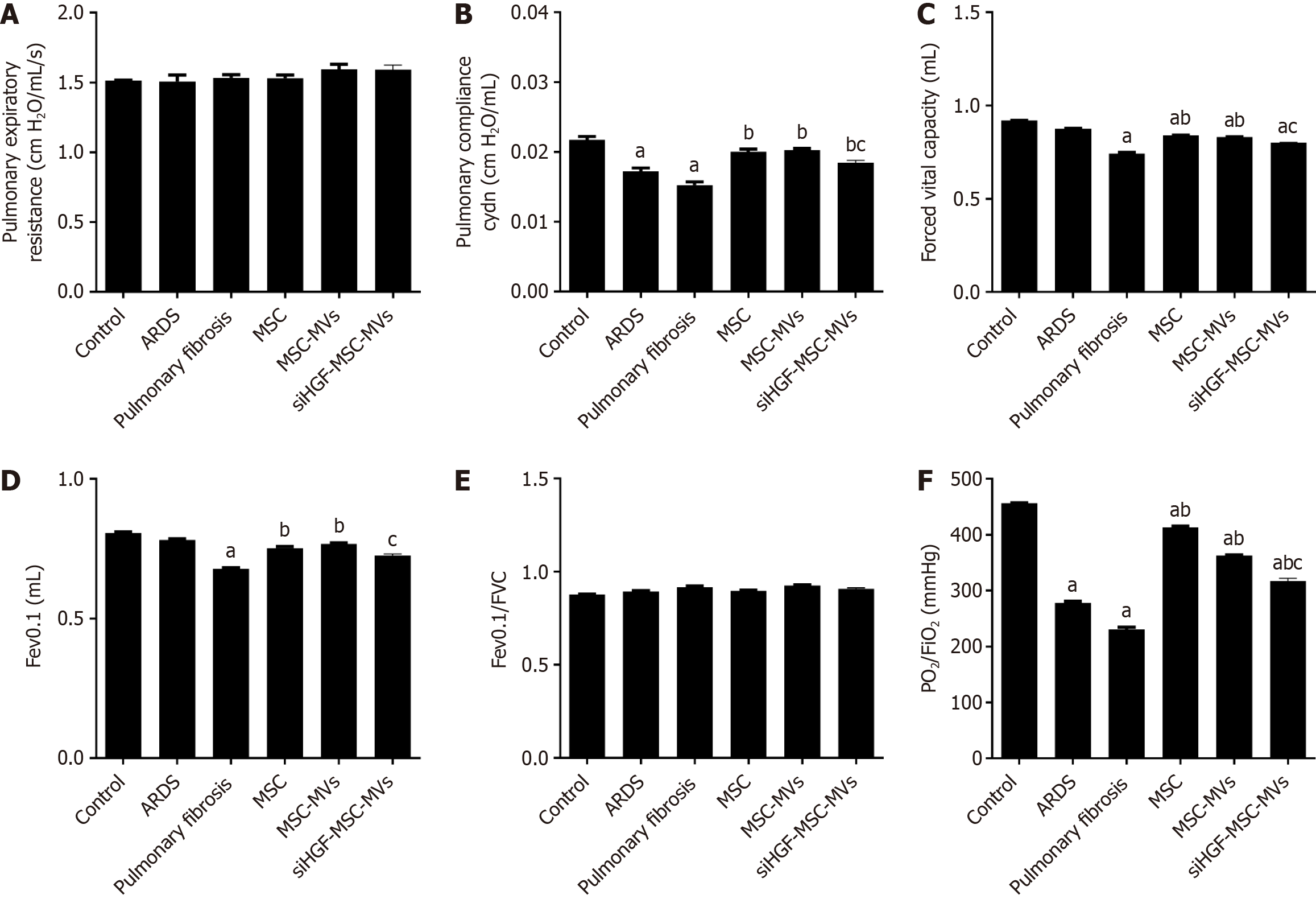
Figure 7 Effects of mesenchymal stromal cell-derived microvesicles on respiratory mechanics and lung functions in acute respiratory distress syndrome pulmonary fibrosis mouse models.
A: Pulmonary expiratory resistance; B: Pulmonary compliance; C: Forced expiratory volume in 0.1 seconds (Fev0.1); D: Forced vital capacity (FVC), E: Fev0.1/FVC; F: Pressure of oxygen (PO2)/oxygen inhalation (FiO2); G: Transmission and scanning electron microscopy were performed on purified mesenchymal stromal cell-derived microvesicles (MSC-MVs) to reveal their spheroid morphologies and confirm their sizes. aP < 0.05, vs the control group; bP < 0.05, vs the pulmonary fibrosis group; cP < 0.05, vs the MSC-MVs group (n = 6). Control, acute respiratory distress syndrome (ARDS), pulmonary fibrosis, MSC, MSC-MVs and low hepatocyte growth factor (HGF)-MSC-MVs represent the control group, ARDS group, pulmonary fibrosis group, MSC group, MSC-MVs group and low HGF-MSC-MVs group, respectively.
- Citation: Chen QH, Zhang Y, Gu X, Yang PL, Yuan J, Yu LN, Chen JM. Microvesicles derived from mesenchymal stem cells inhibit acute respiratory distress syndrome-related pulmonary fibrosis in mouse partly through hepatocyte growth factor. World J Stem Cells 2024; 16(8): 811-823
- URL: https://www.wjgnet.com/1948-0210/full/v16/i8/811.htm
- DOI: https://dx.doi.org/10.4252/wjsc.v16.i8.811


