Copyright
©The Author(s) 2024.
World J Stem Cells. May 26, 2024; 16(5): 486-498
Published online May 26, 2024. doi: 10.4252/wjsc.v16.i5.486
Published online May 26, 2024. doi: 10.4252/wjsc.v16.i5.486
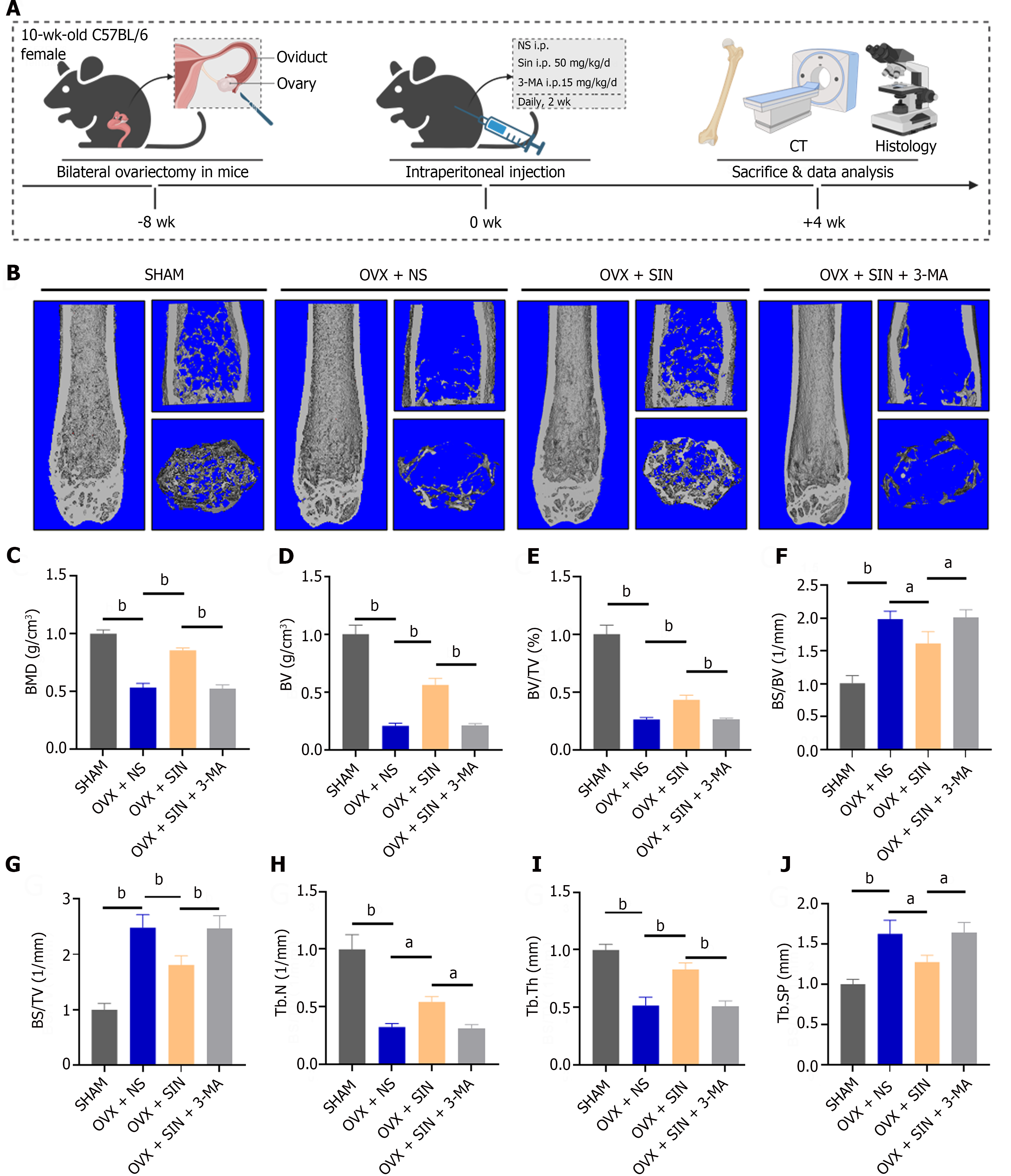
Figure 1 Sinomenine ameliorates bone loss in ovariectomy mice.
A: Schematic diagram of the experimental design for assessing the effects of sinomenine (SIN) and 3-methyladenine (3-MA) in ovariectomized (OVX) mice; B: Representative 3D micron-scale computed tomography reconstruction images of trabecular bone under the distal femur growth plate in the sham group, OVX + NS group, OVX + SIN group, and OVX + SIN + 3-MA group; C-J: Quantitative analysis of bone parameters. The region of interest selected for trabecular analysis started 100 sections below the proximal end of the distal femur growth plate, and 150 slices (6 μm each) were read per sample. Bone mineral density (g/cm3) (C), bone volume (BV) (mm3) (D), bone volume fractions (%) (E), bone surface/volume ratio (1/mm) (F), bone surface/volume ratio (1/mm) (G), trabecular number (1/mm) (H), trabecular thickness (mm) (I), trabecular separation (mm) (J). aP < 0.05, bP < 0.01, n = 6, biologically independent mice per group, all data were normalized relative to the sham group. OVX: Ovariectomized; SIN: Sinomenine; 3-MA: 3-methyladenine; BMD: Bone mineral density; BV: Bone volume; BV/TV: bone volume fractions; BS/BV: Bone surface/volume ratio; BS/TV: Bone surface/volume ratio; Tb.N: Trabecular number; Tb.Th: Trabecular thickness; Tb.Sp: Trabecular separation.
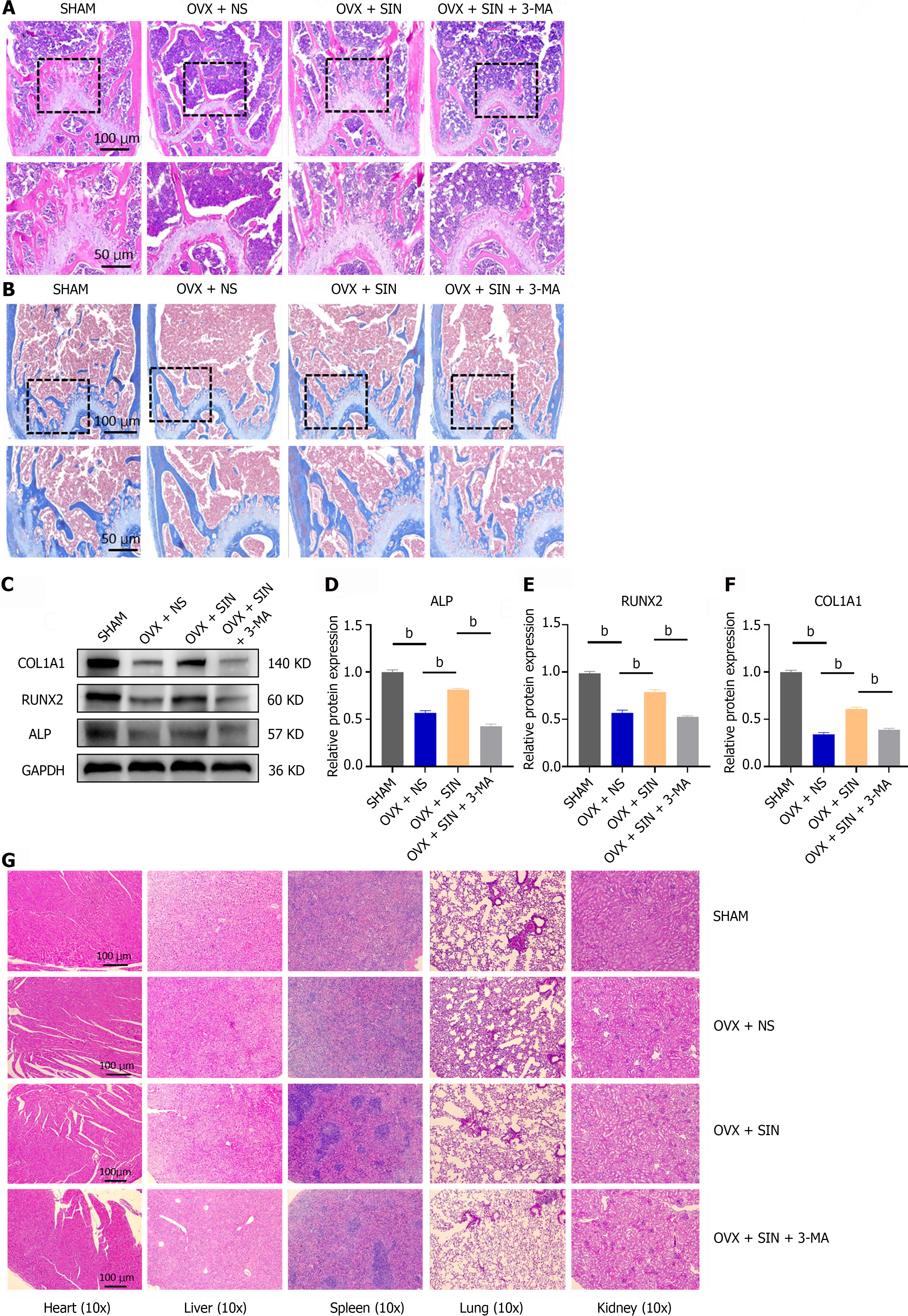
Figure 2 Morphology, quantitative protein assay of femur and visceral toxicity assays.
A: Representative hematoxylin and eosin (H&E) staining images of trabecular bone under the distal femur growth plate from the sham group, ovariectomized (OVX) + NS group, OVX + sinomenine (SIN) group, and OVX + SIN + 3-methyladenine (3-MA) group; B: Representative Masson staining images of trabecular bone under the distal femur growth plate from each group; C-F: Protein levels of ALP, RUNX2 and COL1A1 in mouse bone tissues; G: Effects of SIN and 3-MA on organ tissues. H&E staining was performed on heart, lung, liver, spleen and kidney sections. No changes in organ structure were observed, indicating that the administration of SIN and 3-MA had no harmful effects on organ tissues in this study. Three fields were randomly selected for each sample, and 5 biologically independent mouse samples were used in each group. n = 3 per group. Values are shown as the mean ± SD. bP < 0.01, all data were normalized relative to the sham group. OVX: Ovariectomized; SIN: Sinomenine; 3-MA: 3-methyladenine.
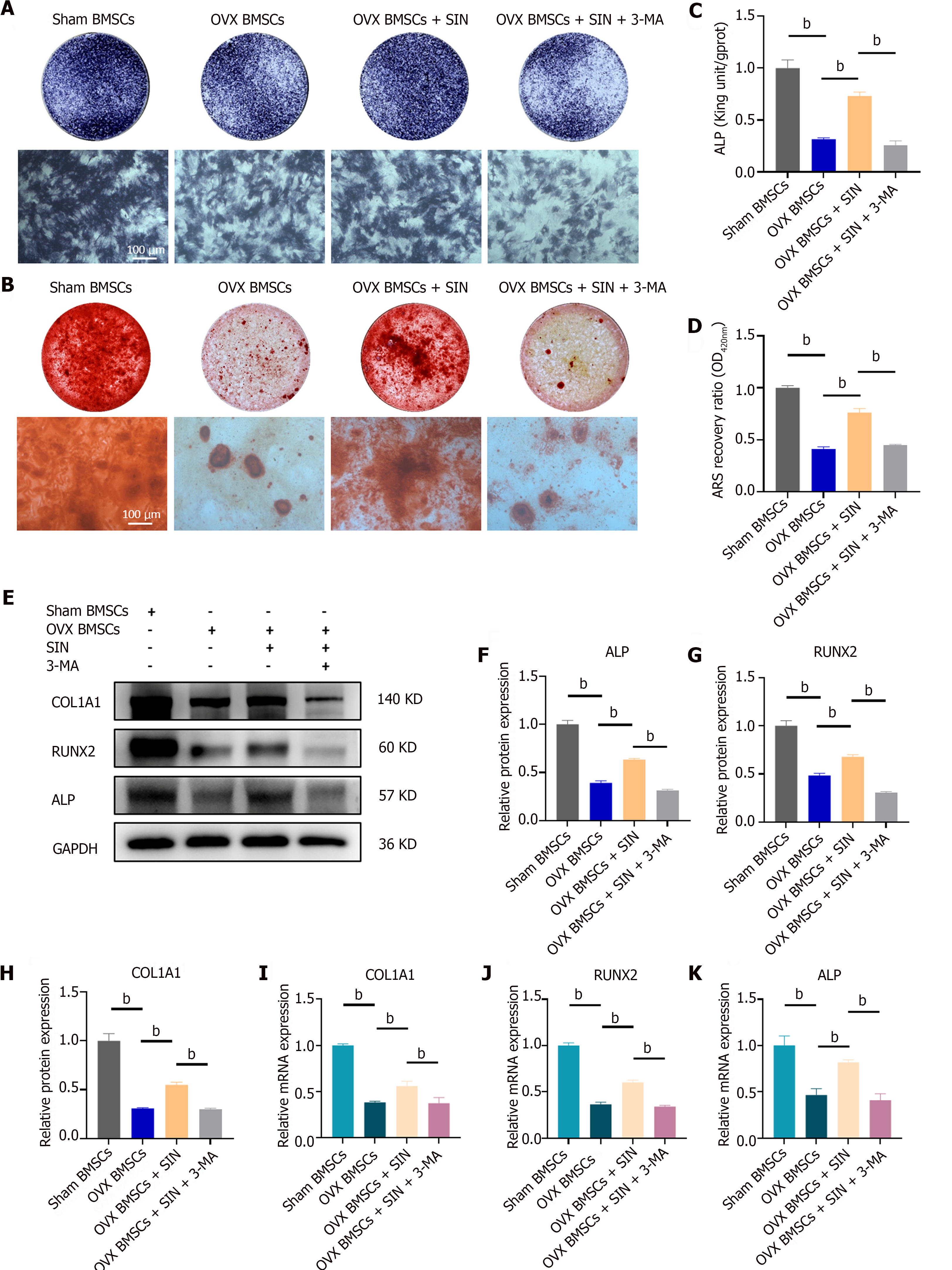
Figure 3 Sinomenine promotes osteogenic differentiation of bone marrow mesenchymal stromal cells but can be reversed by 3-methyladenine.
A: Representative images of ALP staining at 7 d; B: Representative images of Alizarin red S (ARS) staining at 21 d; C and D: Quantitative analysis of ALP activity and the ARS recovery ratio; E-H: Protein levels of ALP, RUNX2 and COL1A1 after sinomenine (SIN) and 3-methyladenine treatment for five days; I-K: Runx2, ALP and COL1A1 mRNA levels after SIN treatment for five days. n = 3 per group. The three groups were cultured in osteogenic medium, and the values are shown as the mean ± SD. aP < 0.01, all data were normalized relative to the sham bone marrow mesenchymal stromal cell group. OVX: Ovariectomized; SIN: Sinomenine; 3-MA: 3-methyladenine; BMSC: Bone marrow mesenchymal stromal cell.
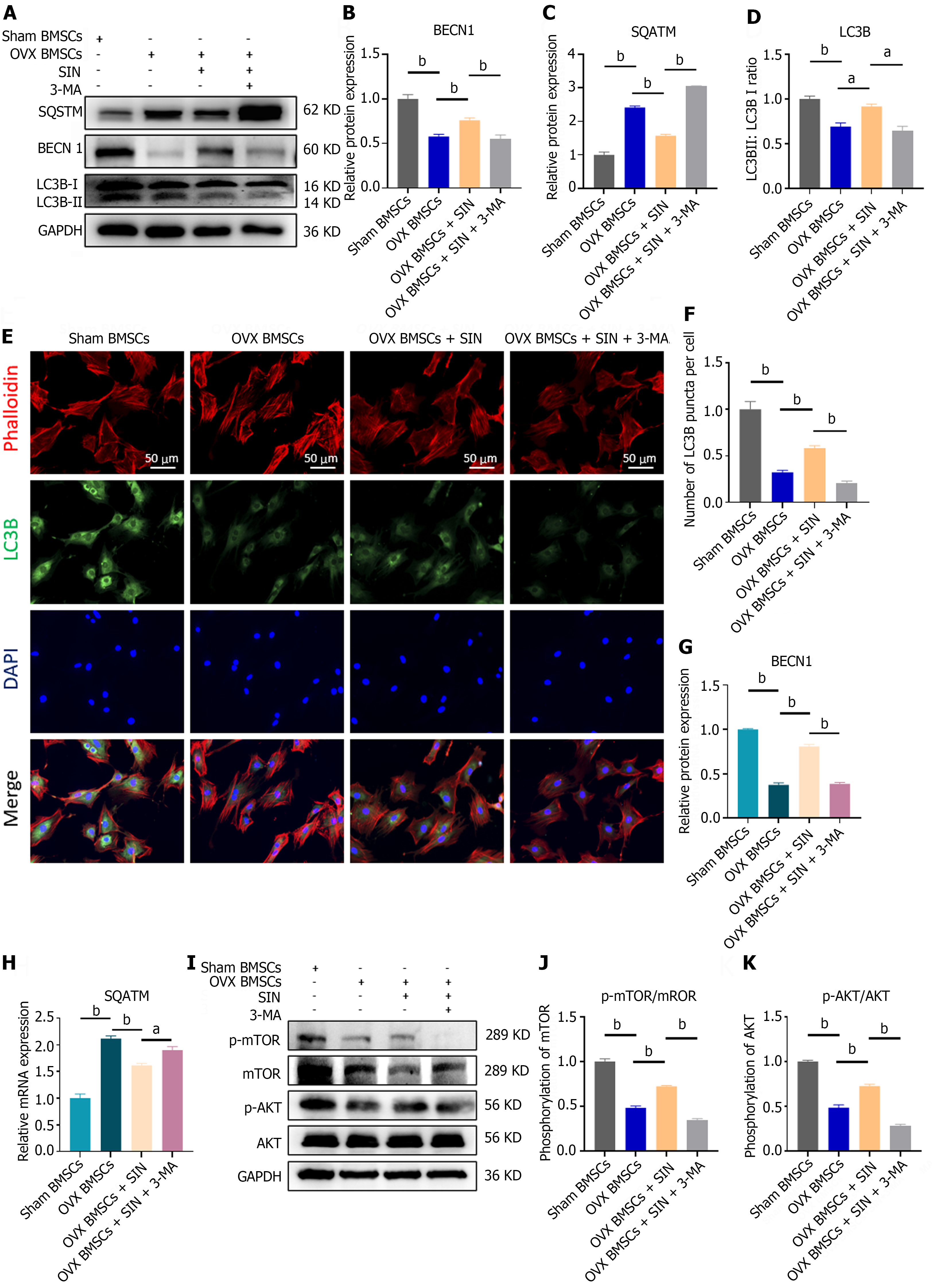
Figure 4 Sinomenine upregulates autophagy in bone marrow mesenchymal stromal cells through the phosphatidylinositol 3-kinase/AKT/mammalian target of the rapamycin pathway.
A-D: Protein levels of SQSTM, BECN1 and LC3B II/LC3B I after sinomenine (SIN) treatment (12 h); E and F: Quantitative analysis of the average fluorescence intensity of LC3B; G and H: BECN1 and SQSTM mRNA levels after SIN and 3-methyladenine (3-MA) treatment (12 h); I-K: Protein levels of phosphatidylinositol 3-kinase and AKT and mammalian target of the rapamycin phosphorylation levels after SIN and 3-MA treatment (12 h). n = 3 per group. Values are shown as the means ± SD. aP < 0.05, bP < 0.01, all data were normalized relative to the sham bone marrow mesenchymal stromal cell group. OVX: Ovariectomized; SIN: Sinomenine; 3-MA: 3-methyladenine; BMSC: Bone marrow mesenchymal stromal cell; mTOR: Mammalian target of the rapamycin.
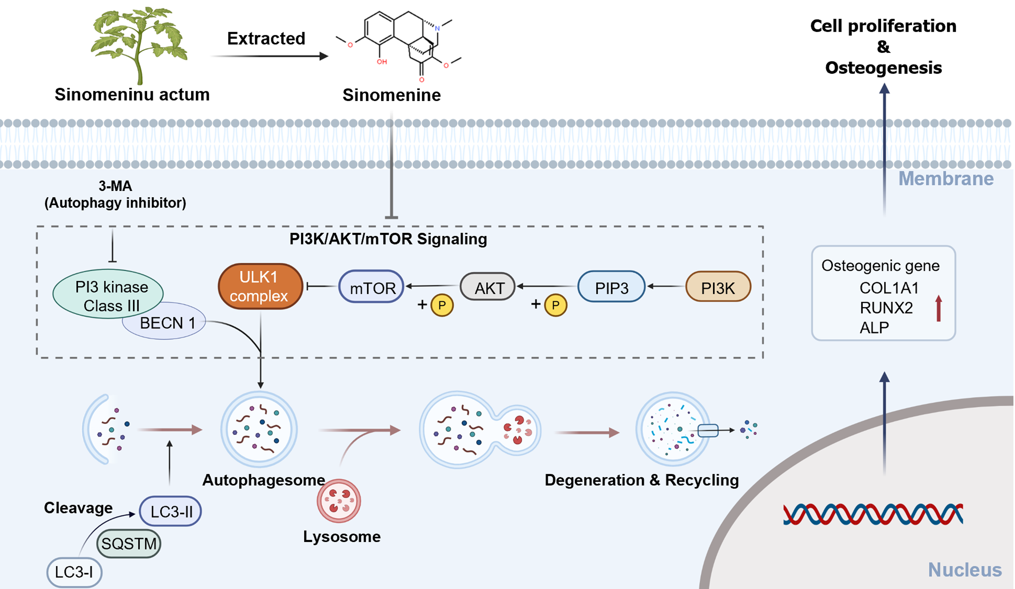
Figure 5 Schematic illustration showing the mechanism by which sinomenine promotes osteogenic differentiation of bone marrow mesenchymal stromal cells through modulation of autophagy.
Sinomenine decreased the levels of AKT and mammalian target of the rapamycin (mTOR) phosphorylation in the phosphatidylinositol 3-kinase/AKT/mTOR signaling pathway, and reduced mTOR activation lifted the inhibition of downstream autophagy and increased cellular autophagosomes, which increased the viability and osteogenic differentiation of bone marrow mesenchymal stromal cells. 3-MA: 3-methyladenine; PI3K: Phosphatidylinositol 3-kinase; mTOR: Mammalian target of the rapamycin.
- Citation: Xiao HX, Yu L, Xia Y, Chen K, Li WM, Ge GR, Zhang W, Zhang Q, Zhang HT, Geng DC. Sinomenine increases osteogenesis in mice with ovariectomy-induced bone loss by modulating autophagy. World J Stem Cells 2024; 16(5): 486-498
- URL: https://www.wjgnet.com/1948-0210/full/v16/i5/486.htm
- DOI: https://dx.doi.org/10.4252/wjsc.v16.i5.486


