Copyright
©The Author(s) 2024.
World J Stem Cells. Feb 26, 2024; 16(2): 137-150
Published online Feb 26, 2024. doi: 10.4252/wjsc.v16.i2.137
Published online Feb 26, 2024. doi: 10.4252/wjsc.v16.i2.137
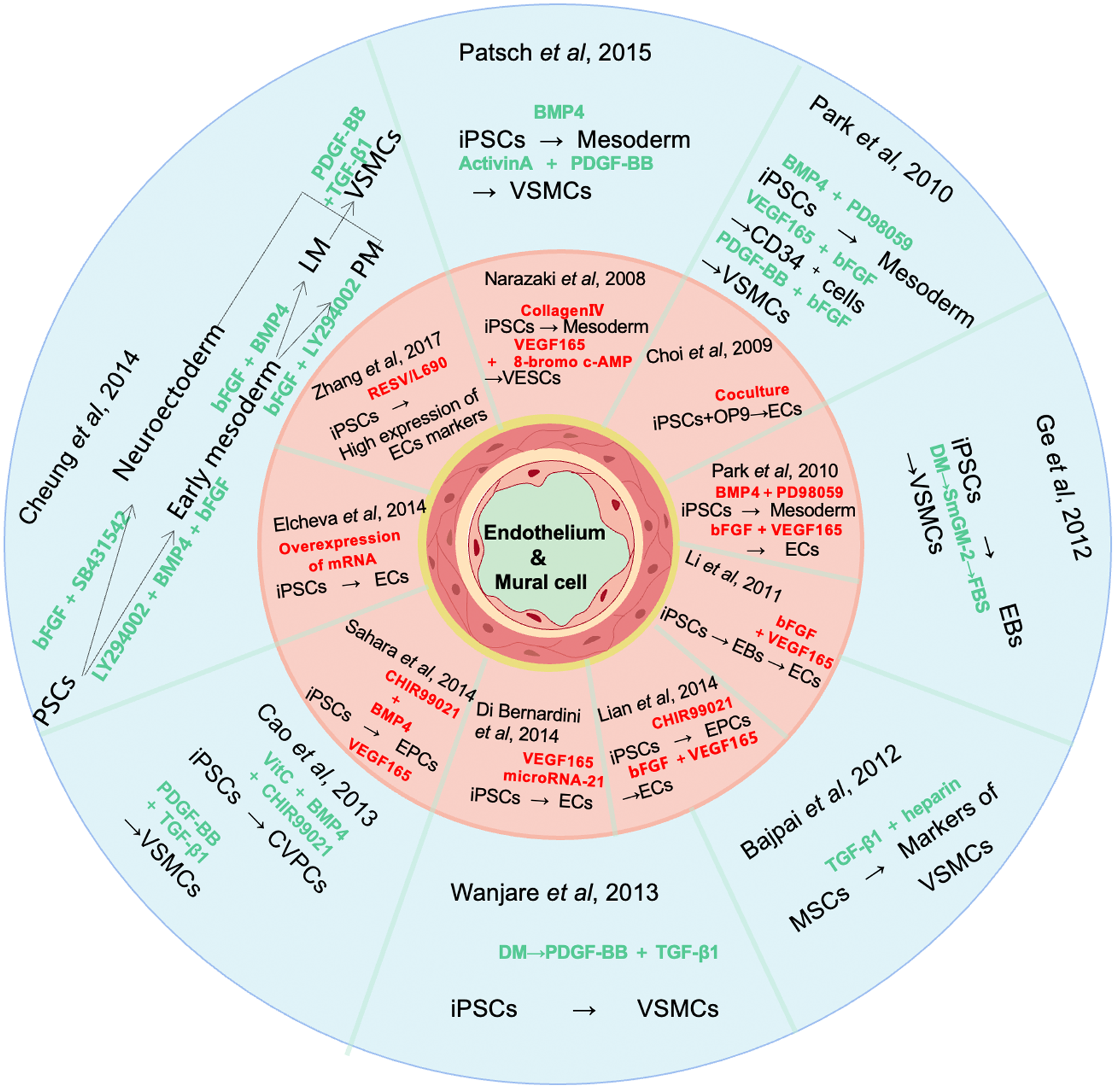
Figure 1 Modeling methods for endothelial cells and mural cells.
Several methods for differentiating induced pluripotent cell into endothelial cells and mural cells have been developed. The procedures of these modeling strategies are presented here as a schematic drawing. The inner ring shows the modeling strategies for endothelial cells, while the outer ring shows those for mural cells. The complementary factors and differentiation nodes are the focus of this graph. VEGF: Vascular endothelial growth factor; TGF: Transforming growth factor; iPSC: Induced pluripotent cell; VSMC: Vascular smooth muscle cell; EB: Embryoid body; MSC: Mesenchymal stem cell; DM: Differentiation medium; CVPC: Cardiovascular progenitor cell; LM: Lateral mesoderm; PM: Paraxial mesoderm; VESC: Vascular endothelial stem cells; EC: Endothelial cell; EPC: Endothelial progenitor cell; BMP4: Bone morphogenetic protein 4; PDGF-BB: Platelet-derived growth factor-BB; FBS: Fetal bovine serum; bFGF: Basic fibroblast growth factor.
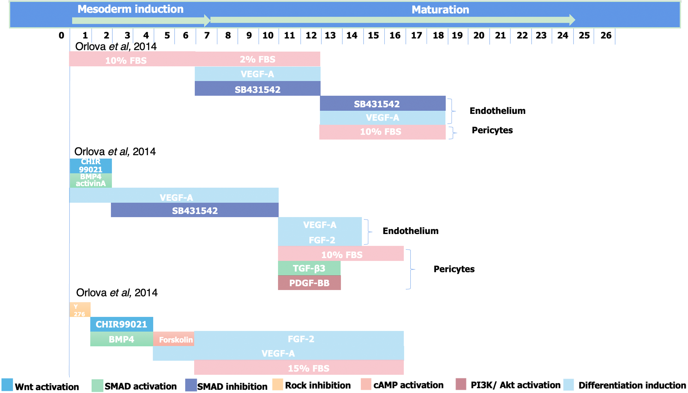
Figure 2 Methods for modeling three-dimensional vascular organoid or pericyte-endothelium co-culture.
Both co-differentiation or co-culture of endothelium and pericyte and three-dimensional vascular organoids can model different cell types of vascular cells at the same time. This figure shows the main procedures of these methods, revealing the similarities and differences in the order of activation and inhibition of key pathways related to differentiation. FBS: Foetal bovine serum; VEGF: Vascular endothelial growth factor; BMP4: Bone morphogenetic protein 4; FGF-2: Fibroblast growth factor 2; TGF: Transforming growth factor; PDGF-BB: Platelet-derived growth factor-BB.
Figure 3 Summary of induced pluripotent cells-derived vascular derivatives.
Various human vascular models based on induced pluripotent cell technology have been developed, including two-dimensional vascular cell culture, tissue-engineered vascular grafts, vascular organ chips, and three-dimensional vascular organoid construction. Alongside the increasing complexity and the ability to faithfully represent the in vivo process, the ease of handling and scalability of these human-based vascular models usually decrease. 3D: Three-dimensional.
- Citation: Jiao YC, Wang YX, Liu WZ, Xu JW, Zhao YY, Yan CZ, Liu FC. Advances in the differentiation of pluripotent stem cells into vascular cells. World J Stem Cells 2024; 16(2): 137-150
- URL: https://www.wjgnet.com/1948-0210/full/v16/i2/137.htm
- DOI: https://dx.doi.org/10.4252/wjsc.v16.i2.137


