Copyright
©The Author(s) 2024.
World J Stem Cells. Nov 26, 2024; 16(11): 926-943
Published online Nov 26, 2024. doi: 10.4252/wjsc.v16.i11.926
Published online Nov 26, 2024. doi: 10.4252/wjsc.v16.i11.926
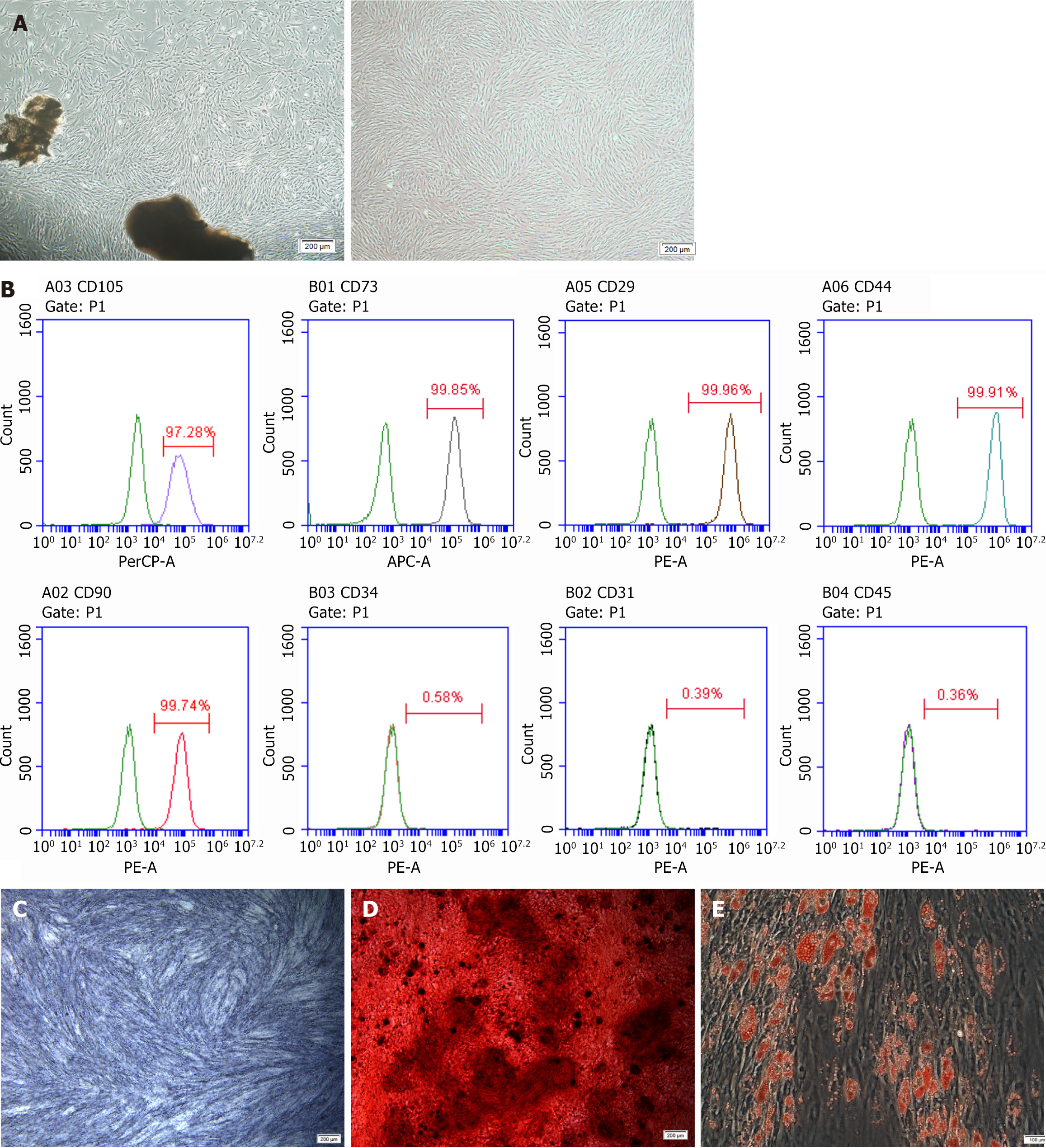
Figure 1 Characterization of periodontal ligament stem cells.
A: Periodontal ligament stem cells (PDLSCs) were cultured (scale bar = 200 μm); B: PDLSCs expressed CD105, CD73, CD29, CD44 and CD90 positively and expressed CD34, CD31 and CD45 negatively; C: Alkaline phosphatase staining of PDLSCs; D: Alizarin Red staining of PDLSCs; E: Oil Red O staining of PDLSCs. Data are presented as mean ± SD, n = 3.
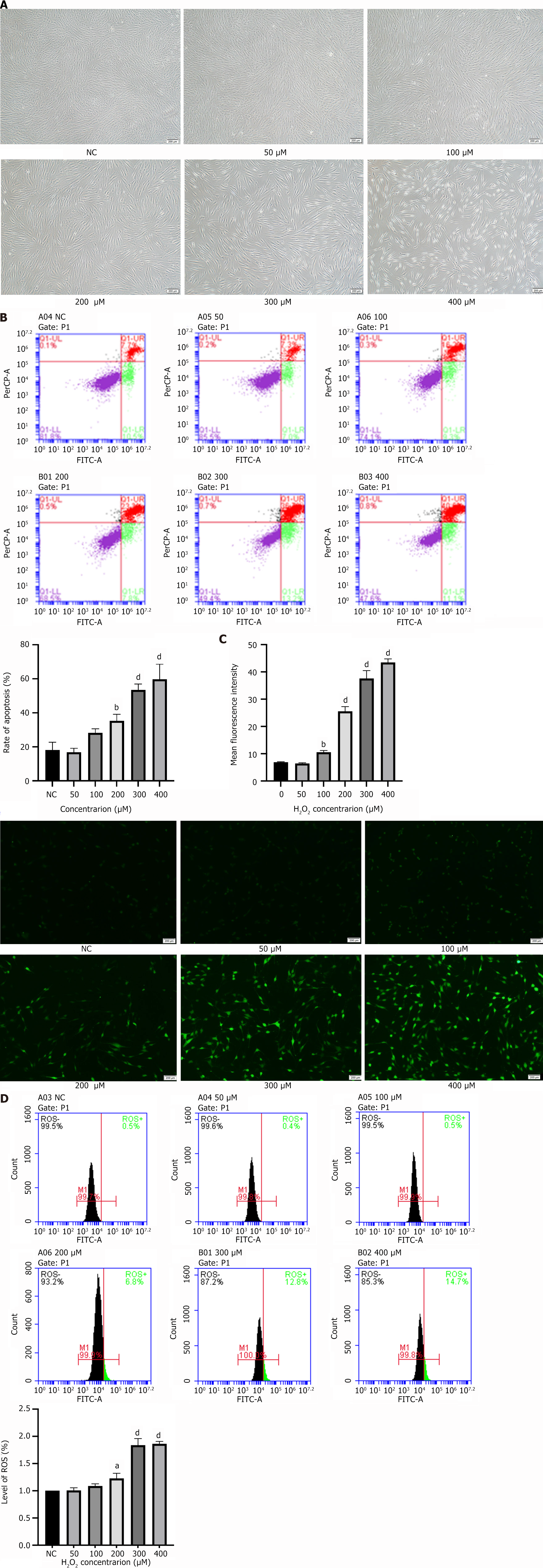
Figure 2 Hydrogen peroxide-induced oxidative stress in periodontal ligament stem cells.
A: Hydrogen peroxide (H2O2) affected the growth state and cell morphology of periodontal ligament stem cells (PDLSCs); B: H2O2 promoted apoptosis of PDLSCs; C: H2O2 increased the reactive oxygen species fluorescence intensity of PDLSCs; D: H2O2 increased the proportion of reactive oxygen species-positive cells in PDLSCs. Data are presented as mean ± SD, n = 3. aP < 0.05; bP < 0.01; dP < 0.0001. NC: Negative control; ROS: Reactive oxygen species; H2O2: Hydrogen peroxide.
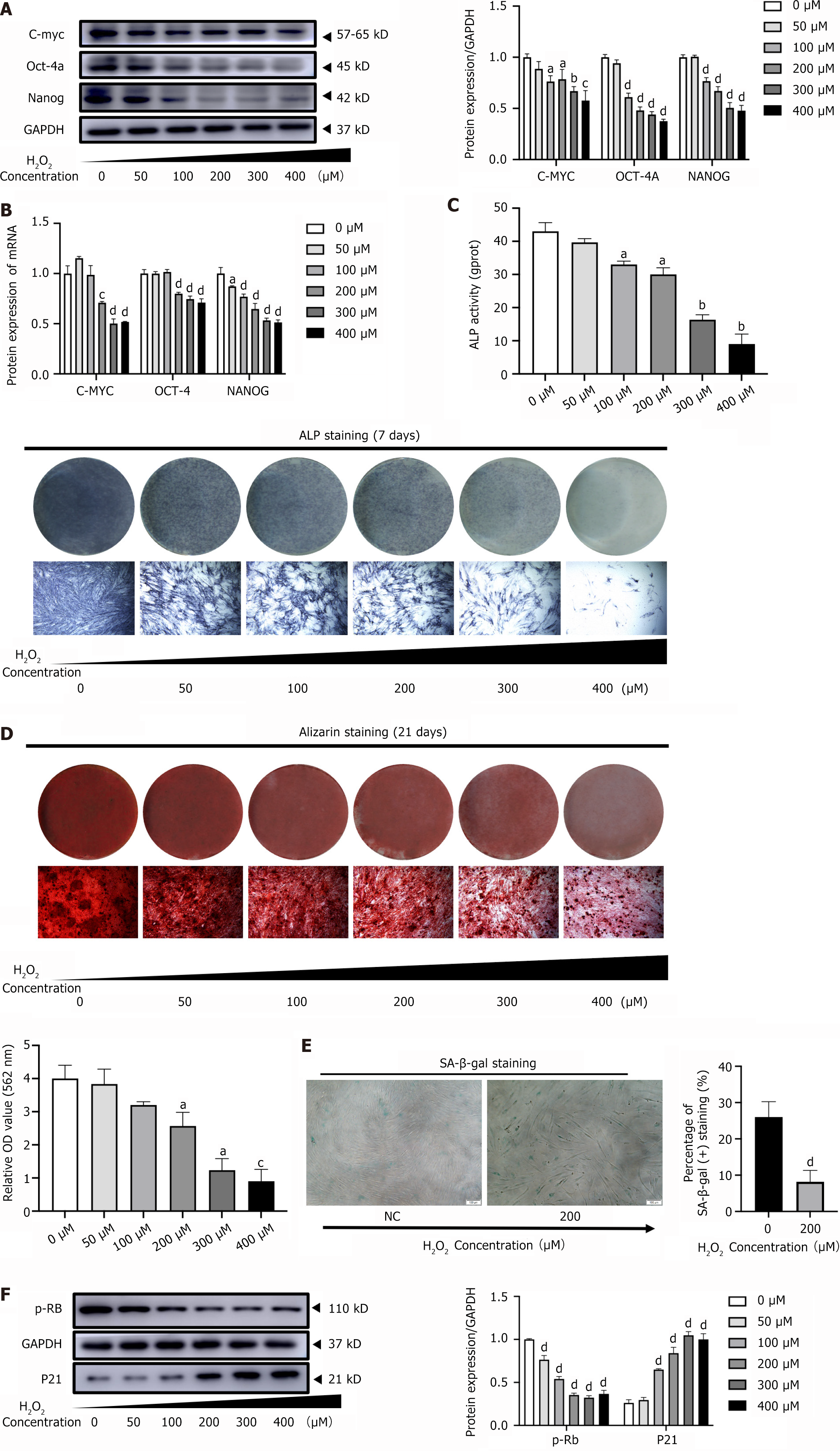
Figure 3 Hydrogen peroxide inhibited stemness and promoted senescence in periodontal ligament stem cells.
A: Hydrogen peroxide (H2O2) inhibited the expression level of stemness-related proteins C-MYC, OCT-4, and NANOG in periodontal ligament stem cells (PDLSCs); B: H2O2 suppressed the expression levels of stemness-related genes C-MYC, OCT-4, and NANOG in PDLSCs; C: Alkaline phosphatase staining and alkaline phosphatase activity showed that the osteogenic differentiation ability of the H2O2-induced PDLSCs was weaker than that of the control group; D: Alizarin Red staining and quantitative analysis showed that the osteogenic differentiation ability of the H2O2-induced PDLSCs was weaker than that of the control group; E: Senescence-associated β-galactosidase staining indicated significantly more senescence-associated β-galactosidase-positive cells in H2O2-induced PDLSCs compared with the control group; F: Western blot results showed that H2O2 inhibited the expression of p-RB but increased the expression of p21 in PDLSCs. Data are presented as mean ± SD, n = 3. aP < 0.05; bP < 0.01; cP < 0.001; dP < 0.0001. NC: Negative control; H2O2: Hydrogen peroxide; ALP: Alkaline phosphatase; SA β-gal: Senescence-associated β-galactosidase.
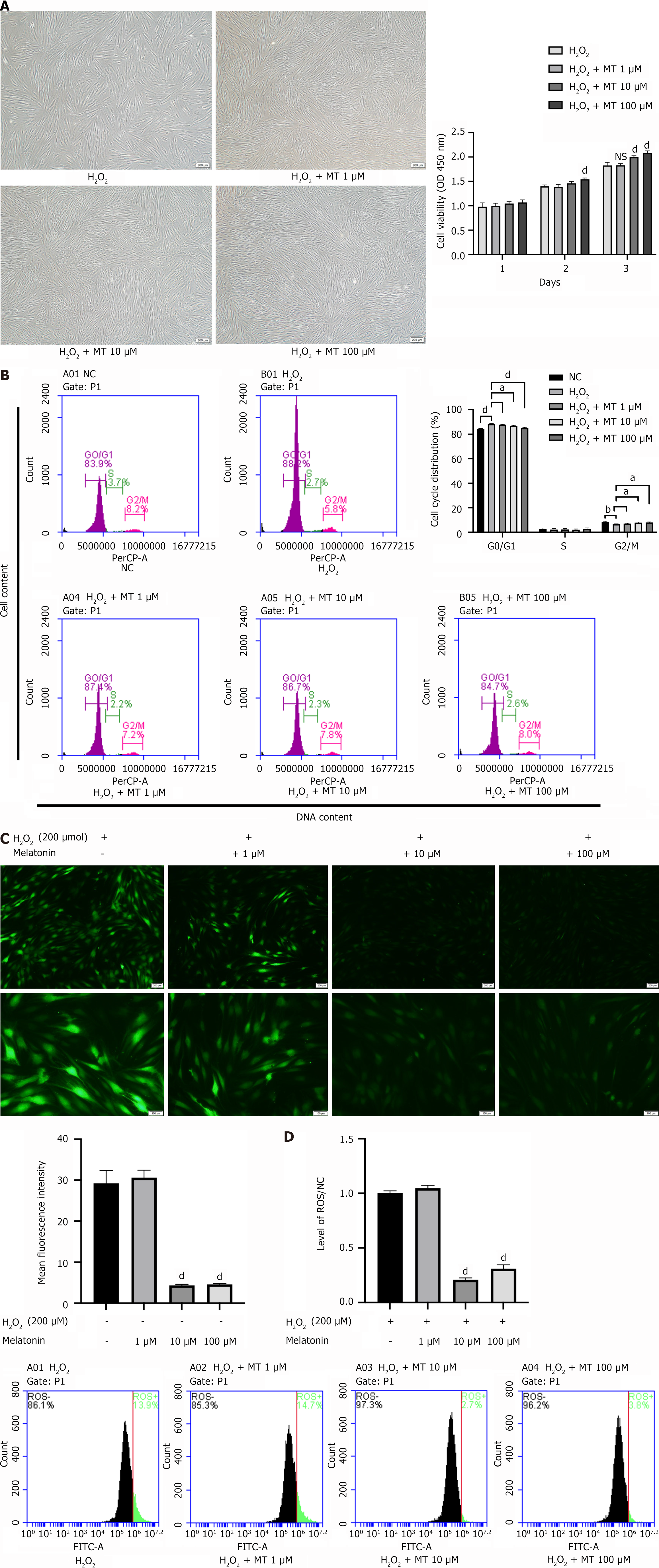
Figure 4 Melatonin improved proliferative capacity and reduced oxidative stress levels in hydrogen peroxide-induced periodontal ligament stem cells.
A: Melatonin (MT) restored the growth state and enhanced the proliferative capacity of hydrogen peroxide (H2O2)-induced periodontal ligament stem cells (PDLSCs); B: MT reduced the proportion of G0/G1 phase and increased the proportion of G2/M phase in H2O2-induced PDLSCs; C: MT decreased the reactive oxygen species fluorescence intensity of H2O2-induced PDLSCs; D: MT decreased the proportion of reactive oxygen species-positive cells in H2O2-induced PDLSCs. Data are presented as mean ± SD, n = 3. aP < 0.05; bP < 0.01; dP < 0.0001. NS: No significance; H2O2: Hydrogen peroxide; MT: Melatonin; NC: Negative control.
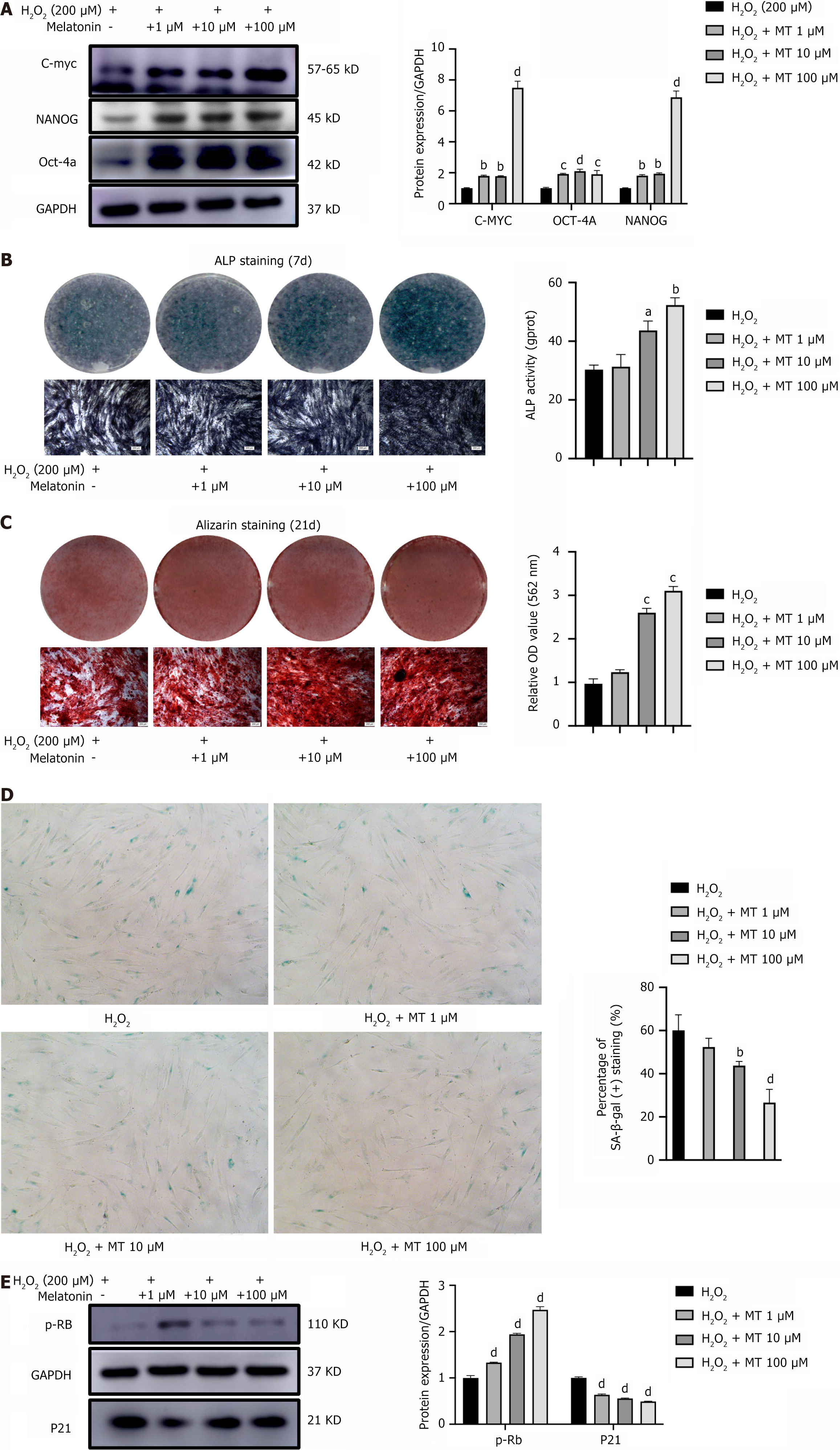
Figure 5 Melatonin increased stemness and delayed senescence in hydrogen peroxide-induced periodontal ligament stem cells.
A: Melatonin (MT) restored the expression levels of stemness-related proteins C-MYC, OCT-4, and NANOG in hydrogen peroxide (H2O2)-induced periodontal ligament stem cells (PDLSCs); B: Alkaline phosphatase staining and alkaline phosphatase activity showed that MT enhanced the osteogenic differentiation ability of the H2O2-induced PDLSCs; C: Alizarin Red staining and quantitative analysis showed that MT enhanced the osteogenic differentiation ability of the H2O2-induced PDLSCs; D: Senescence-associated β-galactosidase staining indicated MT decreased senescence-associated β-galactosidase-positive cells in H2O2-induced PDLSCs; E: Western blot results showed that MT increased the expression of p-RB but inhibited the expression of p21 in H2O2-induced PDLSCs. Data are presented as mean ± SD, n = 3. aP < 0.05; bP < 0.01; cP < 0.001; dP < 0.0001. H2O2: Hydrogen peroxide; ALP: Alkaline phosphatase; ARS: Alizarin Red staining; MT: Melatonin; SA β-gal: Senescence-associated β-galactosidase.
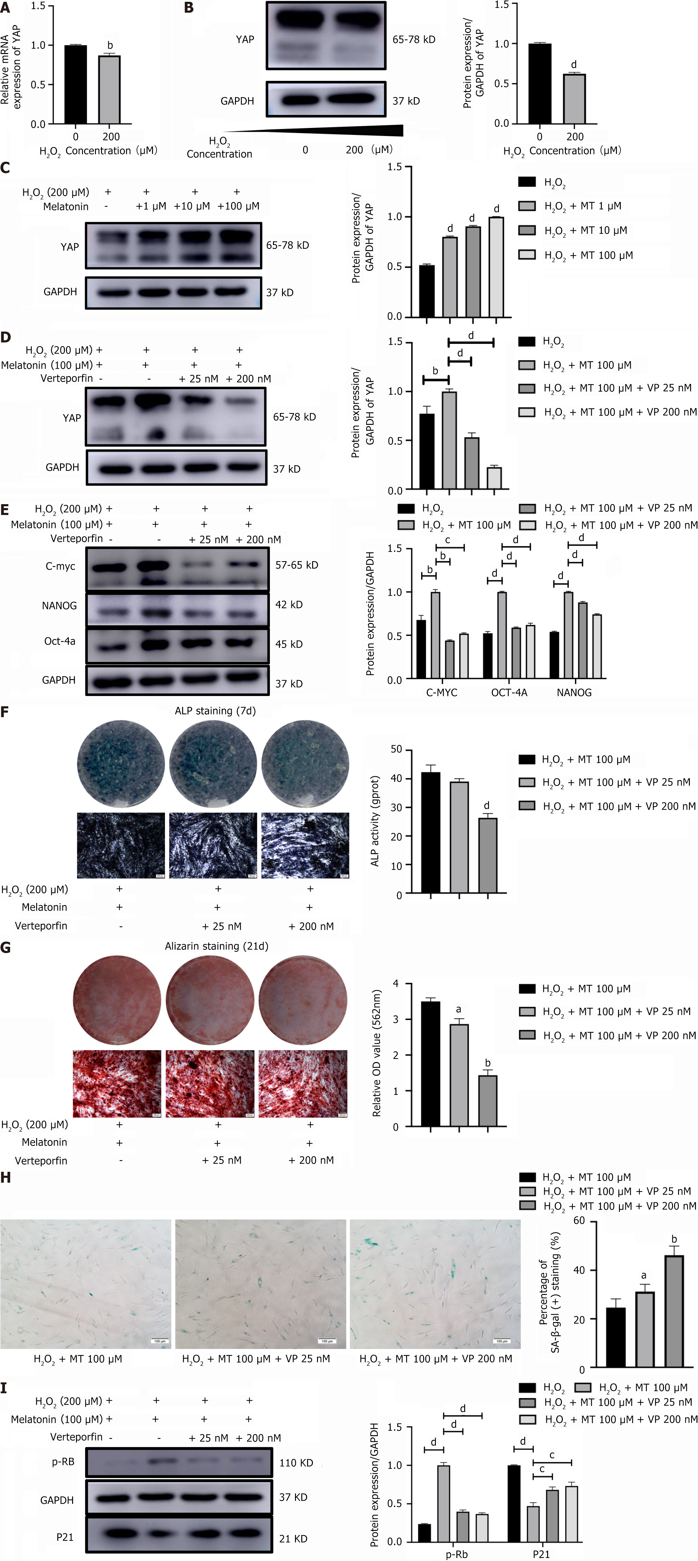
Figure 6 Yes-associated protein mediated melatonin to enhance stemness and reduce senescence in hydrogen peroxide-induced periodontal ligament stem cells.
A: Hydrogen peroxide (H2O2) reduced the mRNA expression level of Yes-associated protein (YAP) in periodontal ligament stem cells (PDLSCs); B: H2O2 reduced the protein expression level of YAP in PDLSCs; C: Melatonin (MT) increased the protein expression level of YAP in H2O2-induced PDLSCs; D: Verteporfin reduced the protein expression level of YAP in MT-increased PDLSCs; E: Verteporfin decreased the protein expression levels of stemness-related proteins C-MYC, OCT-4 and NANOG in MT-increased PDLSCs; F: Alkaline phosphatase staining and alkaline phosphatase activity showed that verteporfin reduced the osteogenic differentiation ability in MT-increased PDLSCs; G: Alizarin Red staining and quantitative analysis showed that verteporfin reduced the osteogenic differentiation ability in MT-increased PDLSCs; H: Senescence-associated β-galactosidase staining indicated verteporfin increased senescence-associated β-galactosidase-positive cells in MT-increased PDLSCs; I: Western blot results showed that verteporfin decreased the expression of p-RB but increased the expression of p21 in MT-treated PDLSCs. Data are presented as mean ± SD, n = 3. aP < 0.05; bP < 0.01; cP < 0.001; dP < 0.0001. H2O2: Hydrogen peroxide; MT: Melatonin; YAP: Yes-associated protein; VP: Verteporfin; ALP: Alkaline phosphatase; ARS: Alizarin Red staining.
- Citation: Gu K, Feng XM, Sun SQ, Hao XY, Wen Y. Yes-associated protein-mediated melatonin regulates the function of periodontal ligament stem cells under oxidative stress conditions. World J Stem Cells 2024; 16(11): 926-943
- URL: https://www.wjgnet.com/1948-0210/full/v16/i11/926.htm
- DOI: https://dx.doi.org/10.4252/wjsc.v16.i11.926


