Copyright
©The Author(s) 2023.
World J Stem Cells. Jun 26, 2023; 15(6): 617-631
Published online Jun 26, 2023. doi: 10.4252/wjsc.v15.i6.617
Published online Jun 26, 2023. doi: 10.4252/wjsc.v15.i6.617
Figure 1 Mesenchymal stem cells pretreated with calycosin enhance the protective effect of mesenchymal stem cells on podocyte injury in adriamycin-induced focal segmental glomerulosclerosis mice.
A: Mice received adriamycin injections through the tail vein at week 6, were injected with Dulbecco’s modified eagle medium, mesenchymal stem cells (MSCs), calycosin (CA), and MSCs pretreated with CA (MSCsCA) at week 10, respectively, and were sacrificed at week 14; B: Levels of albumin/creatinine ratio in urine (n = 6), aP < 0.05, bP < 0.001; C: Pathological changes in the kidneys of mice examined by hematoxylin-eosin staining. Typical glomeruli are indicated by black boxes and enlarged to the next row. Bar = 50 μm; D: Changes in the kidneys of mice examined by podocin immunofluorescence staining. Glomeruli are indicated by white boxes and enlarged to the next row. Bar = 50 μm; E: Relative (podocin/GAPDH) mRNA expression analyzed by real-time quantitative polymerase chain reaction. Data are expressed as the mean ± SD (n = 6). aP < 0.05, bP < 0.001; F and G: Relative protein levels (podocin/GAPDH) detected by Western blot. Data are expressed as the mean ± SD (n = 3). aP < 0.05, bP < 0.001. NC: Normal control; ADR: Adriamycin; DMEM: Dulbecco’s modified eagle medium; MSCs: Mesenchymal stem cells; CA: Calycosin; MSCsCA: Mesenchymal stem cells pretreated with calycosin.
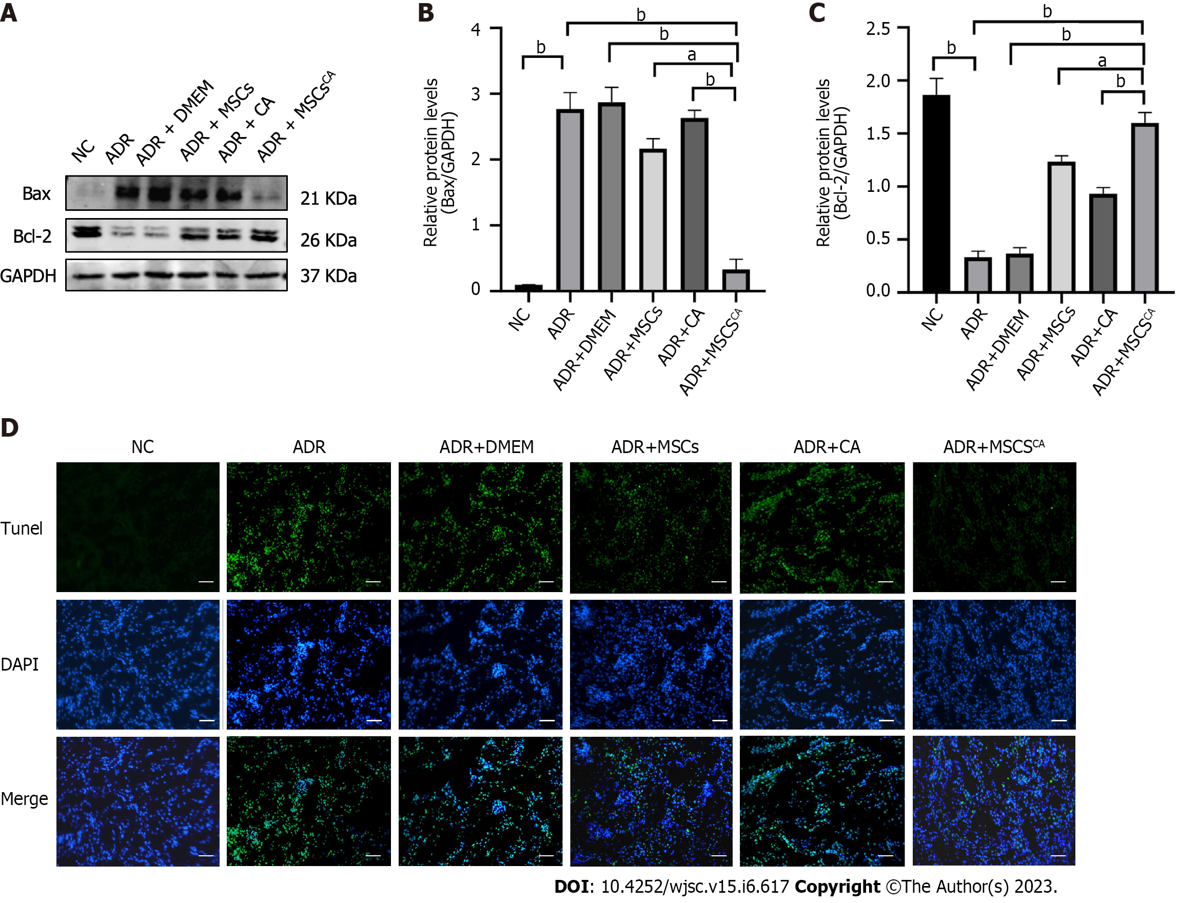
Figure 2 Calycosin pretreatment enhances the ability of mesenchymal stem cells to inhibit apoptosis in adriamycin-induced focal segmental glomerulosclerosis mice.
A-C: Protein expression levels of Bax and Bcl-2 in the kidneys measured by Western blot and normalized to control. Data are expressed as the mean ± SD (n = 3). aP < 0.05, bP < 0.001; D: Apoptosis in each group as determined by TUNEL assay. Bar = 50 μm. NC: Normal control; ADR: Adriamycin; MSCs: Mesenchymal stem cells; CA: Calycosin; MSCsCA: Mesenchymal stem cells pretreated with calycosin.
Figure 3 P-Smad3 is upregulated in podocytes of adriamycin-induced focal segmental glomerulosclerosis mice and reversed after treatment with mesenchymal stem cells pretreated with calycosin.
A and B: Protein expression levels of p-Smad3 and Smad3 detected using Western blot and normalized to control. Data are expressed as the mean ± SD (n = 3). bP < 0.001; C: Immunohistochemistry staining for p-Smad3 in mouse glomeruli, which are indicated by red boxes and enlarged to the next row. Bar = 50 μm. NC: Normal control; ADR: Adriamycin; MSCs: Mesenchymal stem cells; CA: Calycosin; MSCsCA: Mesenchymal stem cells pretreated with calycosin.
Figure 4 Calycosin pretreatment enhances the ability of mesenchymal stem cells to ameliorate the injury of adriamycin-stimulated mouse podocyte cells in vitro.
A: Podocin expression in each group as determined by immunofluorescence staining. Bar = 50 μm; B: Analysis of relative (podocin/GAPDH) mRNA expression by real-time quantitative polymerase chain reaction. Data are expressed as the mean ± SD (n = 3). bP < 0.001; C and D: Protein expression levels of podocin detected by Western blot and normalized to control. Data are expressed as the mean ± SD (n = 3). aP < 0.05, bP < 0.001. NC: Normal control; ADR: Adriamycin; MSCs: Mesenchymal stem cells; CA: Calycosin; MSCsCA: Mesenchymal stem cells pretreated with calycosin.
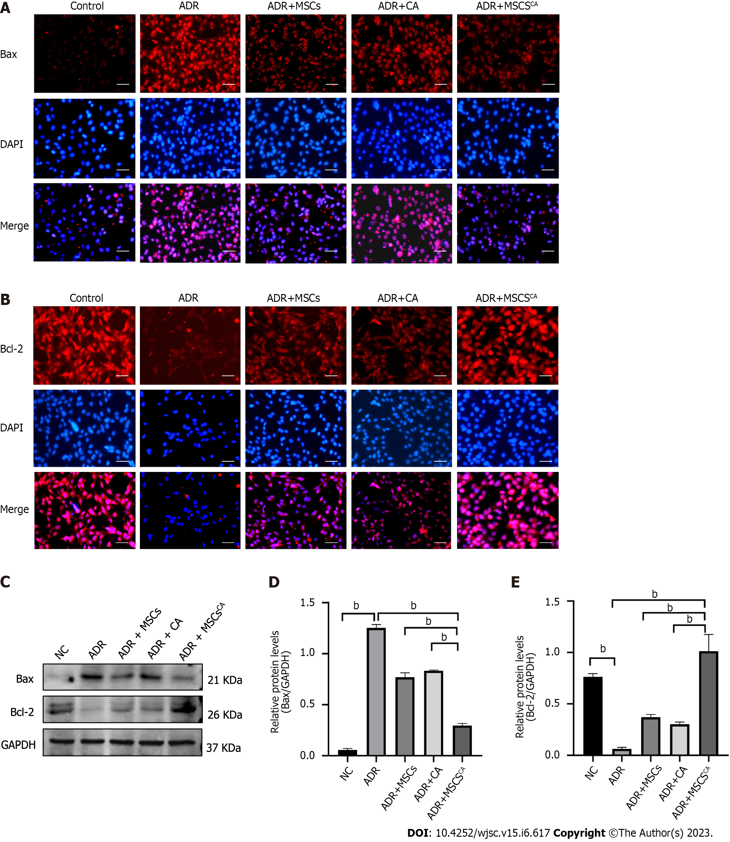
Figure 5 Calycosin pretreatment enhances the ability of mesenchymal stem cells to inhibit apoptosis in adriamycin-stimulated mouse podocyte cells.
A and B: Expression of Bax and Bcl-2 in each group as determined by immunofluorescence staining. Bar = 50 μm; C-E: Protein expression levels of Bax and Bcl-2 detected by Western blot and normalized to control. Data are expressed as the mean ± SD (n = 3). bP < 0.001. NC: Normal control; ADR: Adriamycin; MSCs: Mesenchymal stem cells; CA: Calycosin; MSCsCA: Mesenchymal stem cells pretreated with calycosin.
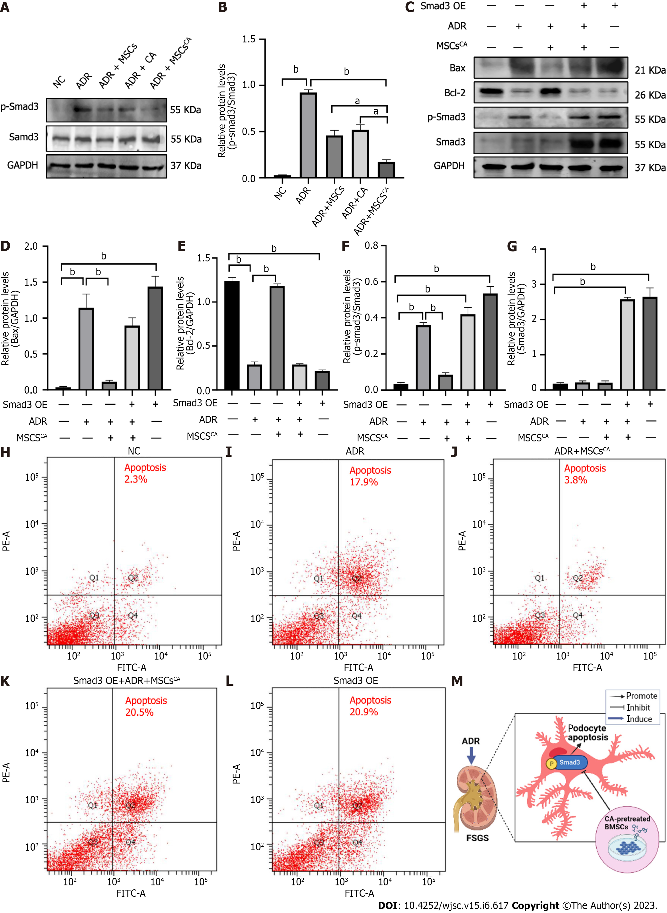
Figure 6 Calycosin-pretreated mesenchymal stem cells improve adriamycin-induced podocyte apoptosis by targeting p-Smad3 expression.
A and B: Protein expression levels of p-Smad3 and Smad3 in mouse podocyte cells (MPC5) detected by Western blot and normalized to control. Data are expressed as the mean ± SD (n = 3). aP < 0.05, bP < 0.001; C-G: Protein expression levels of Bax, Bcl-2, p-Smad3, and Smad3 in Smad3-overexpressing MPC5 cells detected by Western blot and normalized to control. Data are expressed as the mean ± SD (n = 3). bP < 0.001; H-L: Cell apoptosis detected by flow cytometry; M: Graphical abstract (created in BioRender.com). NC: Normal control; ADR: Adriamycin; MSCs: Mesenchymal stem cells; CA: Calycosin; MSCsCA: Mesenchymal stem cells pretreated with calycosin; FSGS: Focal segmental glomerulosclerosis.
- Citation: Hu QD, Tan RZ, Zou YX, Li JC, Fan JM, Kantawong F, Wang L. Synergism of calycosin and bone marrow-derived mesenchymal stem cells to combat podocyte apoptosis to alleviate adriamycin-induced focal segmental glomerulosclerosis. World J Stem Cells 2023; 15(6): 617-631
- URL: https://www.wjgnet.com/1948-0210/full/v15/i6/617.htm
- DOI: https://dx.doi.org/10.4252/wjsc.v15.i6.617


