Copyright
©The Author(s) 2022.
World J Stem Cells. Jul 26, 2022; 14(7): 539-555
Published online Jul 26, 2022. doi: 10.4252/wjsc.v14.i7.539
Published online Jul 26, 2022. doi: 10.4252/wjsc.v14.i7.539
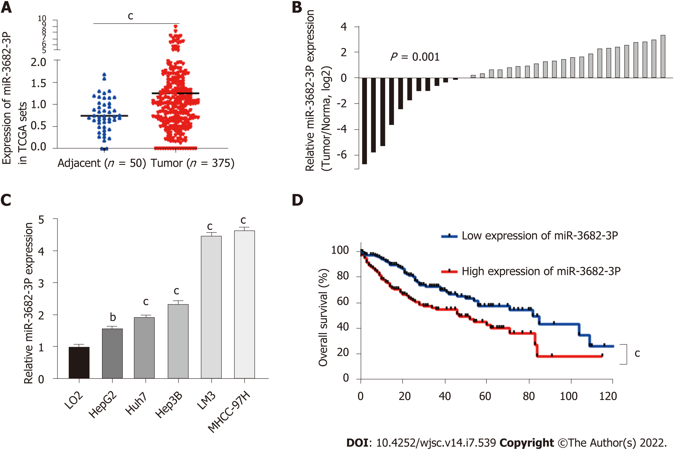
Figure 1 High expression of miR-3682-3p reflected poor prognosis of hepatocellular carcinoma.
A: The expression levels of miR-3682-3p in normal and hepatocellular carcinoma (HCC) tissues in The Cancer Genome Atlas (TCGA) dataset; B: The expression of miR-3682-3p in 34 HCC and paired paracancerous tissue samples as determined by RT-qPCR; C: miR-3682-3p expression in HCC cells (HepG2, Huh7, Hep3B, LM3, and MHCC-97H) and normal liver epithelial (LO2) cells as determined by RT-qPCR; D: Analysis of Kaplan-Meier overall survival curves for HCC patients based on miR-3682-3p expression in TCGA dataset. bP < 0.01, cP < 0.001. Experiments were repeated three times.
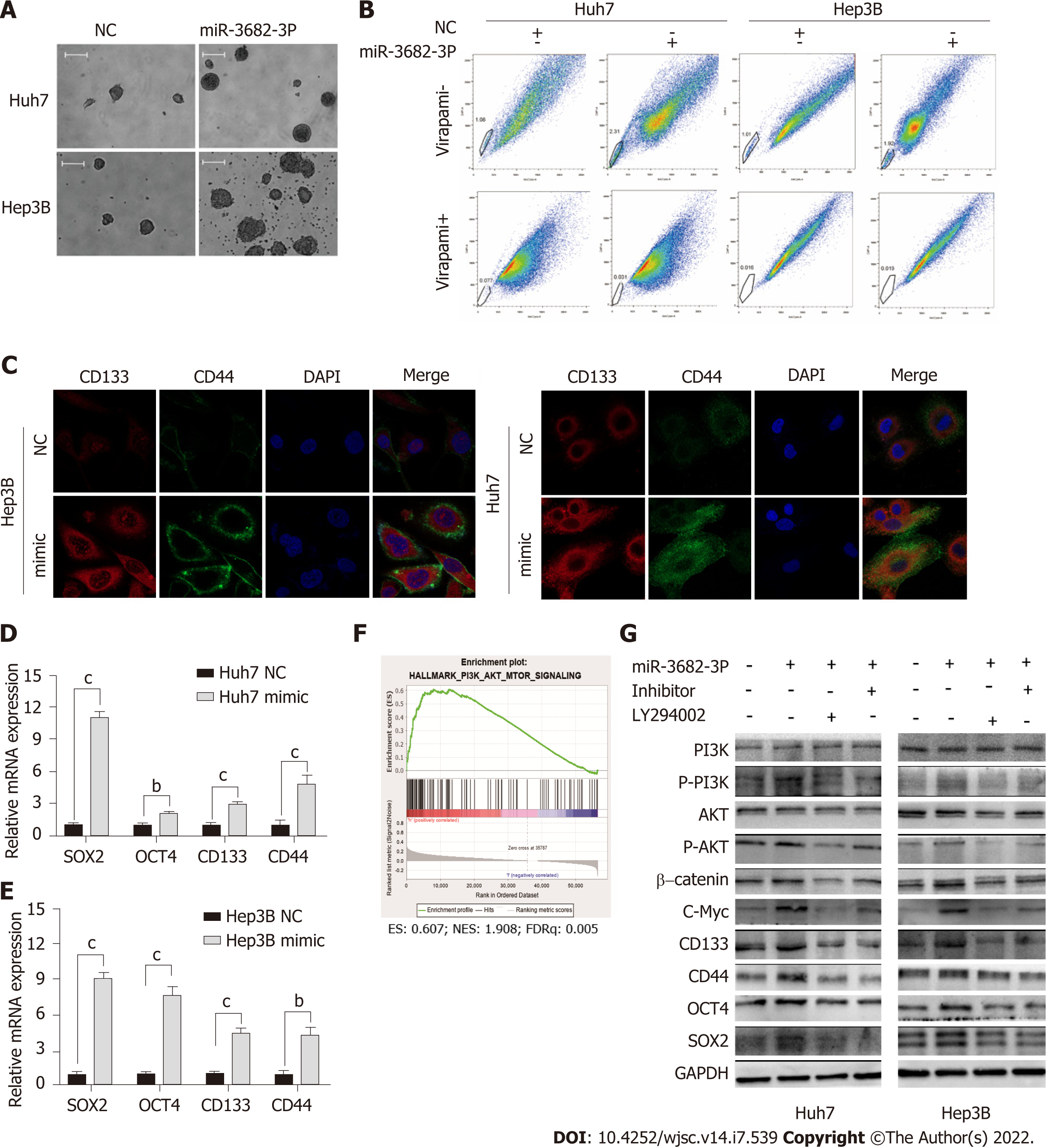
Figure 2 miR-3682-3p promotes the stem cell properties of hepatocellular carcinoma cells via the PI3K/AKT/c-Myc signaling pathway.
A: Representative images of formed hepatospheres (× 100 magnification); scale bar: 200 μm; B: Flow cytometric analysis of the proportion of side population cells; C: Immunofluorescence staining was performed to assess the expression levels of CD44 and CD133 in hepatocellular carcinoma cells (scale bar: 5 μm); D and E: RT-qPCR analysis of SOX2, OCT4, CD133, and CD44 expression; F: Gene set enrichment analysis showing that miR-3682-3p regulates the PI3K/AKT signaling axis; G: Western blotting analysis of PI3K/AKT/c-Myc signaling- and stemness-related protein expression levels. bP < 0.01; cP < 0.001. Experiments were repeated three times. NC: Negative control.
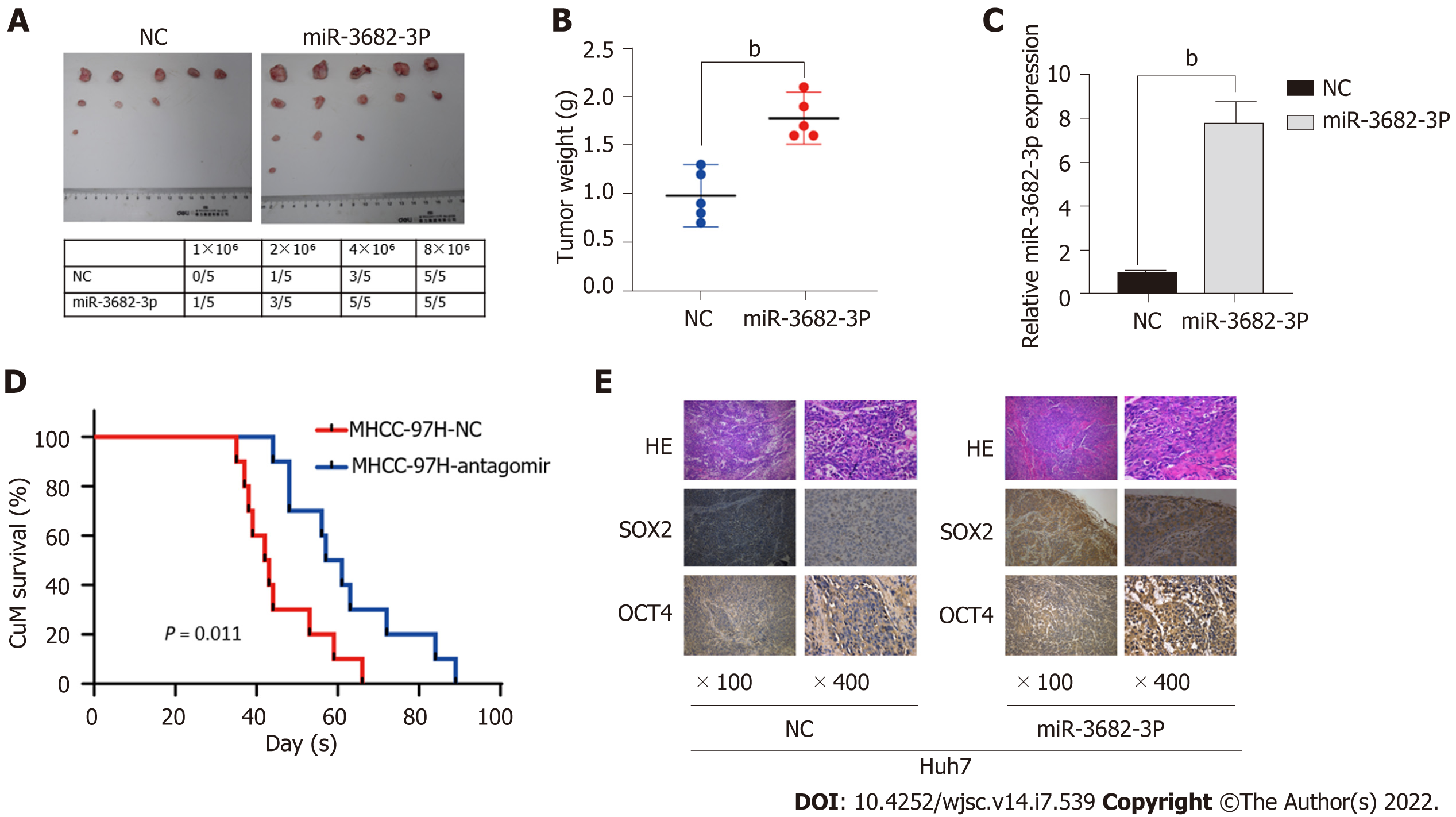
Figure 3 miR-3682-3p promotes the tumorigenicity of hepatocellular carcinoma cells in vivo.
A: The tumorigenic rate of different concentrations of hepatocellular carcinoma cells (n = 5); B: Tumors in the miR-3682-3p overexpression group weighed significantly more than those of the control group (n = 5); C: Detection of miR-3682-3p expression in tumor tissues by RT-qPCR; D: Survival analysis revealed that miR-3682-3p antagomir prolongs the survival time of mice (n = 10, log-rank test); E: Immunohistochemical staining for stem cell-associated markers (SOX2 and OCT4) in xenograft tumors (n = 5) (× 100 magnification scale bar: 200 µm; × 400 magnification scale bar: 50 µm). bP < 0.01. NC: Negative control; HE: Hematoxylin and eosin.
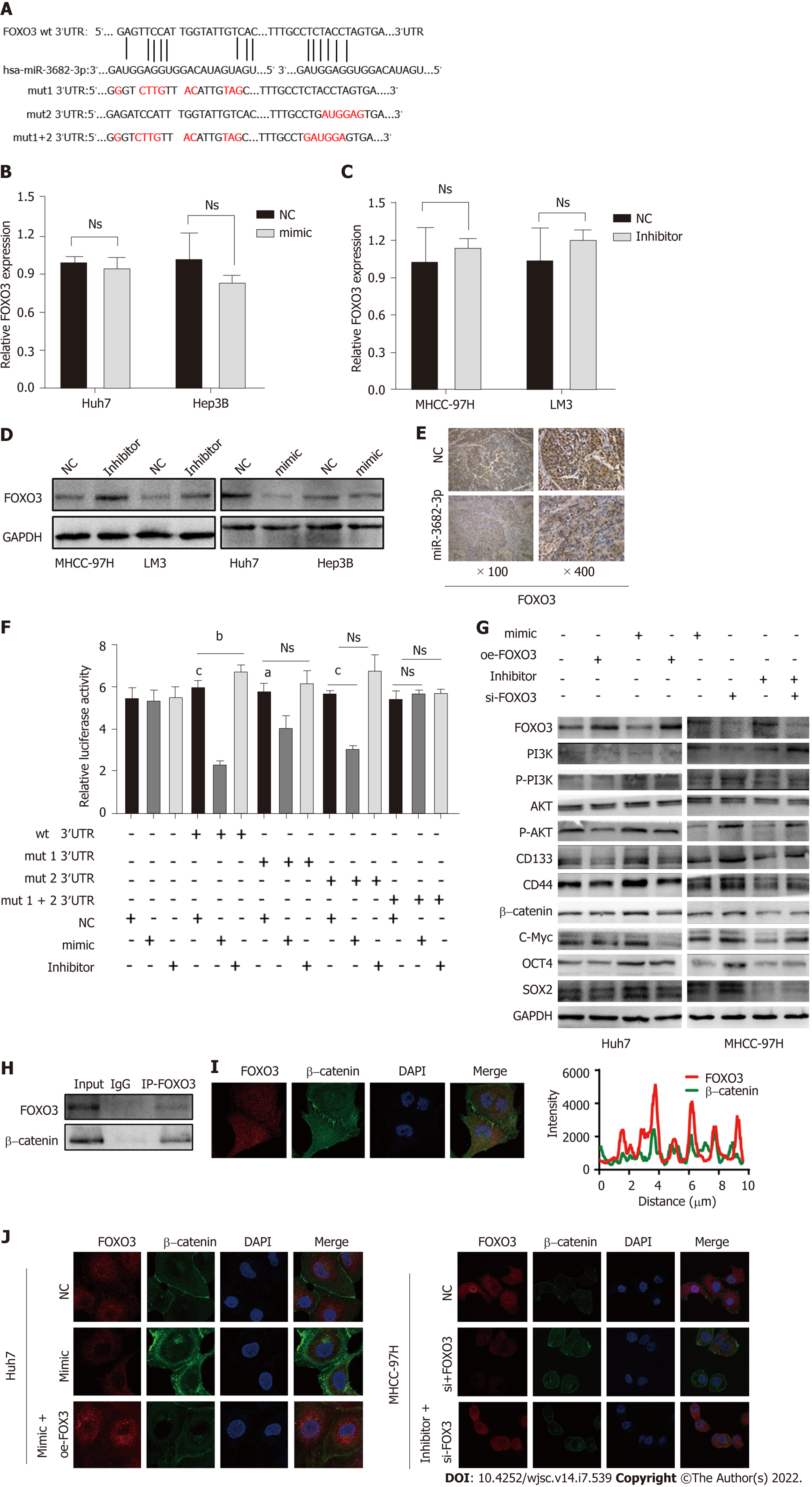
Figure 4 FOXO3 is a direct target of miR-3682-3p.
A: Binding sites for miR-3682-3p in the 3′-UTR of FOXO3 were predicted using bioinformatics tools; B-D: The effect of miR-3682-3p on FOXO3 mRNA and protein levels as determined by RT-qPCR and western blot, respectively; E: Dual-luciferase reporter assay was used to detect the interaction between miR-3682-3p and the 3′-UTR of FOXO3; F: Immunohistochemical analysis of FOXO3 expression in xenograft tumors (n = 5) (× 100 magnification scale bar: 200 µm, × 400 magnification scale bar: 50 µm); G: Western blot-based analysis of the effects of FOXO3 on stemness- and PI3K/AKT/c-Myc pathway-related proteins; H: Immunoprecipitation assays for the interaction between endogenous FOXO3 and β-catenin; I: Immunofluorescence staining-based assessment of FOXO3/β-catenin co-localization; J: Immunofluorescence staining of FOXO3 and β-catenin in hepatocellular carcinoma cells (scale bar: 5 μm). aP < 0.05, cP < 0.001. Experiments were repeated three times. NC: Negative control; wt: Wild-type; mut: Mutant.
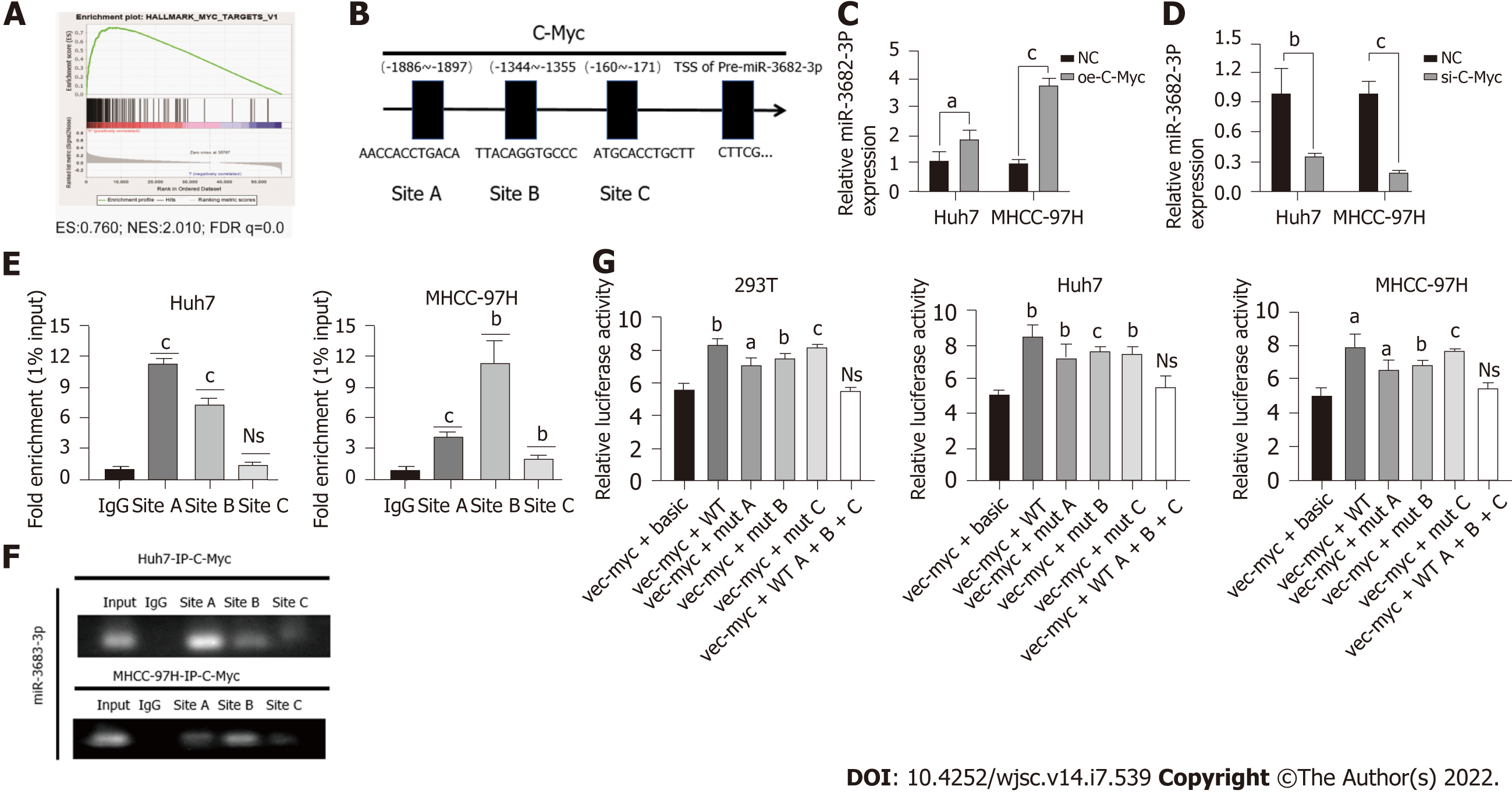
Figure 5 c-Myc binds to the miR-3682-3p promoter region and promotes its transcriptional activation.
A: Gene set enrichment analysis revealed that miR-3682-3p is positively associated with c-Myc; B: Bioinformatic-based prediction of c-Myc binding sites in the miR-3682-3p promoter region; C and D: RT-qPCR analysis of miR-3682-3p levels after c-Myc silencing and overexpression; E and F: Chromatin immunoprecipitation validated the binding of c-Myc to the miR-3682-3p promoter; G: Dual-luciferase reporter assay confirmed the binding of c-Myc to the miR-3682-3p promoter. aP < 0.05, bP < 0.01, cP < 0.001. Experiments were repeated three times. NC: Negative control.
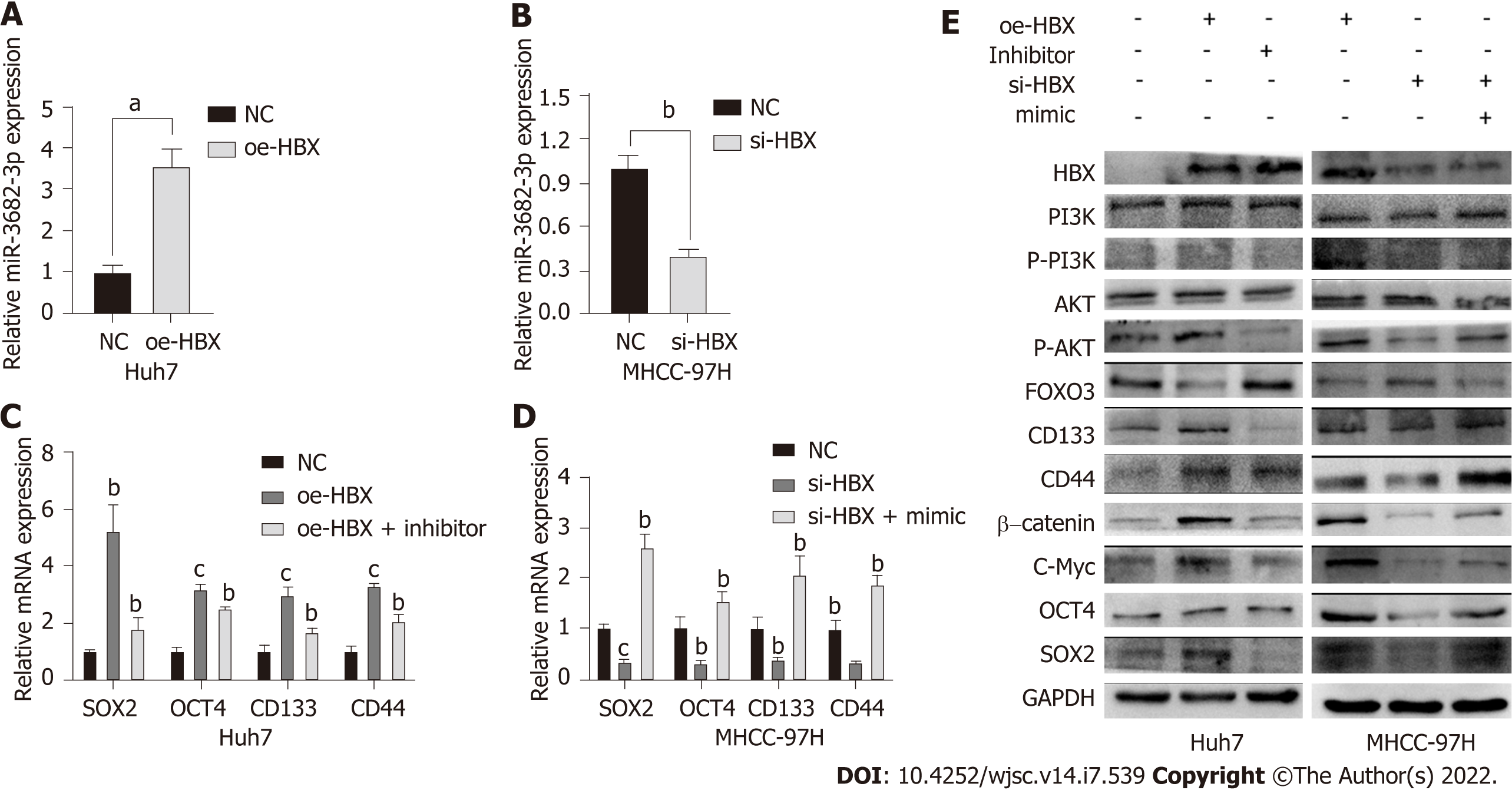
Figure 6 HBx induces miR-3682-3p expression, thereby enhancing the cancer stem cell-like properties of hepatocellular carcinoma cells.
A and B: The levels of miR-3682-3p after the overexpression and silencing of HBx were detected by RT-qPCR; C and D: RT-qPCR and western blot analysis of the effect of HBx on stemness markers. aP < 0.05, bP < 0.01, cP < 0.001. Experiments were repeated three times. NC: Negative control.
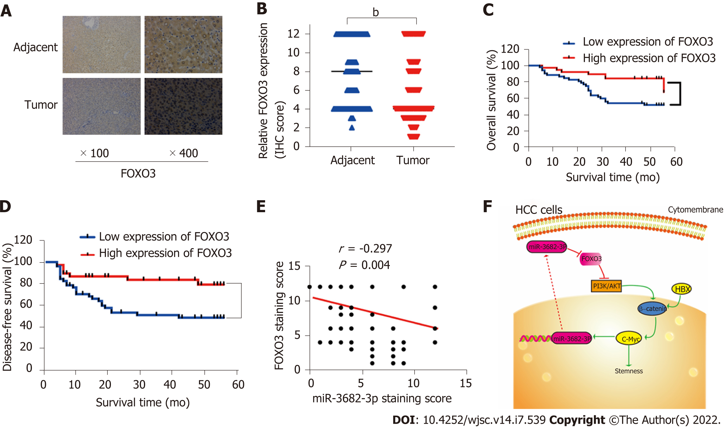
Figure 7 Pathoclinical features of FOXO3 expression.
A: FOXO3 expression in hepatocellular carcinoma (HCC) tissue microarray (× 100 magnification scale bar: 200 µm; × 400 magnification scale bar: 50 µm); B: Immunohistochemical scoring of FOXO3 staining; C: Kaplan-Meier survival analysis showing the FOXO3 expression level-related overall survival in HCC; D: Kaplan-Meier survival analysis showing the FOXO3 expression level-related disease-free survival in HCC; E: Correlation between FOXO1 and miR-3682-3p expression (Spearman’s rank correlation test); F: Schematic representation of the HBx-induced miR-3682-3p/FOXO3/PI3K/AKT/c-Myc feedback loop that promotes HCC stemness. bP < 0.01.
- Citation: Chen Q, Yang SB, Zhang YW, Han SY, Jia L, Li B, Zhang Y, Zuo S. miR-3682-3p directly targets FOXO3 and stimulates tumor stemness in hepatocellular carcinoma via a positive feedback loop involving FOXO3/PI3K/AKT/c-Myc. World J Stem Cells 2022; 14(7): 539-555
- URL: https://www.wjgnet.com/1948-0210/full/v14/i7/539.htm
- DOI: https://dx.doi.org/10.4252/wjsc.v14.i7.539


