Copyright
©The Author(s) 2022.
World J Stem Cells. Jul 26, 2022; 14(7): 473-489
Published online Jul 26, 2022. doi: 10.4252/wjsc.v14.i7.473
Published online Jul 26, 2022. doi: 10.4252/wjsc.v14.i7.473
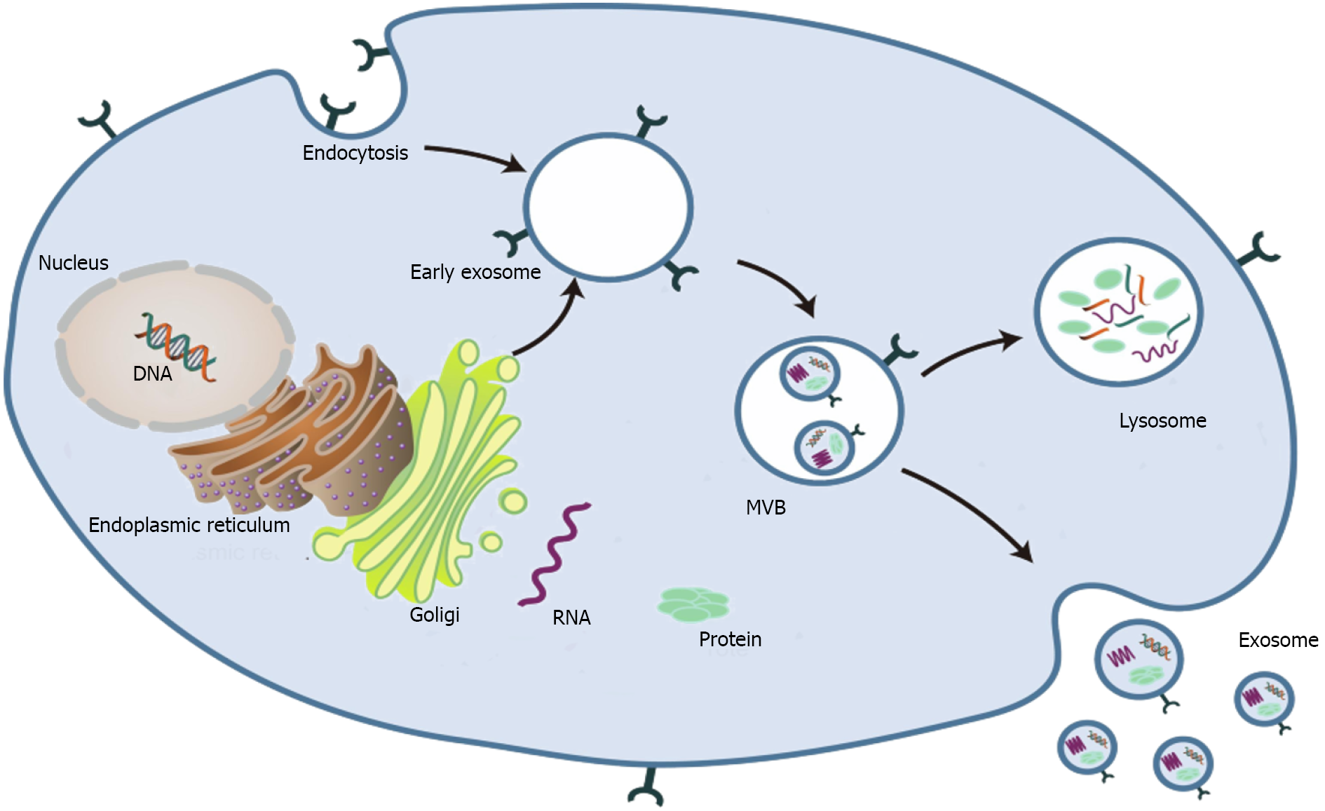
Figure 1 Schematic profile of the biogenesis of exosomes[2].
MVB: Multivesicular body. Citation: Liu Y, Wang Y, Lv Q, Li X. Exosomes: From garbage bins to translational medicine. Int J Pharm 2020; 583: 119333. Copyright© The Authors 2020. Published by Elsevier B.V.
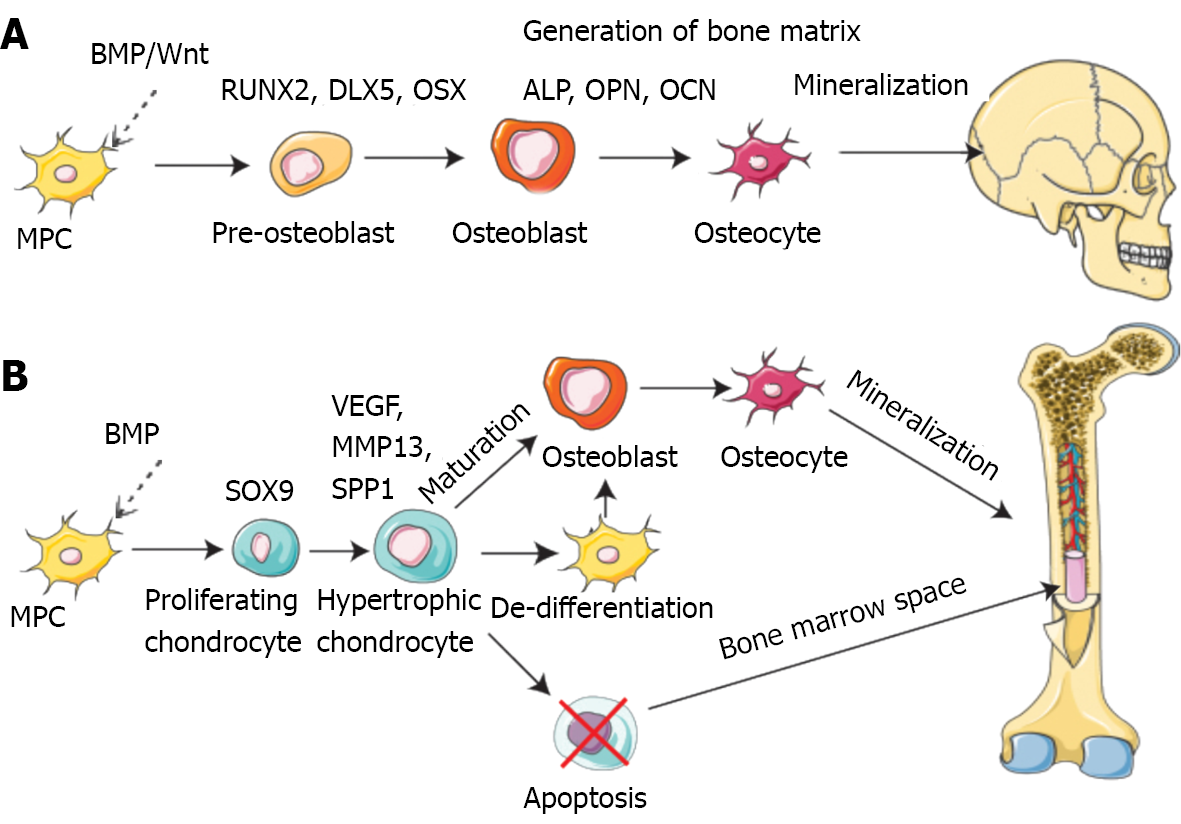
Figure 2 Pathways of bone formation during development[46].
A: Direct (intramembranous); B: Indirect (endochondral). BMP: Bone morphogenetic protein; MPC: Muscle precursor cell; VEGF: Vascular endothelial growth factor; RUNX2: Runt-related transcription factor 2; DLX5: Distal-less homeobox gene 5; ALP: Alkaline phosphatase; OPN: Osteopontin; OCN: Osteocalcin; MMP13: Matrix metallopeptidase-13. Citation: Schott NG, Friend NE, Stegemann JP. Coupling osteogenesis and vasculogenesis in engineered orthopedic tissues. Tissue Eng Part B Rev 2021; 27: 199-214. Copyright© The Authors 2021. Published by Mary Ann Liebert, Inc.
- Citation: Ren YZ, Ding SS, Jiang YP, Wen H, Li T. Application of exosome-derived noncoding RNAs in bone regeneration: Opportunities and challenges. World J Stem Cells 2022; 14(7): 473-489
- URL: https://www.wjgnet.com/1948-0210/full/v14/i7/473.htm
- DOI: https://dx.doi.org/10.4252/wjsc.v14.i7.473


