Copyright
©The Author(s) 2022.
World J Stem Cells. May 26, 2022; 14(5): 347-361
Published online May 26, 2022. doi: 10.4252/wjsc.v14.i5.347
Published online May 26, 2022. doi: 10.4252/wjsc.v14.i5.347
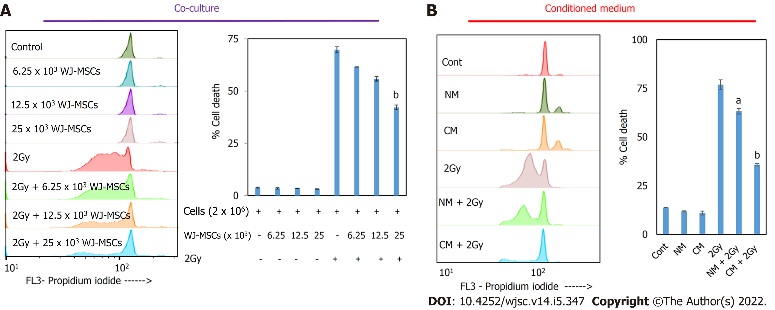
Figure 1 In vitro radioprotective property of Wharton’s jelly-derived mesenchymal stromal/stem cells and its conditioned medium to murine splenic lymphocytes.
Irradiated splenic lymphocytes were either co-cultured with Wharton’s jelly-derived mesenchymal stromal/stem cells (WJ-MSCs) or cultured with conditioned media collected from WJ-MSCs culture for 24 h. Cells were stained with propidium iodide, and cell death was enumerated in lymphocytes. A: Flow cytometry data along with bar graph showed therapeutic radioprotection to lymphocytes when cultured in the presence of WJ-MSCs; B: Flow cytometry data along with bar graph showed radioprotection to lymphocytes when cultured in the presence of conditioned medium. aP < 0.05; bP < 0.005 compared to radiation control. NM: Non-conditioned media; CM: Conditioned media.
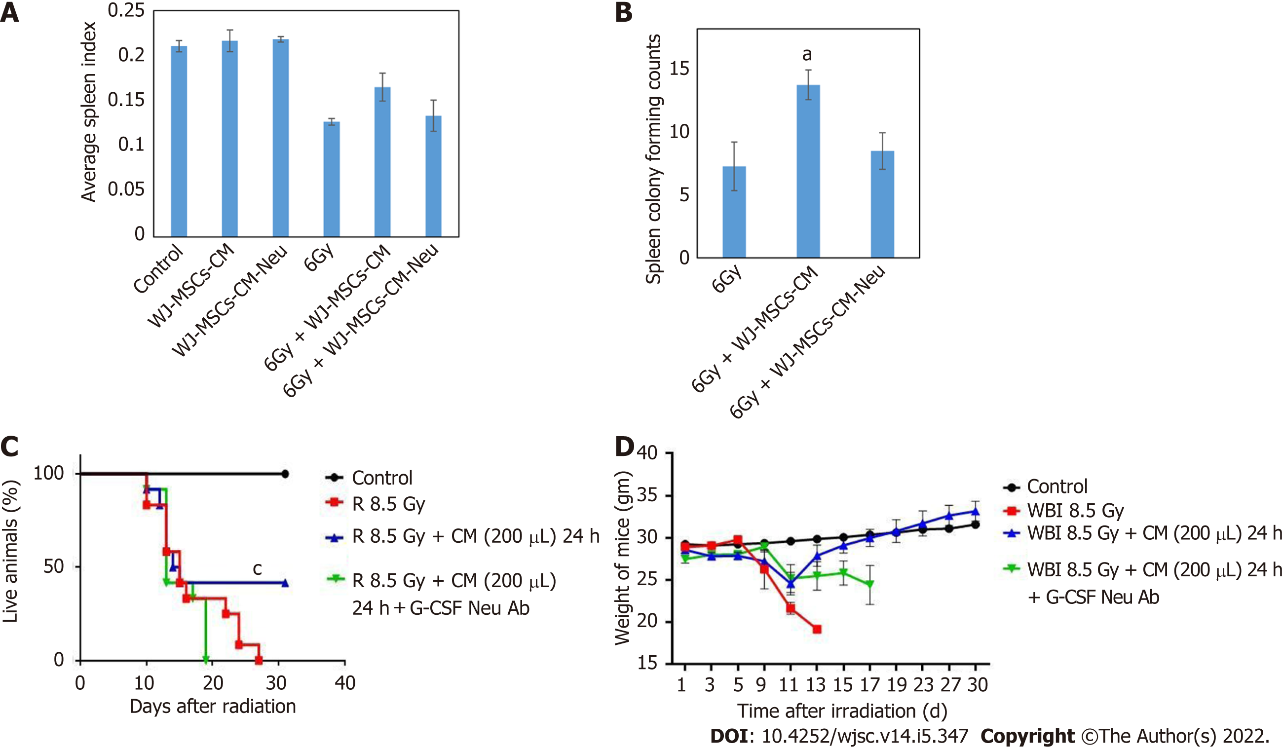
Figure 2 Role of granulocyte-colony stimulating factor in Wharton’s jelly-derived mesenchymal stromal/stem cells-conditioned medium-mediated radioprotection.
Wharton’s jelly-derived mesenchymal stromal/stem cells-conditioned medium (WJ-MSCs-CM) was neutralized with anti-granulocyte-colony stimulating factor (G-CSF) antibody and was then infused to irradiated (6 Gy or 8.5 Gy) or control mice. A: Mice were euthanized on day 15 to calculate the average spleen index. Each bar represents mean ± standard error of the mean from 5 mice; B: Bar chart showed the number of colony forming units in the spleen in different groups; C: Mice were exposed to 8.5 Gy whole-body irradiation and injected intravenously with PBS or WJ-MSC-CM or WJ-MSC-CM neutralized with G-CSF 24 h after irradiation (n = 12 mice per group) and monitored for survival; D: Plot showing changes in body weight for 30 d. aP < 0.05; bP < 0.0001 as compared to whole body irradiation. CM: Conditioned media; G-CSF Neu Ab: Granulocyte-colony stimulating factor neutralizing antibody; WBI: Whole body irradiation.
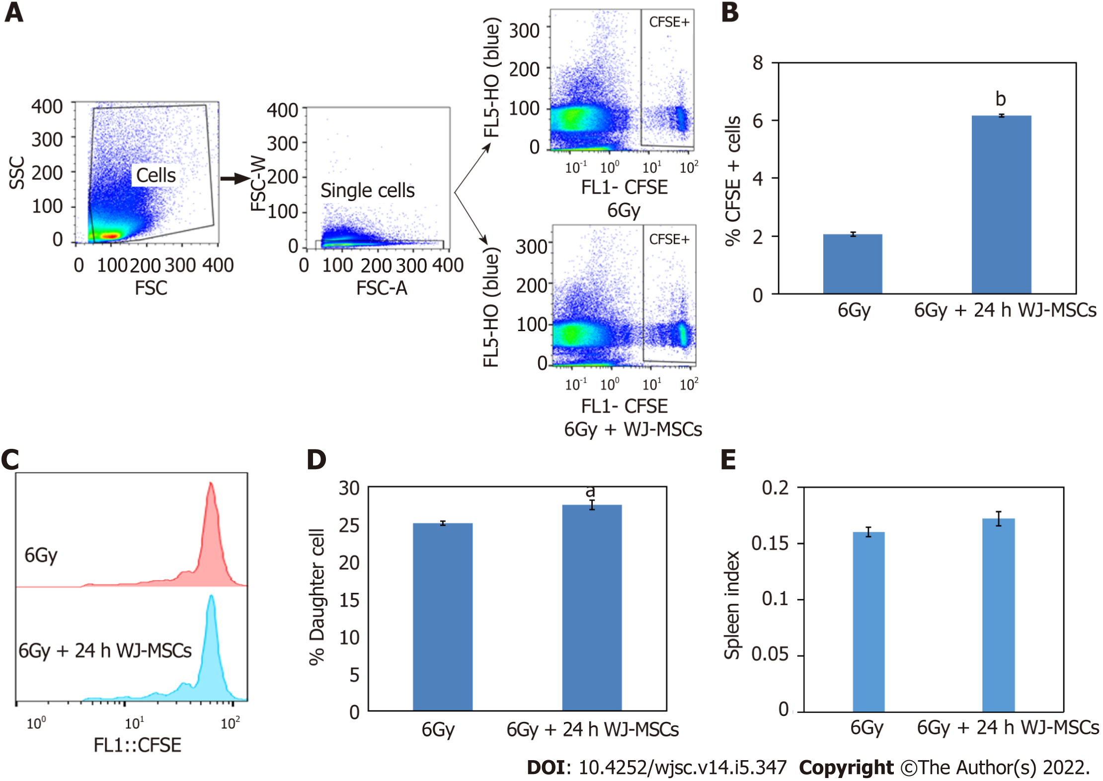
Figure 3 Effect of Wharton's jelly-derived mesenchymal stromal/stem cells on homeostasis-driven proliferation of lymphocytes in lymphopenic mice.
Wharton's jelly-derived mesenchymal stromal/stem cells (WJ-MSCs) were mixed with carboxyfluorescein diacetate succinimidyl ester (CFSE) labeled lymphocytes were injected into 6 Gy exposed mice. In another group, only CFSE labeled lymphocytes were injected into irradiated mice. A: Dot plot shows gating strategy to monitor the proliferation of lymphocytes after acquiring the samples on flow cytometer; B: Bar graph showing % of CFSE positive lymphocytes with and without WJ-MSCs; C: Flow cytometric histograms showing lymphocyte proliferation; D: Bar graph showing percent daughter cells; E: Bar graph showing spleen index (n = 4 mice per group). aP < 0.05; bP < 0.005 as compared to radiation alone.
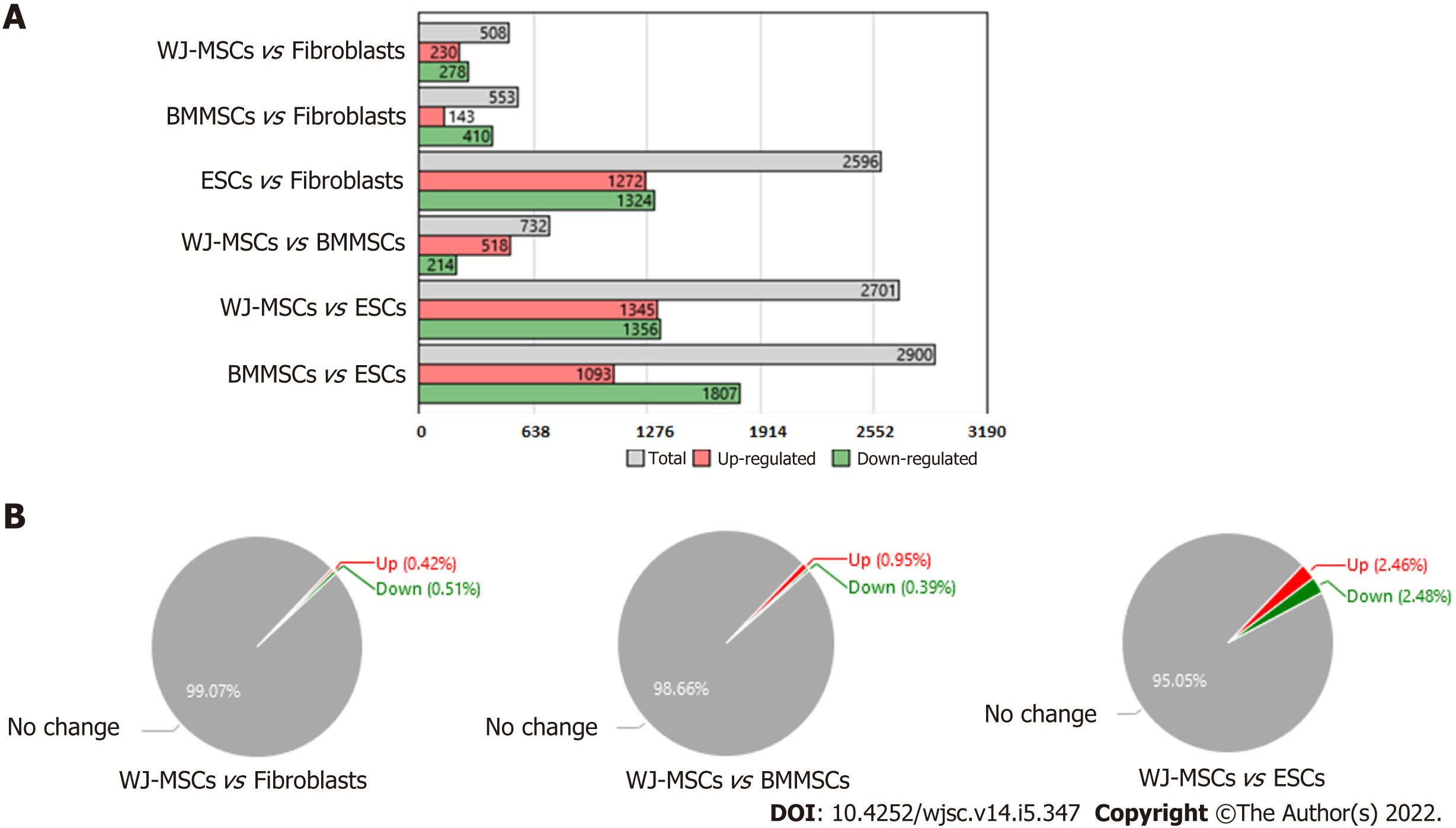
Figure 4 Comparative differential gene expression analysis of stem cells (Wharton’s jelly-derived mesenchymal stromal/stem cells, bone marrow mesenchymal stromal/stem cells and embryonic stem cells) vs fibroblasts.
A: Bar graph showed differential gene expression among different stem cells as well as with fibroblasts. Expression Analysis Settings: Fold change in gene expression < -5 or > 5 and P < 0.05, Anova Method: Ebayes; B: Pie chart showed percentage of gene expression in Wharton’s jelly-derived mesenchymal stromal/stem cells (MSCs), bone marrow MSCs, and embryonic stem cells as compared to fibroblasts. WJ-MSC: Wharton’s jelly-derived mesenchymal stromal/stem cells; BMMSC: Bone marrow mesenchymal stem cells; ESC: Embryonic stem cells.
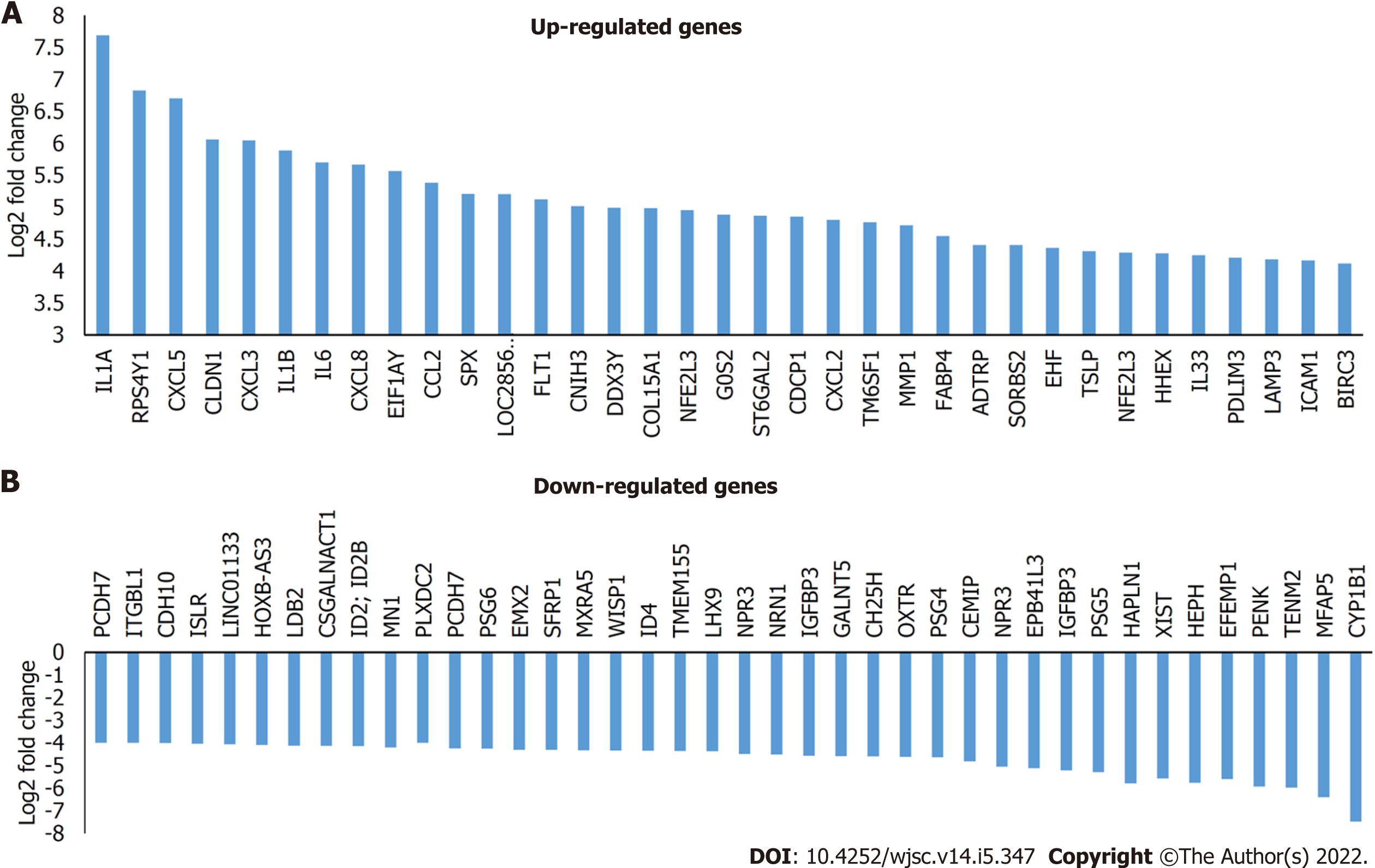
Figure 5 Differential gene expression in Wharton’s jelly-derived mesenchymal stromal/stem cells compared to fibroblasts.
A: Bar graph showed some of the highly upregulated genes in Wharton’s jelly-derived mesenchymal stromal/stem cells (WJ-MSCs); B: Bar graph showed some of the highly downregulated genes in WJ-MSCs.
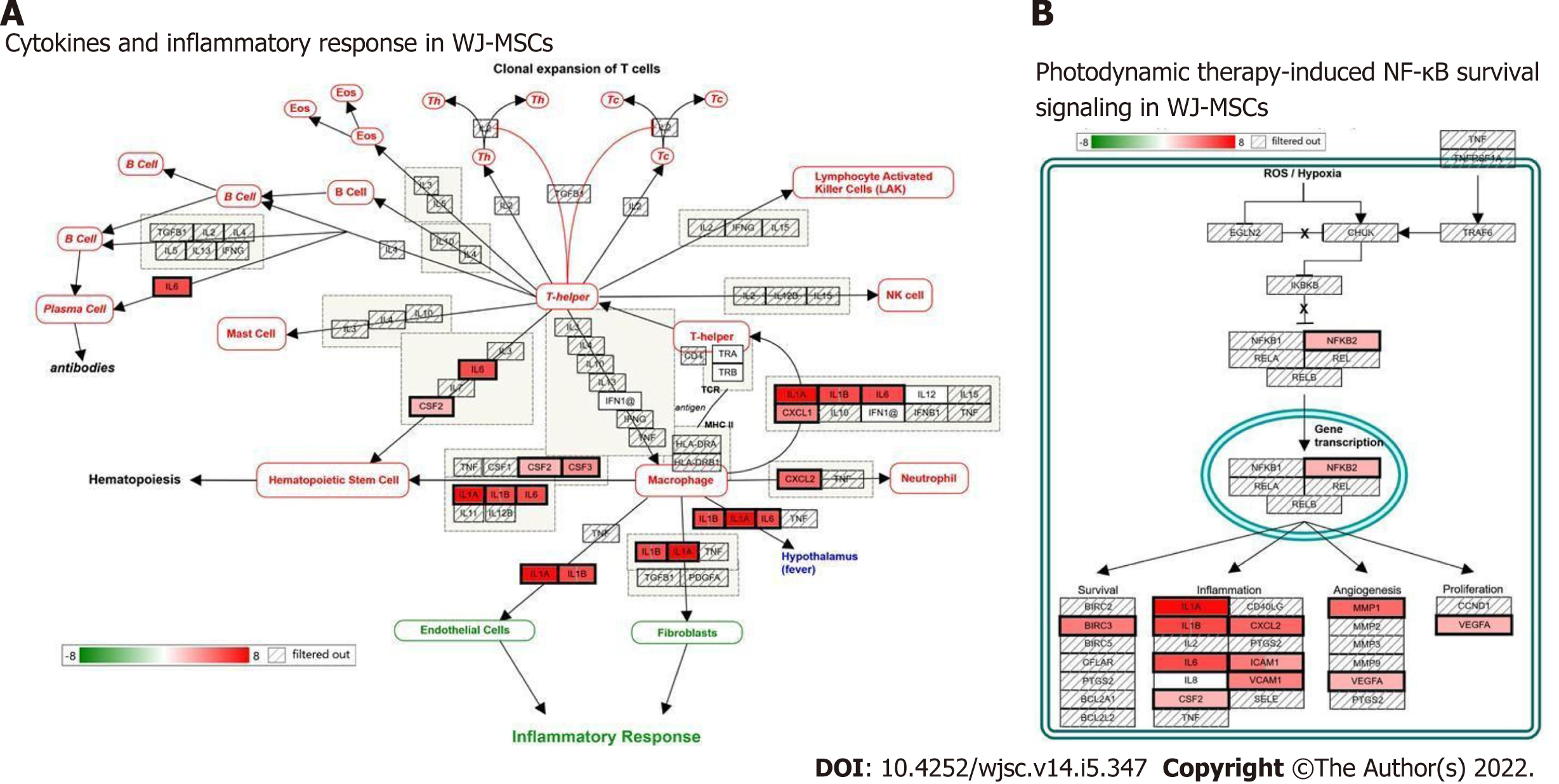
Figure 6 Two of the highly modulated pathways by differentially expressed genes of Wharton’s jelly-derived mesenchymal stromal/stem cells.
A: Cytokines and inflammatory response pathway; B: Photodynamic induced nuclear factor kappa B survival pathway. WJ-MSC: Wharton’s jelly-derived mesenchymal stromal/stem cells; NF-κB: Nuclear factor kappa B; NK cell: Natural killer cell; ROS: Reactive oxygen species.
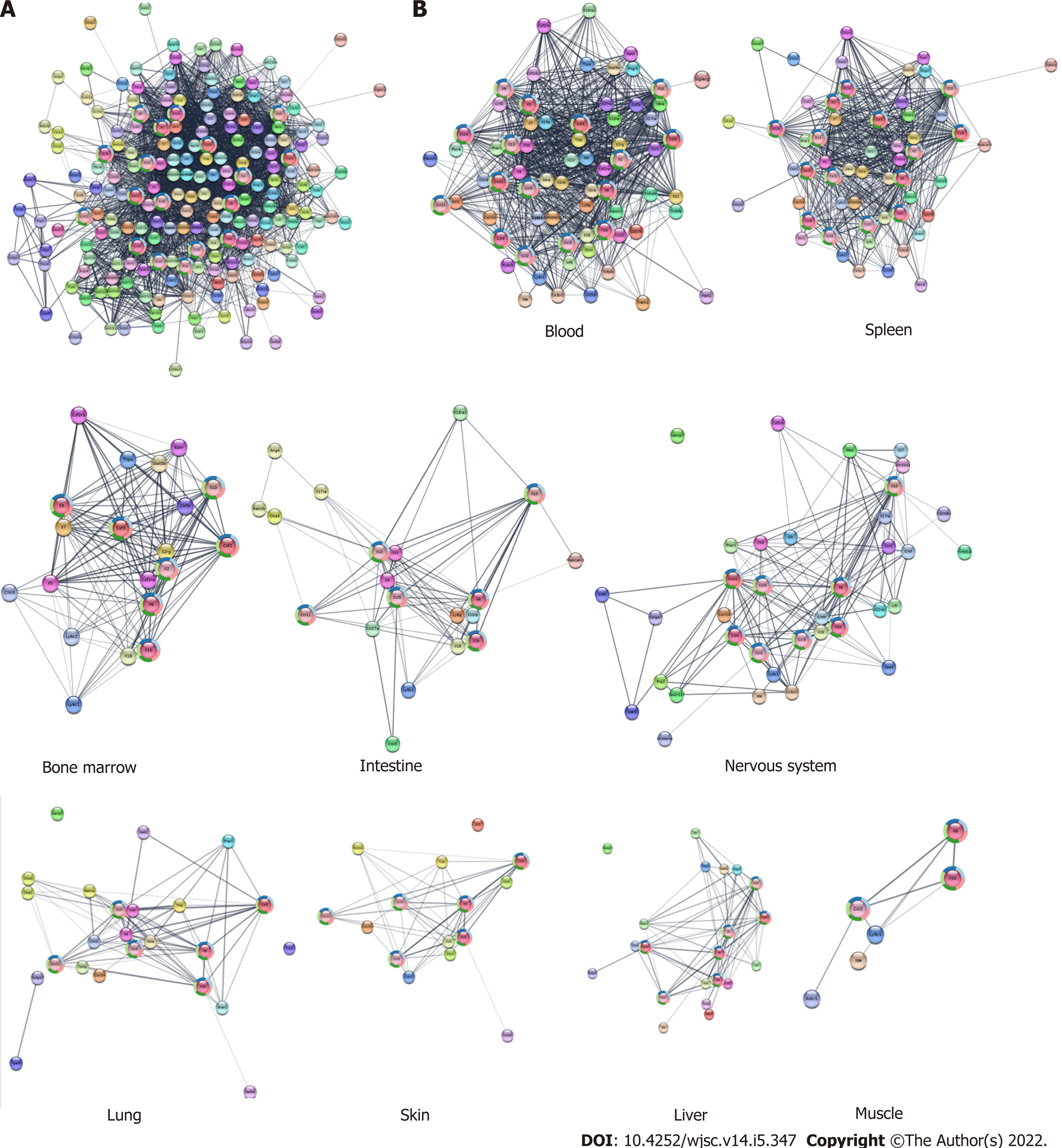
Figure 7 Network of protein-protein interaction obtained using serum cytokines/chemokines in mice after Wharton’s jelly-derived mesenchymal stromal/stem cell infusion.
A: Extended protein-protein interaction (PPI) network was constructed using data from serum collected from mice injected with Wharton’s jelly-derived mesenchymal stromal/stem cells; B: Tissue specific extended PPI network created from the mouse serum cytokine data.
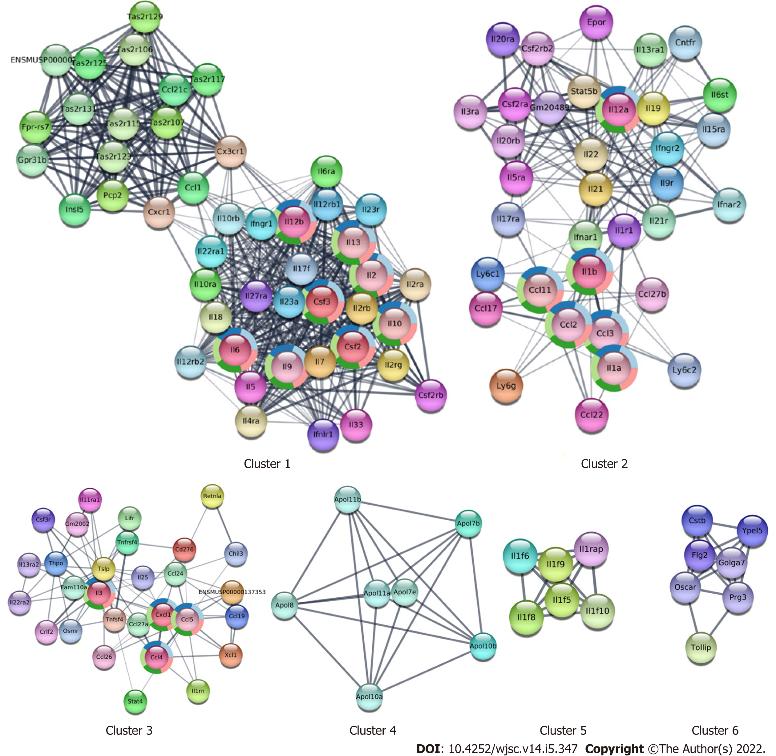
Figure 8 Cluster analysis of the protein-protein interaction network.
Different clustered nodes and edges derived from the extended protein-protein interaction from the secreted mouse cytokine/chemokine data.
- Citation: Maurya DK, Bandekar M, Sandur SK. Soluble factors secreted by human Wharton’s jelly mesenchymal stromal/stem cells exhibit therapeutic radioprotection: A mechanistic study with integrating network biology. World J Stem Cells 2022; 14(5): 347-361
- URL: https://www.wjgnet.com/1948-0210/full/v14/i5/347.htm
- DOI: https://dx.doi.org/10.4252/wjsc.v14.i5.347


