Copyright
©The Author(s) 2021.
World J Stem Cells. Jan 26, 2021; 13(1): 78-90
Published online Jan 26, 2021. doi: 10.4252/wjsc.v13.i1.78
Published online Jan 26, 2021. doi: 10.4252/wjsc.v13.i1.78
Figure 1 Beneficial effect of mesenchymal stem cells or iMSCs in neuron/glial cocultures.
A: Mesenchymal stem cells (MSCs) were immunoreactive for green fluorescent protein; B: Diagram of cocultures. Neurons and glia were seeded directly in culture plates, whereas MSCs or iMSCs were seeded on transwell inserts with a 0.4 m pore size; C: Neurons and glia only; D: Coculture of MSCs and neurons/glia; E: Coculture of iMSCs with neurons/glia; F: Quantification of the βIII tubulin density in the cultures shown in C-E. The results are reported as the mean ± SE. aP < 0.05 and cP < 0.001 indicate a significant difference between cocultures and neuron/glia cultures; dP < 0.001 indicates a significant difference between two kinds of cocultures. GFP: Green fluorescent protein; IR: Immunoreactive; MSC: Mesenchymal stem cell.
Figure 2 Purification and identification of adenoviral-glial cell line-derived neurotrophic factor.
A: Adenoviral-glial cell line-derived neurotrophic factor (Ad-GDNF) bands after CsCl ultracentrifugation; B: Identification of the correct 205 bp GDNF gene in Ad-GDNF; C: GDNF mRNA level was enhanced in Ad-GDNF-infected cultures compared to mock (Adpgk)-infected cultures; D: Neuronal connections were increased in Ad-GDNF-infected cultures compared to control cultures. Ad-GDNF: Adenoviral-glial cell line-derived neurotrophic factor; PCR: Polymerase chain reaction; IR: Immunoreactive.
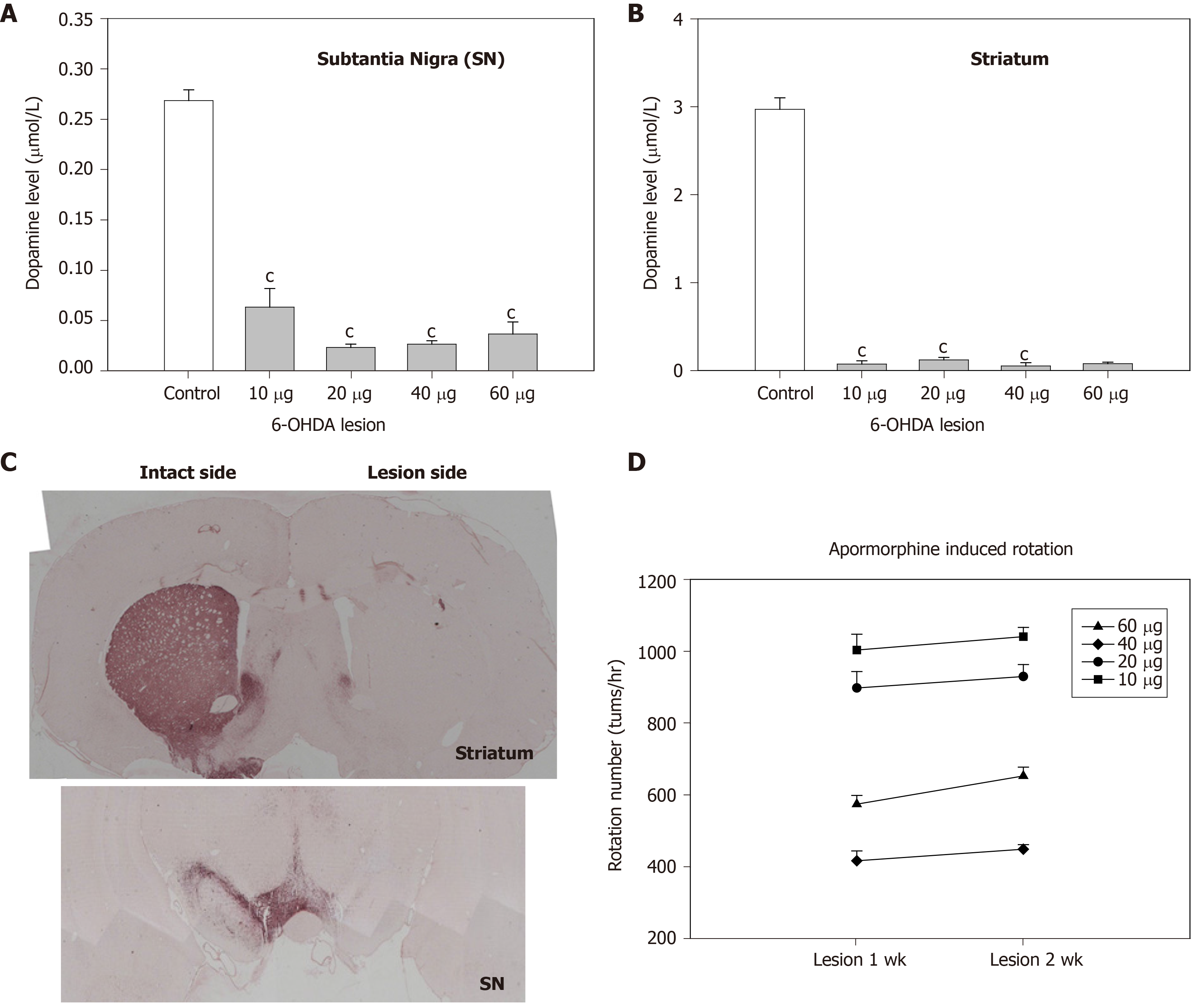
Figure 3 Dose-dependent effect of 6-OHDA infusion into the medial forebrain bundle in Sprague-Dawley rats.
A: Depletion of dopamine levels by different doses of 6-OHDA in the substantia nigra (SN); B: Depletion of dopamine levels by different doses of 6-OHDA in the striatum; C: The tyrosine hydroxylase-immunoreactive area, which indicates dopaminergic neuronal density, in the SN and the striatum of hemiparkinsonian rats 2 wk after unilateral lesion of the nigrostriatal pathway by 20 g of 6-OHDA; D: Apomorphine-induced turning behavior in hemiparkinsonian rats 1 and 2 wk after lesion induction by different doses of 6-OHDA (cP < 0.001). SN: Substantia nigra.
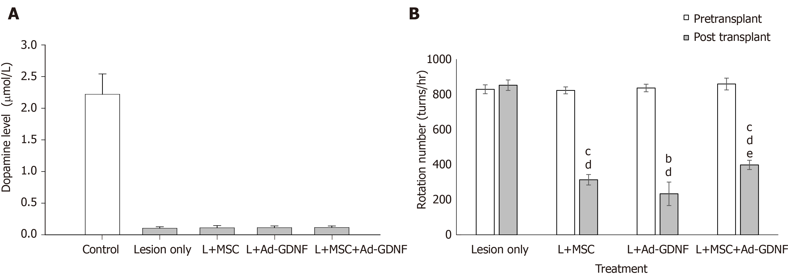
Figure 4 Effect of mesenchymal stem cells and/or adenoviral-glial cell line-derived neurotrophic factor treatment on striatal dopamine levels and apomorphine-induced turning behavior in hemiparkinsonian rats.
A: Dopamine (DA) levels in the striatum of different treatment groups; B: Number of apomorphine-induced rotations by hemiparkinsonian rats before and 4 wk after infusion of mesenchymal stem cells (MSCs) and/or adenoviral-glial cell line-derived neurotrophic factor (Ad-GDNF). L indicates 6-OHDA-lesion. The results are reported as the mean ± SE. aP < 0.05 and cP < 0.001, compared to the pretransplantation level in each group; d P < 0.001, compared to the posttransplantation level of the lesion only group; e P < 0.01, Ad-GDNF compared to the MSC + Ad-GDNF group. L: Lesion; MSC: Mesenchymal stem cell; Ad-GDNF: Adenoviral-glial cell line-derived neurotrophic factor.
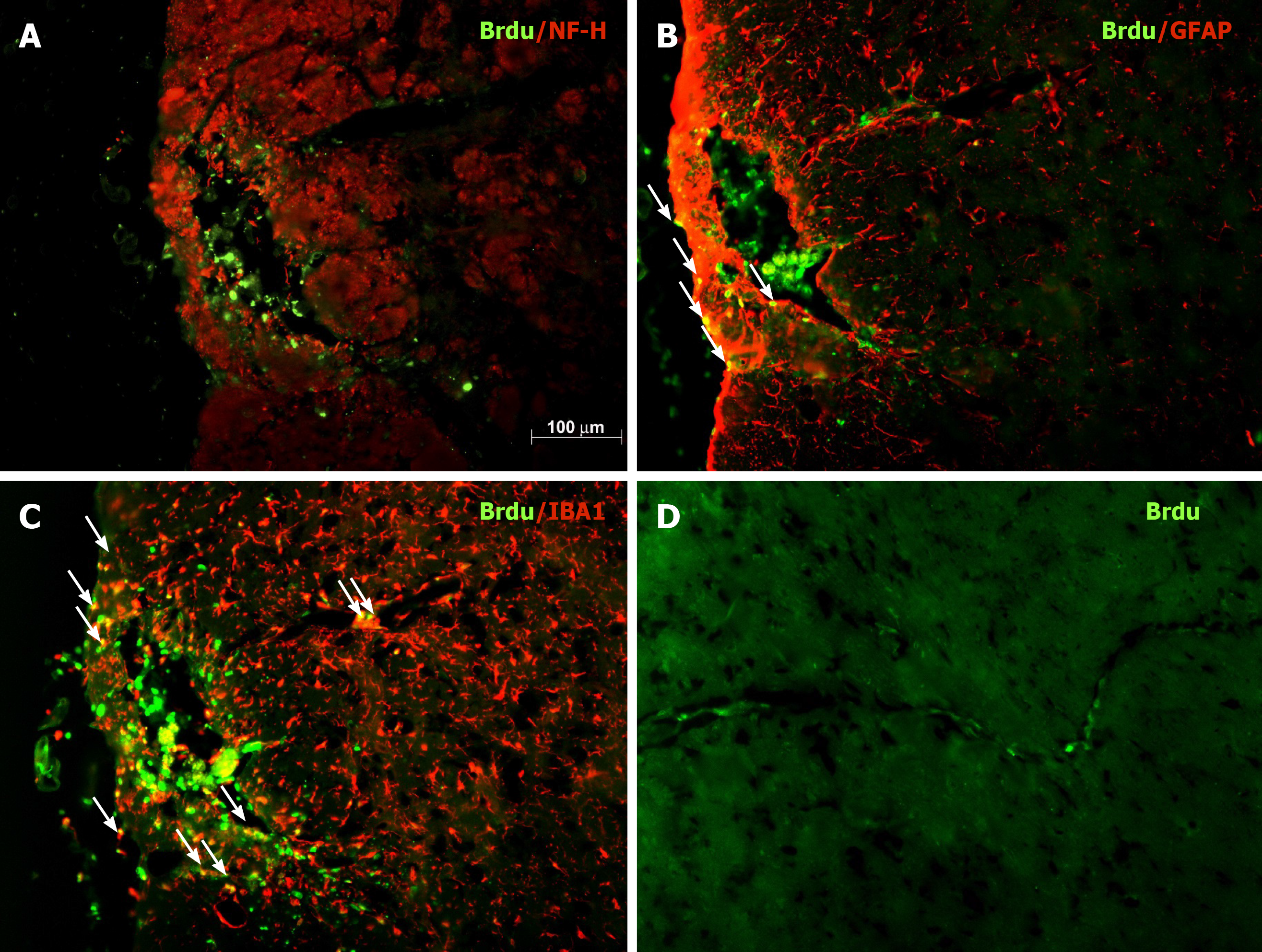
Figure 5 Phenotypic characterization of grafted bone marrow mesenchymal stem cells in the substantia nigra of hemiparkinsonian rats.
A-C: Representative micrographs of BrdU staining of coronal sections of the grafted substantia nigra. Photos of double staining (magnification, 200 ×) are shown. Green: BrdU immunoreactivity; red: NF-H immunoreactivity (neurons, panel A), GFAP immunoreactivity (astroglia, panel B), and IBA1 immunoreactivity (microglia, panel C); D: BrdU-immunoreactive BMSCs along the blood vessels near the graft site.
- Citation: Tsai MJ, Hung SC, Weng CF, Fan SF, Liou DY, Huang WC, Liu KD, Cheng H. Stem cell transplantation and/or adenoviral glial cell line-derived neurotrophic factor promote functional recovery in hemiparkinsonian rats. World J Stem Cells 2021; 13(1): 78-90
- URL: https://www.wjgnet.com/1948-0210/full/v13/i1/78.htm
- DOI: https://dx.doi.org/10.4252/wjsc.v13.i1.78


