Copyright
©The Author(s) 2020.
World J Stem Cells. Mar 26, 2020; 12(3): 203-221
Published online Mar 26, 2020. doi: 10.4252/wjsc.v12.i3.203
Published online Mar 26, 2020. doi: 10.4252/wjsc.v12.i3.203
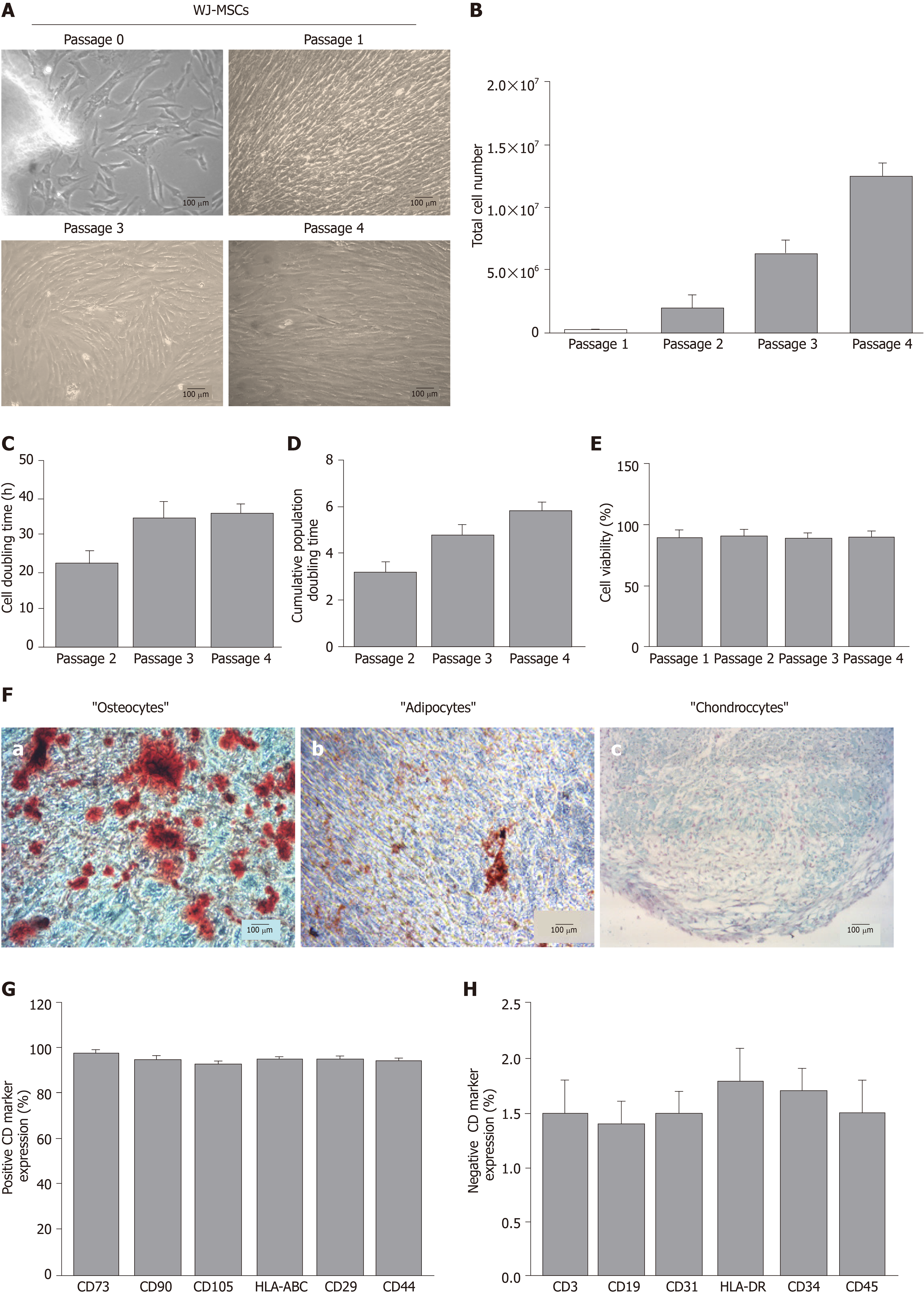
Figure 1 Evaluation of mesenchymal stromal cells derived from the Wharton’s Jelly.
A: Morphological features of mesenchymal stromal cells derived from the Wharton’s Jelly tissue (WJ-MSCs) from P0 to P4 (A-a to A-d); B-F: Determination of total cell number (B), cell doubling time (C), cumulative PD (D) and cell viability (E) of WJ-MSCs from P0 to P4. Evaluation of tri-lineage differentiation capability of WJ-MSCs into “osteocytes” (F-a), “adipocytes” (F-b) and “chondrocytes” (F-c) as indicated by Alizarin Red-S, Oil-Red-O and Alcian blue, respectively. G, H: Positive (G) and negative (H) expression of CD markers in WJ-MSCs based on flow cytometric analysis. Images A-a to A-d and F-a to F-c were obtained with original magnification 10× and 100 μm scale bars. WJ-MSCs: Mesenchymal stromal cells derived from the Wharton’s Jelly tissue.
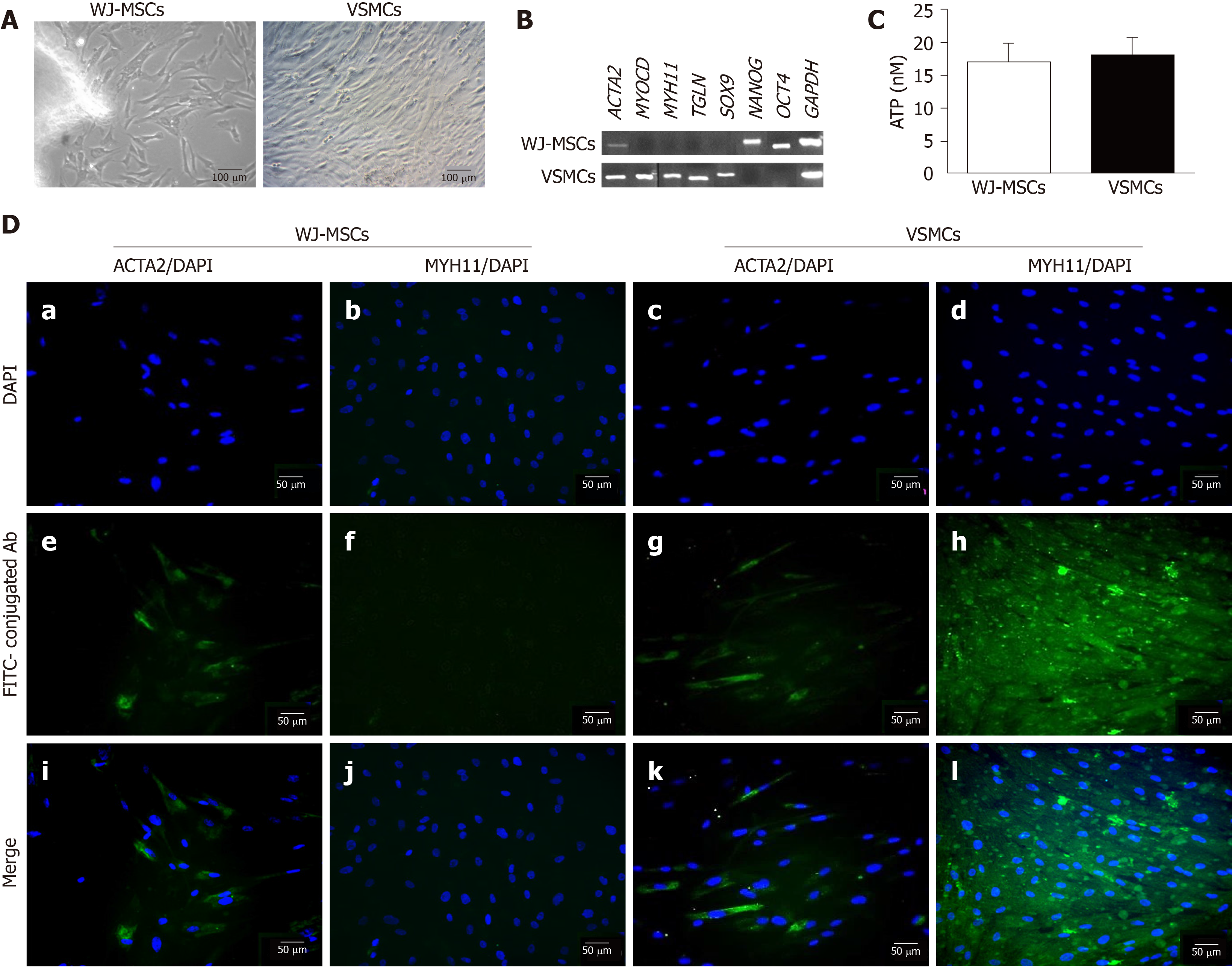
Figure 2 Differentiation of mesenchymal stromal cells derived from the Wharton’s Jelly tissue into vascular smooth muscle cells.
A: Morphological features of untreated mesenchymal stromal cells derived from the Wharton’s Jelly tissue (WJ-MSCs) and differentiated vascular smooth muscle cells (VSMCs); B: Polymerase chain reaction results regarding the expression of VSMC-specific genes, such as ACTA2, MYOCD, MYH11 and TGLN, and pluripotency-related genes, including NANOG and OCT4 in untreated WJ-MSCs and differentiated VSMCs. GAPDH was the desired house-keeping gene for current analysis; C: Determination of WJ-MSC and VSMC proliferation by performing the ATP assay; Indirect immunofluorescence against the early VSMC marker ACTA2 and late VSMC marker MYH11 in untreated WJ-MSCs (D-a, D-e, D-i and D-b, D-f, D-j) and differentiated VSMCs (D-c, D-g, D-k and D-d, D-h, D-l) in combination with DAPI, respectively. Images A-a and A-b were presented with 10× original magnification and 100 μm scale bars. Images D-a to D-l were presented with 20× original magnification and 50 μm scale bars. WJ-MSCs: Mesenchymal stromal cells derived from the Wharton’s Jelly tissue; VSMCs: Vascular smooth muscle cells.
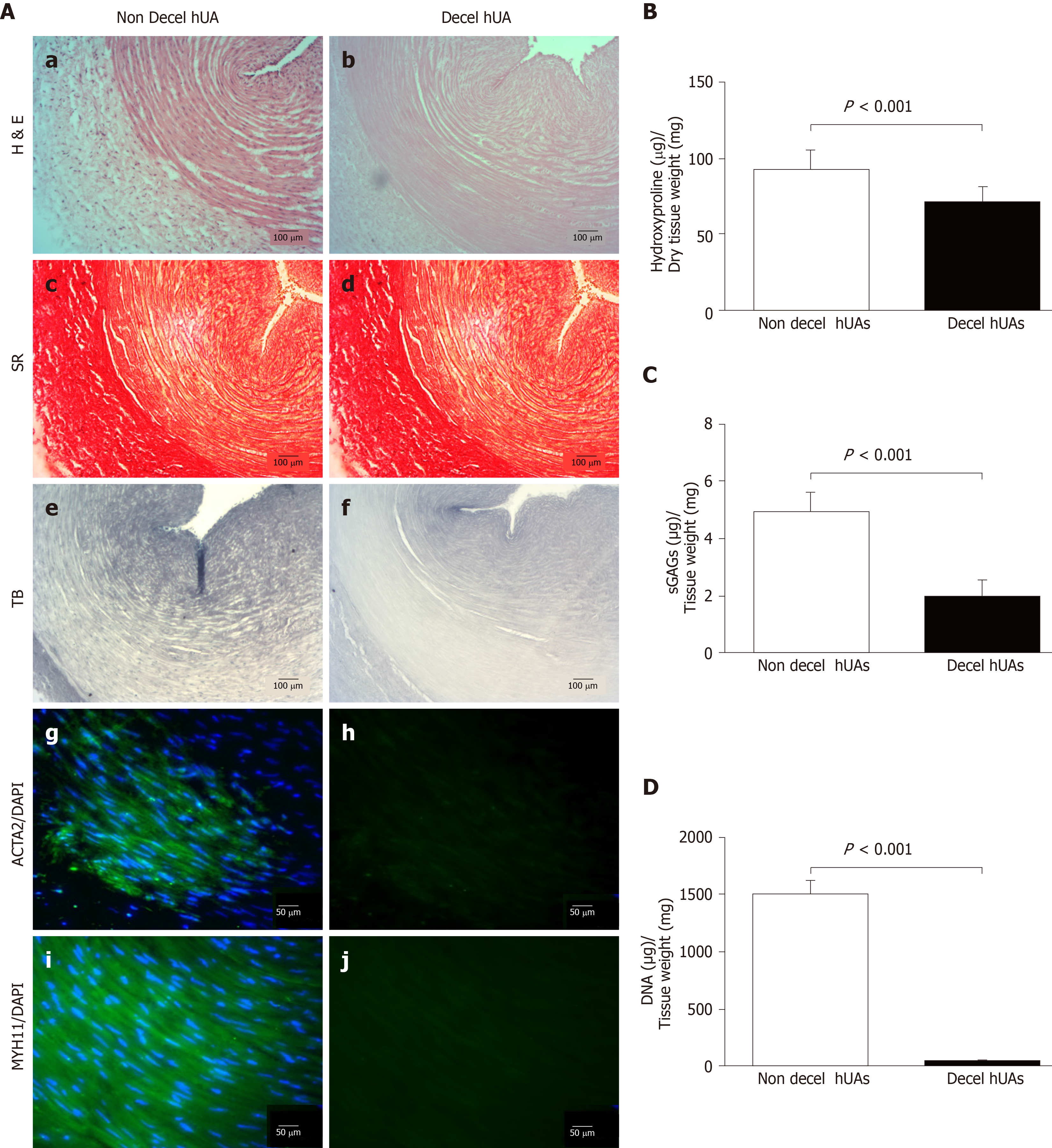
Figure 3 Histological and biochemical analysis of decellularized human umbilical arteries.
A: Histological analysis with H & E (A-a, A-b), SR (A-c, A-d) and TB (A-e, A-f) in non-decellularized and decellularized human umbilical arteries (hUAs). Indirect immunofluorescence against ACTA2 (A-g, A-h) and MYH11 (A9,10) in combination with DAPI was performed in non-decellularized and decellularized hUAs; B-D: Biochemical analysis involved the determination of total hydroxyproline (B), sGAG (C) and DNA content (D) in non-decellularized and decellularized hUAs. Statistically significant differences were observed in total hydroxyproline (P < 0.05), sGAG (P < 0.001) and DNA (P < 0.001) content between non decellularized and decellularized hUAs. Images A-a to A-f were presented with original magnification 10× and 100 μm scale bars. Images A-g to A-j were presented with original magnification 20× and 50 μm scale bars. Non Decel hUA: Non decellularized human umbilical artery; Decell hUA: Decellularized human umbilical artery.
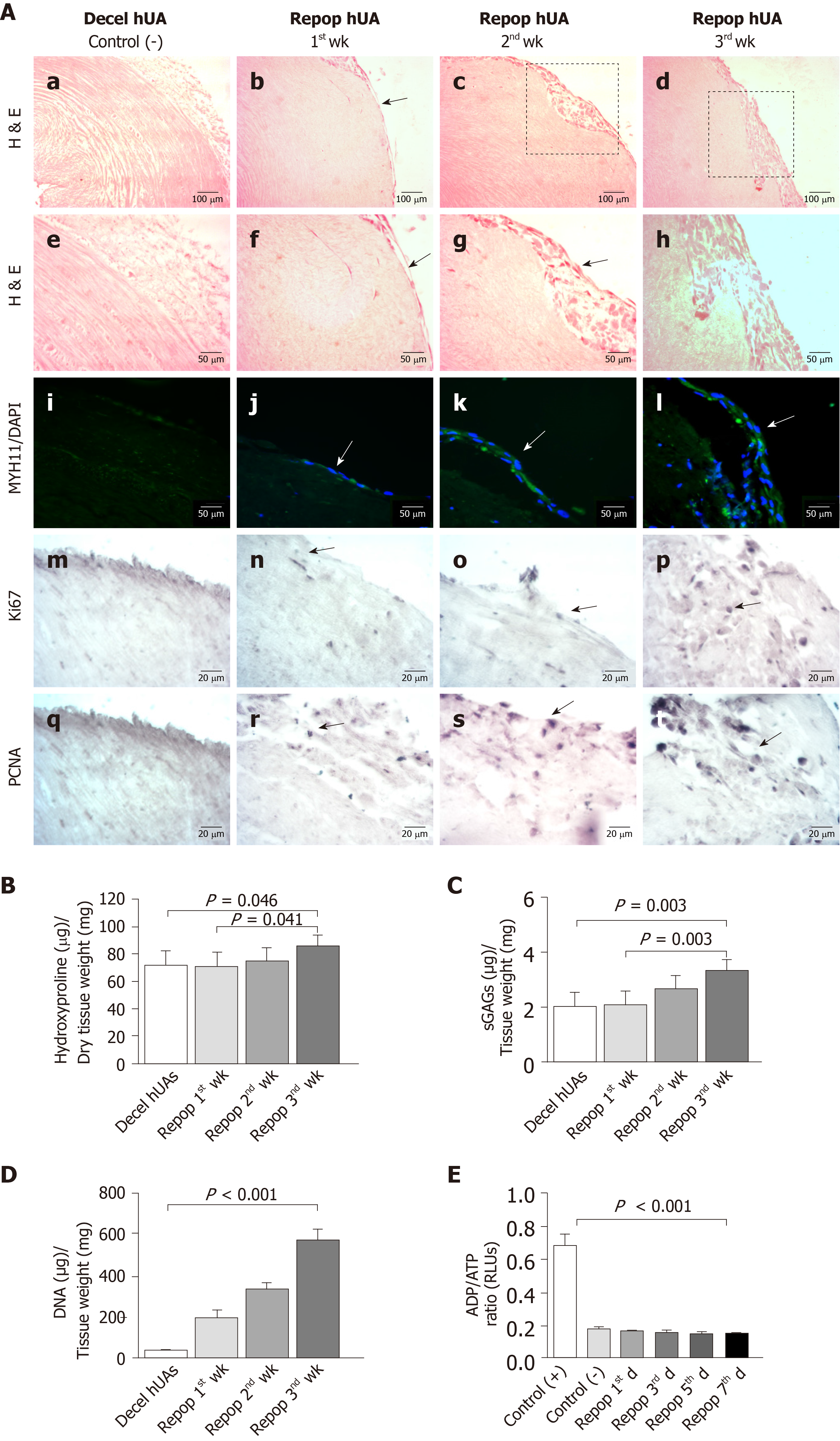
Figure 4 Repopulation of decellularized human umbilical arteries with vascular smooth muscle cells.
A: Histological analysis with H & E of decellularized human umbilical arteries (hUAs) (A-a, A-e), repopulated hUAs after 1st wk (A-b, A-f), 2nd wk (A-c, A-g) and 3rd wk (A-d, A-h). Indirect immunofluorescence against MYH11 in combination with DAPI of decellularized hUAs (A-i), repopulated hUAs after 1st wk (A-j), 2nd wk (A-k) and 3rd wk (A-l). Immunohistochemistry against Ki67 and PCNA of decellularized hUAs (A-m, A-q), and repopulated hUAs after 1st wk (A-n, A-r), 2nd wk (A-o, A-s) and 3rd wk (A-p, A-t). Images A-a to d were presented with original magnification 10×, 100 μm scale bars. Images A-f to l were presented with original magnification 20× and 50 μm scale bars. Images A-i to t were presented with original magnification 40× and 20 μm scale bars; B, C: Total hydroxyproline (B) and sGAG (C) quantification of hUAs before and after repopulation with VSMCs; D, E: Determination of DNA content (D) and ADP/ATP ratio (E). Statistically significant differences in total hydroxyproline, sGAG, DNA content and ADP/ATP ratio were observed between the study groups (P < 0.05). Decel hUA: decellularized human umbilical artery, repop hUA: repopulated human umbilical artery.
- Citation: Mallis P, Papapanagiotou A, Katsimpoulas M, Kostakis A, Siasos G, Kassi E, Stavropoulos-Giokas C, Michalopoulos E. Efficient differentiation of vascular smooth muscle cells from Wharton’s Jelly mesenchymal stromal cells using human platelet lysate: A potential cell source for small blood vessel engineering. World J Stem Cells 2020; 12(3): 203-221
- URL: https://www.wjgnet.com/1948-0210/full/v12/i3/203.htm
- DOI: https://dx.doi.org/10.4252/wjsc.v12.i3.203


