Copyright
©The Author(s) 2020.
World J Stem Cells. Dec 26, 2020; 12(12): 1576-1590
Published online Dec 26, 2020. doi: 10.4252/wjsc.v12.i12.1576
Published online Dec 26, 2020. doi: 10.4252/wjsc.v12.i12.1576
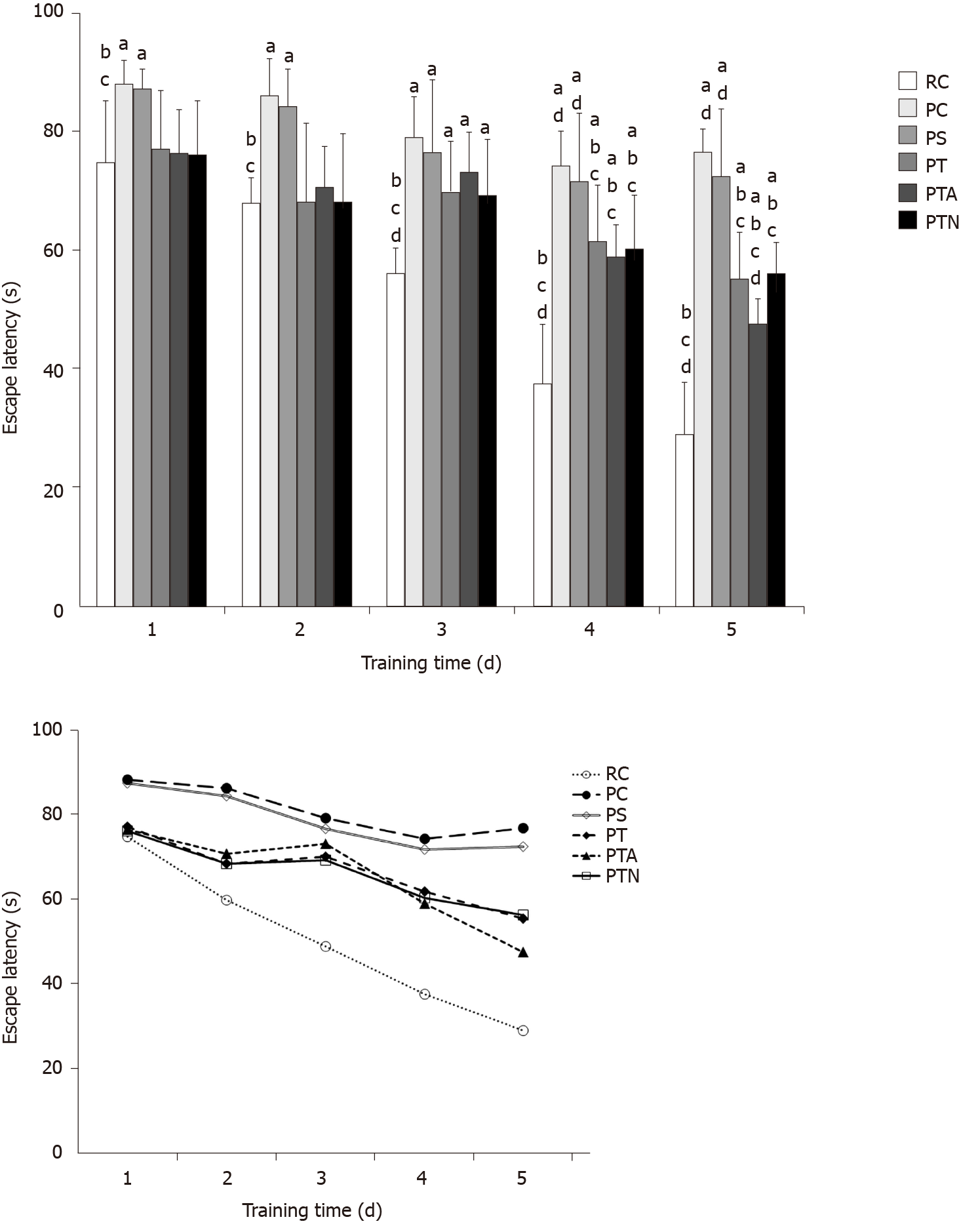
Figure 1 Morris water maze test for behavior of senescence-accelerated mouse.
The time elapsed from entering the water to finding the platform was recorded as escape latency. If the mouse could not find the platform within 90 s, the escape latency was recorded as 90 s. aP < 0.05 when compared to senescence-accelerated mouse resistant 1 control group, bP < 0.05 when compared to senescence-accelerated mouse prone 8 (SAMP8) control group, cP < 0.05 when compared to SAMP8 sham operation group, dP < 0.05 when compared to SAMP8 neural stem cells transplantation with non-acupoint group. RC: Senescence-accelerated mouse resistant 1 control group; PC: Senescence-accelerated mouse prone 8 control group; PS: Senescence-accelerated mouse prone 8 sham operation group; PT: Senescence-accelerated mouse prone 8 neural stem cells transplantation group; PTA: Senescence-accelerated mouse prone 8 neural stem cells transplantation with acupuncture group; PTN: Senescence-accelerated mouse prone 8 neural stem cells transplantation with non-acupoint group.
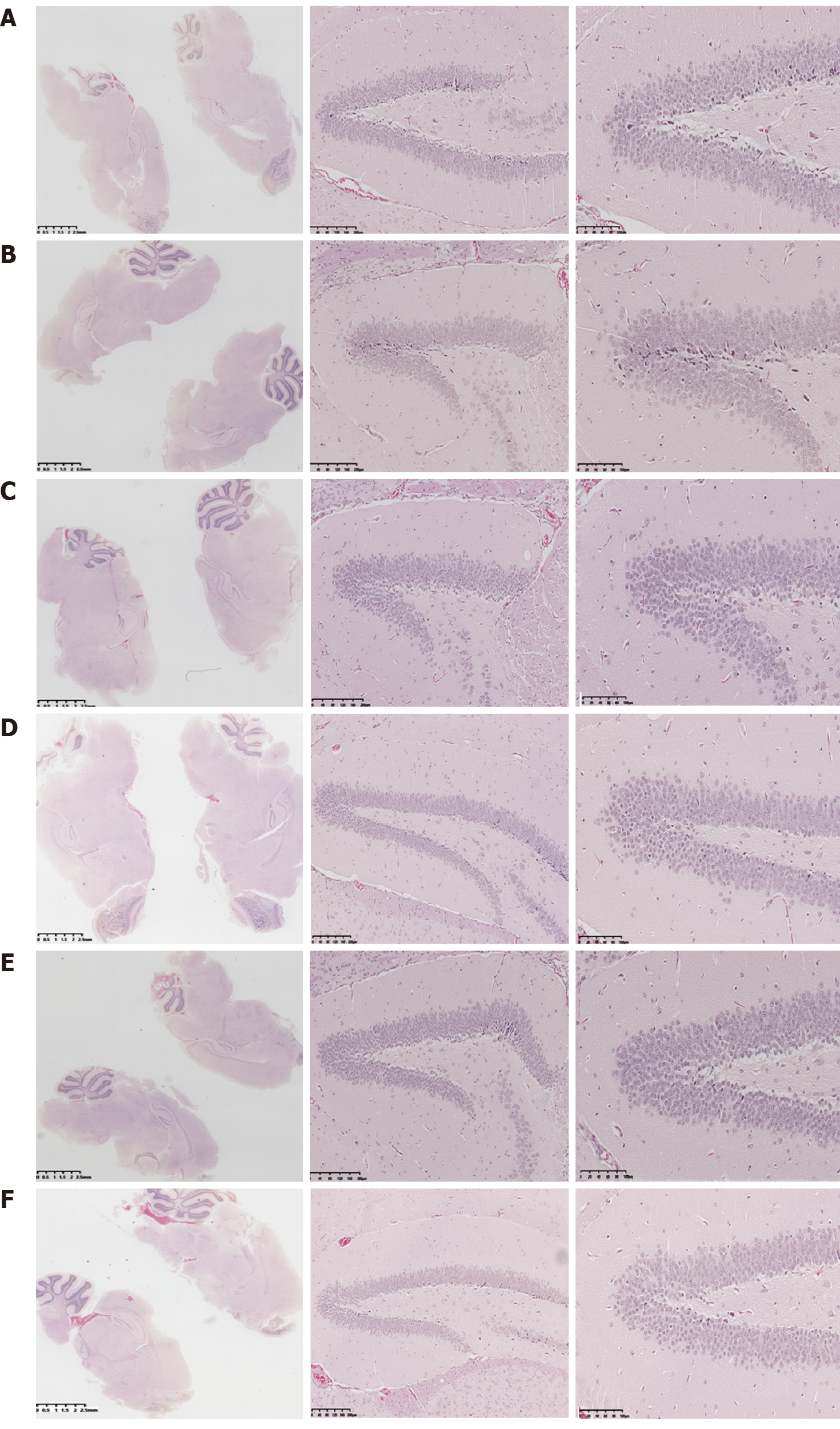
Figure 2 Histopathological changes by hematoxylin-eosin staining.
Coronal paraffin sections of the brain tissue of three mice in each experimental group were prepared. After BrdU-positive staining was confirmed under a microscope, adjacent sections were dewaxed and subjected to hematoxylin-eosin staining. A: Senescence-accelerated mouse resistant 1 control group; B: Senescence-accelerated mouse prone 8 control group; C: Senescence-accelerated mouse prone 8 sham operation group; D: Senescence-accelerated mouse prone 8 neural stem cells transplantation group; E: Senescence-accelerated mouse prone 8 neural stem cells transplantation with acupuncture group; F: Senescence-accelerated mouse prone 8 neural stem cells transplantation with non-acupoint group.
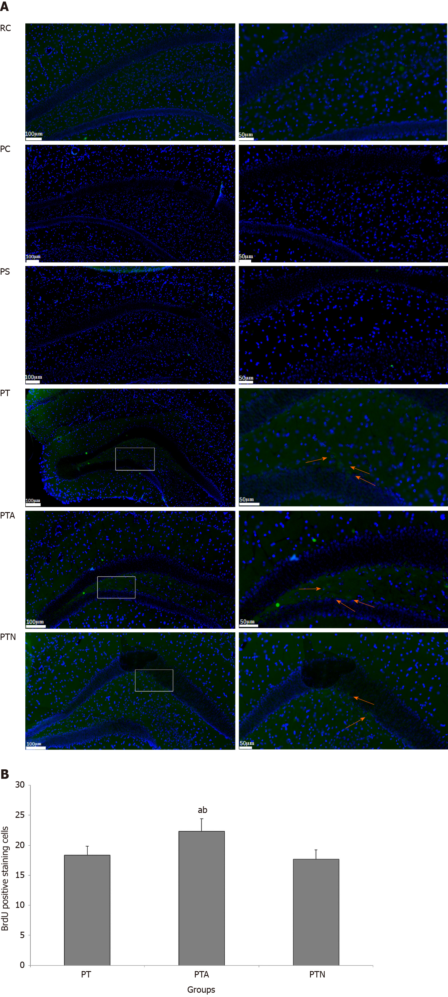
Figure 3 Neural stem cell proliferation by immunofluorescence staining.
Paraffin section of hippocampus tissue of mice was evaluated by indirect immunofluorescence and diaminobenzidine staining. After nuclei were counterstained with hematoxylin, positive BrdU-labeled cells were observed and counted. Arrows indicate positive cells marked by BrdU expression. A: Immunofluorescence staining; B: BrdU positive cell counting. aP < 0.05 when compared to senescence-accelerated mouse prone 8 (SAMP8) neural stem cells (NSCs) transplantation group, bP < 0.05 when compared to SAMP8 NSCs transplantation with non-acupoint group. RC: Senescence-accelerated mouse resistant 1 control group; PC: Senescence-accelerated mouse prone 8 control group; PS: Senescence-accelerated mouse prone 8 sham operation group; PT: Senescence-accelerated mouse prone 8 neural stem cells transplantation group; PTA: Senescence-accelerated mouse prone 8 neural stem cells transplantation with acupuncture group; PTN: Senescence-accelerated mouse prone 8 neural stem cells transplantation with non-acupoint group.
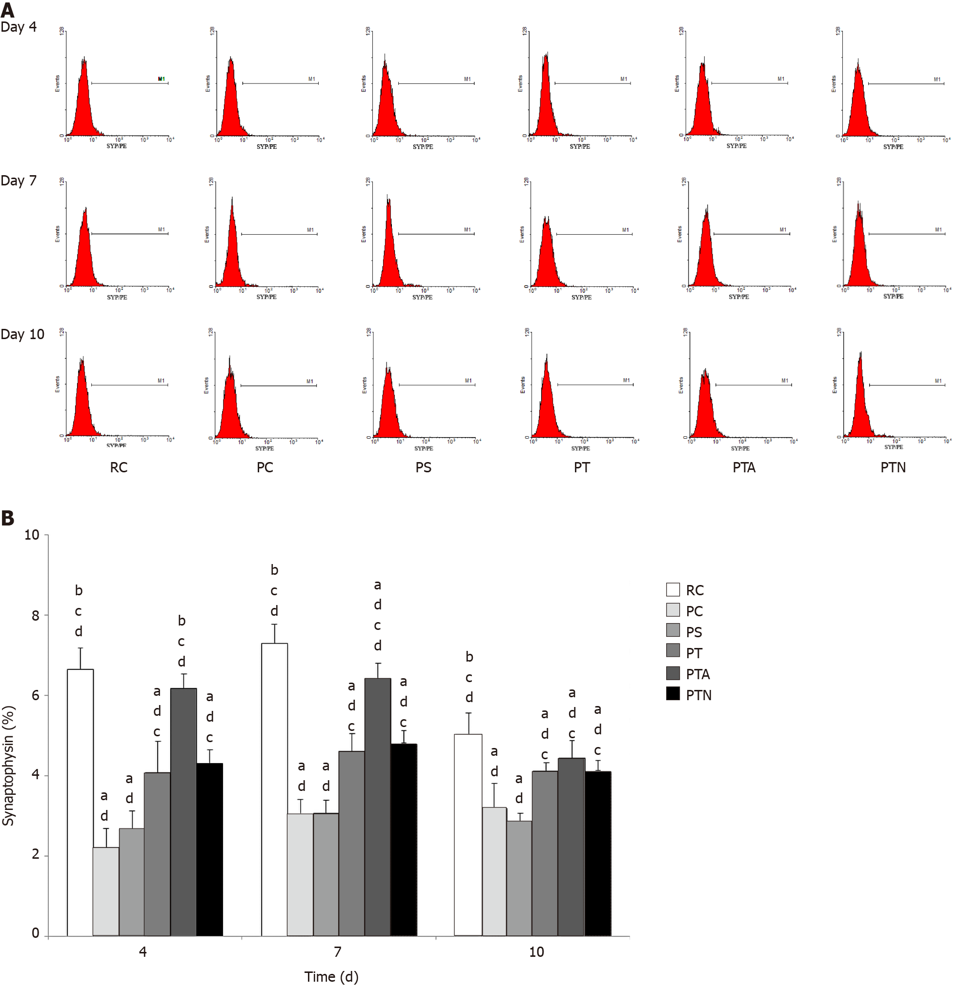
Figure 4 Synaptophysin expression by flow cytometry assay.
Hippocampal brain specimens and neural stem cells were inoculated in the Transwell co-culture system. On the 4th, 7th, and 10th d, flow cytometry was used to detect the expression of synaptophysin-positive cells. A: Synaptophysin expression; B: Proportion of cells with synaptophysin expression. aP < 0.05 when compared to senescence-accelerated mouse resistant 1 control group, bP < 0.05 when compared to senescence-accelerated mouse prone 8 (SAMP8) control group, cP < 0.05 when compared to SAMP8 sham operation group, dP < 0.05 when compared to SAMP8 neural stem cells transplantation with non-acupoint group. RC: Senescence-accelerated mouse resistant 1 control group; PC: Senescence-accelerated mouse prone 8 control group; PS: Senescence-accelerated mouse prone 8 sham operation group; PT: Senescence-accelerated mouse prone 8 neural stem cells transplantation group; PTA: Senescence-accelerated mouse prone 8 neural stem cells transplantation with acupuncture group; PTN: Senescence-accelerated mouse prone 8 neural stem cells transplantation with non-acupoint group.
- Citation: Zhao L, Liu JW, Kan BH, Shi HY, Yang LP, Liu XY. Acupuncture accelerates neural regeneration and synaptophysin production after neural stem cells transplantation in mice. World J Stem Cells 2020; 12(12): 1576-1590
- URL: https://www.wjgnet.com/1948-0210/full/v12/i12/1576.htm
- DOI: https://dx.doi.org/10.4252/wjsc.v12.i12.1576


