Copyright
©The Author(s) 2020.
World J Stem Cells. Dec 26, 2020; 12(12): 1439-1454
Published online Dec 26, 2020. doi: 10.4252/wjsc.v12.i12.1439
Published online Dec 26, 2020. doi: 10.4252/wjsc.v12.i12.1439
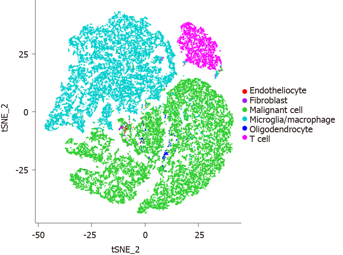
Figure 1 Sing-cell RNA sequencing charts cellular heterogeneity in gliomas.
Sing-cell RNA sequence analysis showing various kinds of cells within glioma tumor microenvironment including tumor cells, microglia/macrophages, T cells, fibroblasts, and endotheliocytes.
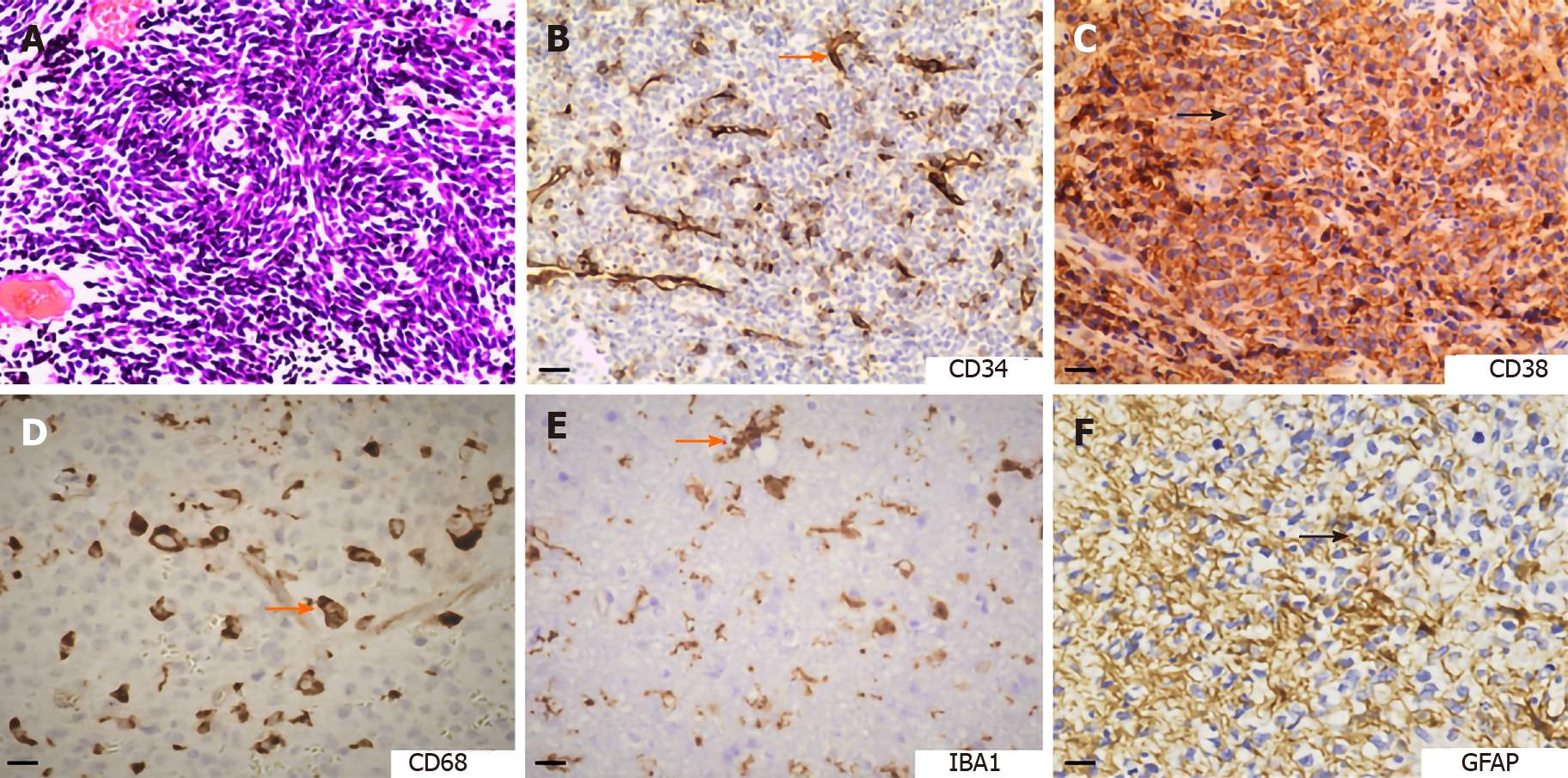
Figure 2 Hematoxylin and eosin staining and immunohistochemical staining of brain tumors.
A: Hematoxylin and eosin staining displaying the pathological vessels distributed in medulloblastoma (bar, 20 µm); B: Expression of CD34 (vascular endothelial cell marker) in chordoma (bar, 20 µm); C: Expression of CD38 (T cell marker) in anaplastic diffuse astrocytoma (bar, 20 µm); D: Expression of CD68 (macrophage marker) in medulloblastoma (bar, 10 µm); E: Expression of IBA1 (microglia marker) in glioblastoma (bar, 10 µm); F: Expression of glial fibrillary acidic protein (astrocyte marker) in medulloblastoma (bar, 10 µm). GFAP: Glial fibrillary acidic protein.
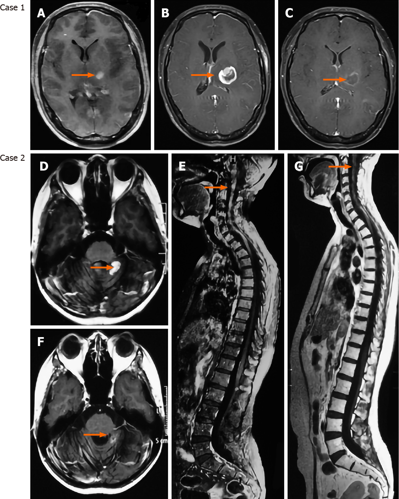
Figure 3 Targeting the vasculature presents promising effects on brain tumor therapy.
Case 1 is a 29-year-old female patient who was diagnosed with IDH1/2 wild-type glioblastoma and received treatment of temozolomide combined with apatinib. Case 2 is a 31-year-old female patient who was diagnosed with recurrent medulloblastoma (local recurrence and dissemination) and received temozolomide + irinotecan + bevacizumab. A: Axial enhanced magnetic resonance imaging (MRI) showing a mass located at the left thalamus; B: Axial enhanced MRI showing that the lesion significantly progressed after 1-mo treatment of temozolomide; C: Axial enhanced MRI showing that the lesion was dramatically reduced after 1-mo treatment of combination of temozolomide and apatinib; D and E: Enhanced cerebrospinal MRI showing the local recurrent medulloblastoma and disseminated lesions along the spinal cord; F and G: Enhanced cerebrospinal MRI showing that the recurrent and disseminated lesions were significantly reduced after one cycle of chemotherapy.
- Citation: Liu HL, Wang YN, Feng SY. Brain tumors: Cancer stem-like cells interact with tumor microenvironment. World J Stem Cells 2020; 12(12): 1439-1454
- URL: https://www.wjgnet.com/1948-0210/full/v12/i12/1439.htm
- DOI: https://dx.doi.org/10.4252/wjsc.v12.i12.1439


