Copyright
©The Author(s) 2019.
World J Stem Cells. Dec 26, 2019; 11(12): 1130-1141
Published online Dec 26, 2019. doi: 10.4252/wjsc.v11.i12.1130
Published online Dec 26, 2019. doi: 10.4252/wjsc.v11.i12.1130
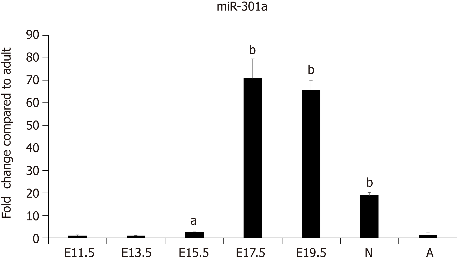
Figure 1 MiR-301a is enriched in the heart of late-stage mouse embryo and neonatal mouse heart tissues.
Tissues were collected from mouse embryos at days 11.5, 13.5, 15.5, and 17.5 and from 3-d-old postnatal and 6-wk-old adult mice. The expression level of miR-301a in the hearts was analyzed using quantitative real-time PCR. 5S rRNA was used for normalization. Data are presented as the mean ± SE (n = 3). aP < 0.05, bP < 0.01.
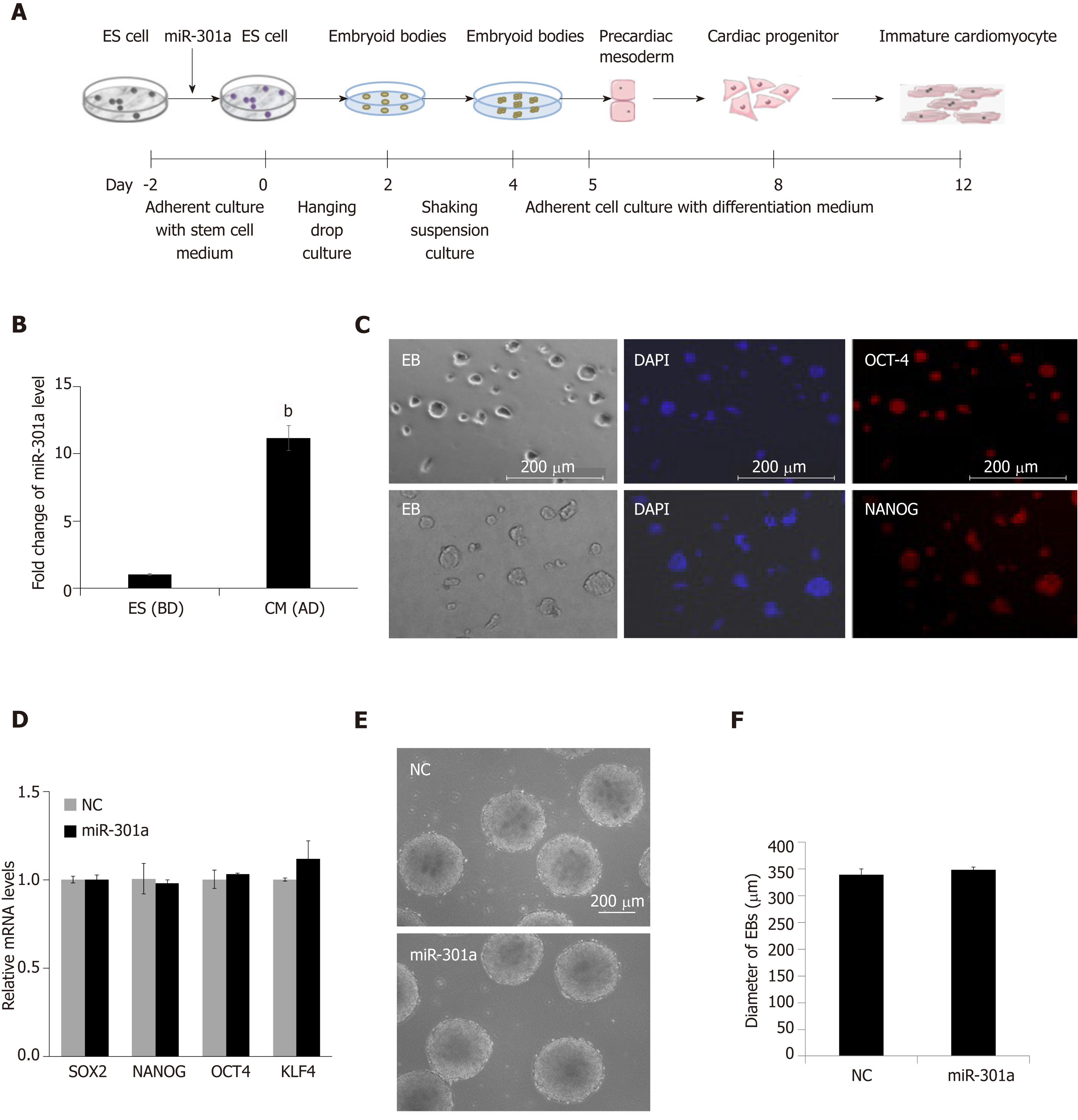
Figure 2 MiR-301a does not affect mouse embryonic stem cell properties or the formation of early embryoid bodies.
A: Schematic representation of the procedure to induce cardiomyocyte differentiation from mouse embryonic stem cells; B: The expression level of miR-301a in mouse embryonic stem cells before differentiation (BD) and cardiomyocytes at day 12 after differentiation (AD). Fold change was calculated by dividing the value at AD by the value at BD. Data are the mean ± SE (n = 3), bP < 0.01; C: Immuno-fluorescence staining for the stem cell markers OCT4 and NANOG in mouse embryonic stem cell clones at day 2 under adherent culture; D: Gene expression detection of stem cell markers, including SOX2, OCT4, NANOG, and KLF4, in embryoid bodies at day 2 with or without overexpression of miR-301a. A quantitative real-time PCR method was applied. Data are presented as the mean ± SEM (n = 3). E: Representative images of embryoid bodies formed from mouse embryonic stem cells at day 4 with or without overexpression of miR-301a; F: Average diameter of embryoid bodies at day 4 with or without overexpression of miR-301a. Data are presented as the mean ± SEM (n = 10).
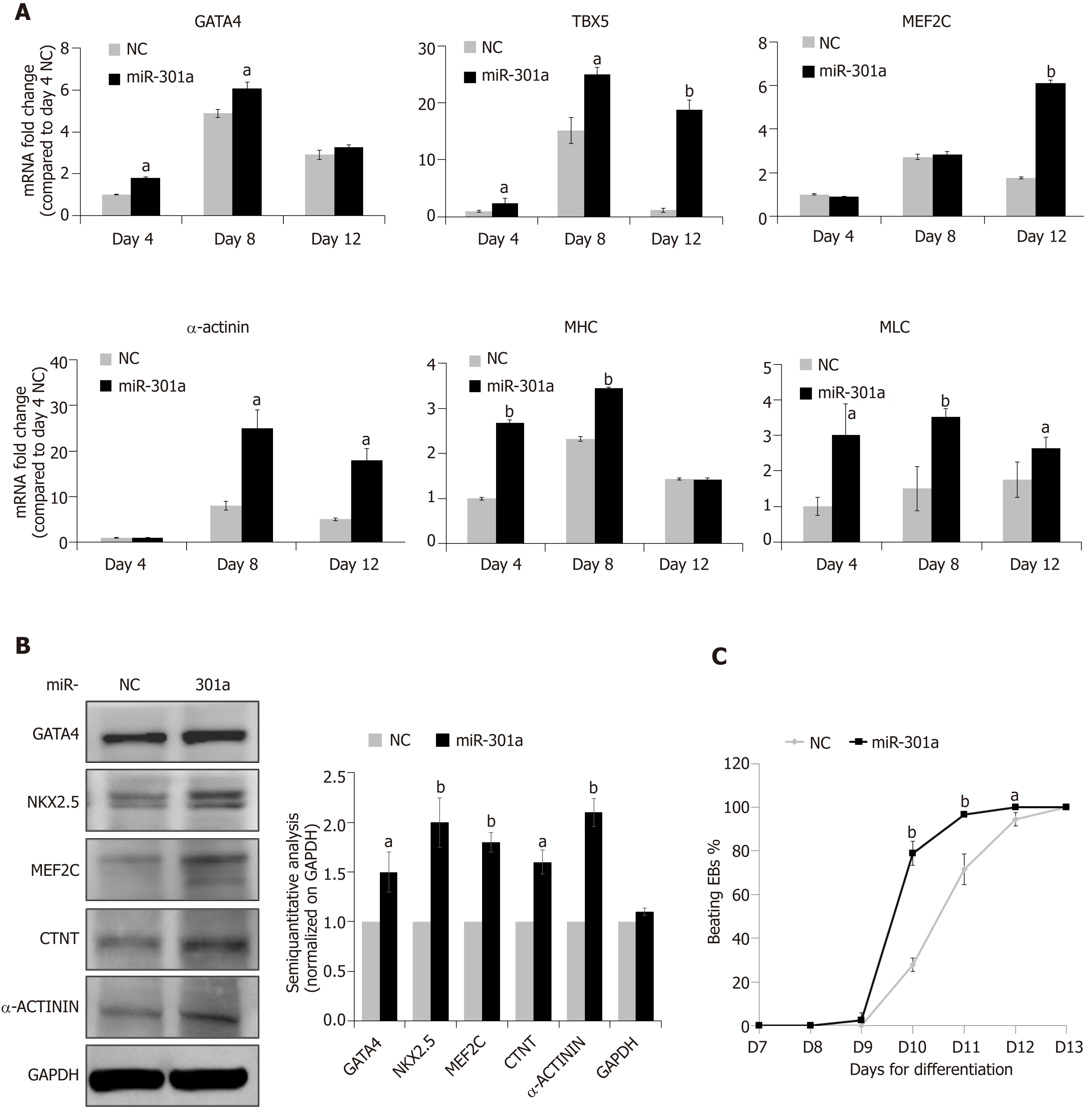
Figure 3 MiR-301a promotes mouse embryonic stem cell differentiation to cardiomyocytes.
A: Quantitative real-time PCR analysis of the gene expression for mid-stage cardiac markers, including GATA-4, TBX5, and MEF2C, and late-stage cardiac markers, including α-actinin, sarcomeric MHC, and MLC, in cells at day 4 (embryoid body formation), day 8 (cardiac differentiation), and day 12 (immature cardiomyocytes) during mouse embryonic stem cell differentiation as indicated in Figure 2A. The gene expression levels are shown as the fold change compared to control cells at day 4. Data are presented as the mean ± SEM (n = 3); B: Western blot analysis of the cardiac markers, including GATA-4, MEF2C, NKX2.5, CTNT, and α-actinin, in cells at day 12 after mouse embryonic stem cell differentiation with or without overexpression of miR-301a. A semiquantitative analysis (cardiac markers normalized on GAPDH, miR-301a group vs control group) was applied and shown; C: Quantitative analysis of the percentage of beating embryoid bodies at different time points demonstrated earlier initiation and more embryoid body beating in the miR-301a group than in the control group. All of the embryoid bodies in three independent dishes in each group (141 embryoid bodies in the control group and 131 embryoid bodies in the miR-301a group) were calculated at each time point. Data are presented as the mean ± SE (n = 3). aP < 0.05, bP < 0.01.
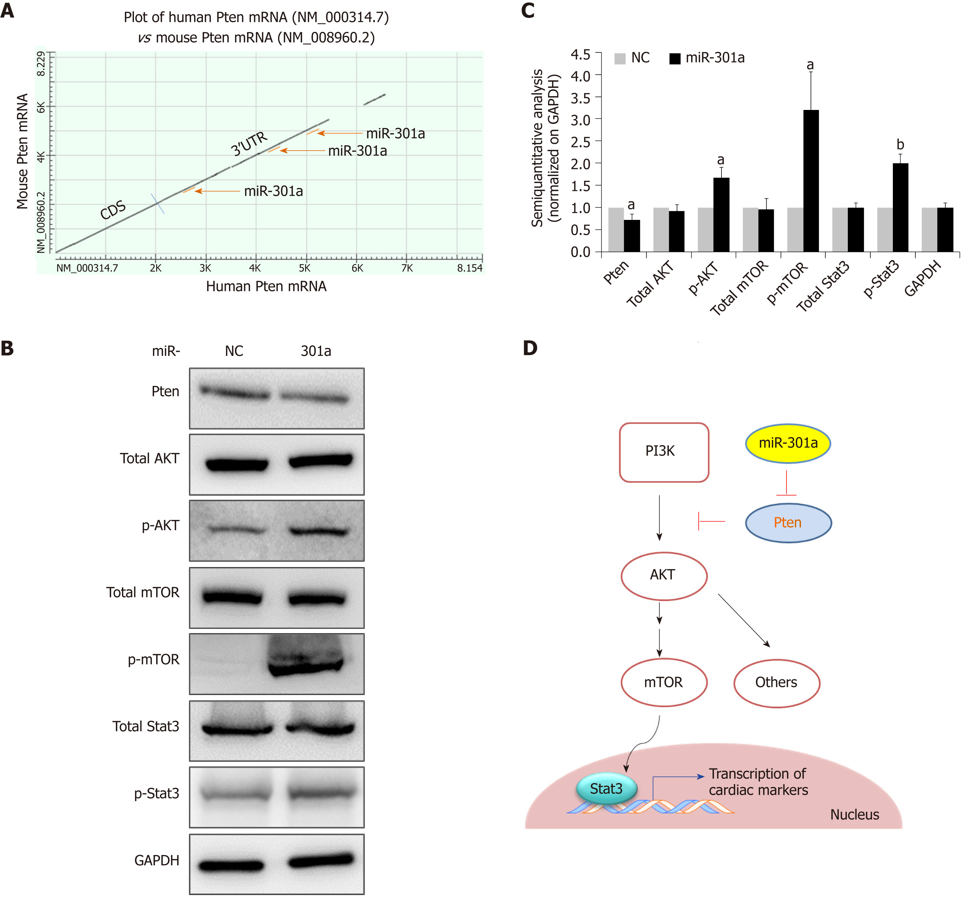
Figure 4 MiR-301a targets PTEN and activates the mTOR-STAT3 signaling pathway in the regulation of mouse embryonic stem cell differentiation.
A: Plot of human PTEN mRNA vs mouse PTEN mRNA showing three predicted binding sites to miR-301a at the highly conserved 3’ untranslated region; B: Western blot analysis showing decreased expression of PTEN and increased expression of p-AKT, p-mTOR, and p-STAT3 by miR-301a overexpression in mouse embryonic stem cell-differentiated cardiomyocytes. Total AKT, total mTOR ,and total STAT3 did not show differences between the two groups; C: A semiquantitative analysis (normalized to GAPDH, miR-301a group vs control group) was applied to protein expression in B; D: Schematic representation of the hypothetical mechanism by which miR-301a regulates mouse embryonic stem cell differentiation to cardiomyocytes by targeting PTEN. Data are presented as the mean ± SE (n = 3). aP < 0.05, bP < 0.01.
- Citation: Zhen LX, Gu YY, Zhao Q, Zhu HF, Lv JH, Li SJ, Xu Z, Li L, Yu ZR. MiR-301a promotes embryonic stem cell differentiation to cardiomyocytes. World J Stem Cells 2019; 11(12): 1130-1141
- URL: https://www.wjgnet.com/1948-0210/full/v11/i12/1130.htm
- DOI: https://dx.doi.org/10.4252/wjsc.v11.i12.1130


