Copyright
©2009 Baishideng.
World J Stem Cells. Dec 31, 2009; 1(1): 43-48
Published online Dec 31, 2009. doi: 10.4252/wjsc.v1.i1.43
Published online Dec 31, 2009. doi: 10.4252/wjsc.v1.i1.43
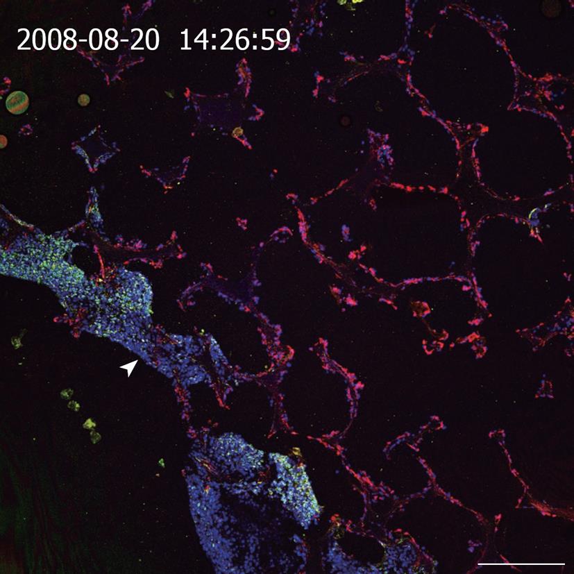
Figure 1 Cross section of a resin embedded co-culture of a hepatoma cell line (C3A) and immortalized BCE in polylactic-co-glycolic acid foam.
Medium inflow from the lower left side into the polymer foam (arrowhead). Staining of cytokeratin 18 (green, C3A cells), vimentin (red, BCE cells), Draq5 nuclear stain (blue, both cell types). Scale bar: 250 μm.
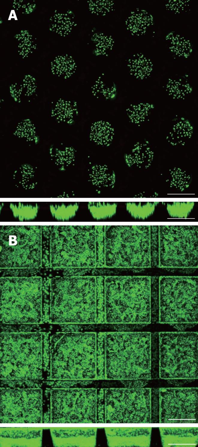
Figure 2 Primary human hepatocytes and Hep G2 hepatoma cells 5 d and 24 h after cell seeding into r- (A) and cf-KITChips (B) respectively (upper panels: top view, lower panels: cross section).
The r-KITChip (20 mm × 20 mm in total) is comprised of up to 625 round microcontainers (diameter up to 300 μm, depth up to 300 μm) or 1156 cubic microcontainers (300 μm × 300 μm × 300 μm in w × l × h) for the cf-KITChip of which 5 × 5 can be seen in 2A and 4 × 4 can be seen in 2B. Live cell staining with Syto 16. Scale bar: 250 μm.
-
Citation: Altmann B, Welle A, Giselbrecht S, Truckenmüller R, Gottwald E. The famous
versus the inconvenient - or the dawn and the rise of 3D-culture systems. World J Stem Cells 2009; 1(1): 43-48 - URL: https://www.wjgnet.com/1948-0210/full/v1/i1/43.htm
- DOI: https://dx.doi.org/10.4252/wjsc.v1.i1.43


