修回日期: 2005-11-21
接受日期: 2005-11-24
在线出版日期: 2006-01-28
目的: 观察血管内皮细胞生长因子A(VEGFA)、血管内皮细胞生长因子C(VEGFC)、Angiopo-ietin-1(Ang-1)、Angiopoietin-2(Ang-2)及其受体在发育第3-5 wk人胚胎肝的表达, 以了解早期人胚发育过程中血管内皮细胞、造血干细胞与肝干细胞之间的相互关系.
方法: 实验用发育3, 4, 5 wk人胚胎标本各8例(共24例), 石蜡切片, 连续切片, 进行CD34、VEGFA、VEGFC、fms样酪氨酸激酶-1(FLT-1)、胚胎肝激酶1(Flk-1)、FLT-4、Ang-1、Ang-2、Tie-2免疫组化染色, 光镜下观察.
结果: 3 wk时靠近肝芽的间充质中的血管内皮细胞多, 而肝芽内未见血管内皮细胞及造血细胞; 4 wk时, 肝索形成处可见较多正在形成的新生血管增多, 肝索间开始出现少量的造血细胞; 5 wk时肝索间出现原始的肝血窦结构, 血窦内造血细胞的数量较4 wk明显增多. 发育的3-5 wk, 部分肝干细胞及血细胞中出现VEGFA阳性反应, 多数肝细胞及内皮细胞中均有VEGFC免疫反应, 而造血细胞中无. 多数肝干细胞和少数血管内皮细胞呈flt-1和flk-1阳性, 造血细胞呈flt-1、flk-1免疫反应阴性, 而呈flt-4阳性. 肝干细胞强表达Ang-2, 而造血细胞表达其受体Tie-2, 与此同时, 内皮细胞表达Ang-1和Ang-2, 而不表达其受体Tie-2.
结论: 来自肝干细胞的VEGFA信号可使内皮细胞分化、增殖并形成血管; 来自内皮细胞的VEGFC、Angiopoietin信号对肝干细胞的生长和分化起作用; 造血细胞分泌的VEGFA可以通过旁分泌途径调节肝干细胞的发育; 造血细胞的发育依赖于肝干细胞产生的VEGFC和Ang-2.
引文著录: 姜红心, 魏志新, 齐安东, 李翠花, 王箐, 李磊. VEGF和Angiopoietin家族在早期人胚肝发育过程中的作用. 世界华人消化杂志 2006; 14(3): 336-340
Revised: November 21, 2005
Accepted: November 24, 2005
Published online: January 28, 2006
AIM: To explore the association among hepatic stem cells, endothelial cells, and hematopoietic stem cells during liver development by observing the expression of vascular endothelial growth factor A (VEGFA), VEGFC, Angiopoietin (Ang) -1, Ang-2 and their receptors in the human emb-ryonic liver of 3-5 wk.
METHODS: Paraffin sections were prepared from 24 embryos of 3, 4, and 5 wk. Immunohistochemistry staining was performed to observe the development of hepatic stem cells, endothelial cells and hematopoietic stem cell as well as the expression of CD34, VEGFA, VEGFC, Ang-1, Ang-2, and their receptors, and flt-1, flk-1, flt-4, and Tie-2 under light microscope.
RESULTS: Endothelial cells predominated in mesenchyme adjacent to the liver bud at 3 wk, but not in the bud. At 4 wk, the cells in the liver bud migrated into the domain of transversum mesenchyme, where endothelial cells began to form vascular structures, and branched to form a number of hepatic cords. A few hematopoietic cells were seen scattered between the liver cords at 5 wk, and primitive sinusoids, which came from vitelline veins, appeared between the hepatic cords. The numbers of hematopoetic cells were increased in comparison with those at 4 wk. VEGFA was expressed by some hepatic stem cells and hematopoetic cells during 3-5 wk. At the same time, most of hepatic stem cells and endothelial cells were positive for VEGFC, while hematopoetic cells were VEGFC-negative. flt-1 and flk-1 positive reaction was found in most of hepatic stem cells and a few endothelial cells. The hematopoetic cells were negative for flt-1 and flk-1, but positive for flt-4. Hepatic stem cells expressed Ang-2, while hematopoetic cells expressed its receptor Tie-2. Meanwhile, endothelial cells expressed Ang-1 and Ang-2, but didn't express Tie-2.
CONCLUSION: Hepatic stem cells may induce the growth and differentiation of vascular endothelial cells through VEGFA signals. The development of hepatic stem cells is affected by VEGFC, angiopoietin secreted by endothelial cells and VEGFA produced by hematopoetic cells. Meanwhile, the development of hematopoietic cells depended on the VEGFC and Ang-2 produced by hepatic stem cells.
- Citation: Jiang HX, Wei ZX, Qi AD, Li CH, Wang Q, Li L. Roles of vascular endothelial growth factor and Angiopoietin family in early stage of human liver development. Shijie Huaren Xiaohua Zazhi 2006; 14(3): 336-340
- URL: https://www.wjgnet.com/1009-3079/full/v14/i3/336.htm
- DOI: https://dx.doi.org/10.11569/wcjd.v14.i3.336
肝干细胞与血管内皮细胞之间存在相互诱导、相互作用的关系[1-3], 肝干细胞可促进造血细胞的生成[4,5], 但这些诱导作用的分子机制目前尚不清楚, 造血细胞反过来对肝干细胞有无影响也未见报道. LeCouter et al[6]发现, 外源性血管内皮细胞生长因子A(VEGFA)可促进成年小鼠内皮细胞和肝细胞的增殖, 从而启动内皮细胞与肝细胞的信息传递. 但目前未见VEGF家族在肝实质细胞中表达的报道, 也不清楚Angiopoietin是否在这一诱导过程中起作用. 为此, 我们对人胚胎肝干细胞的发育与血管内皮细胞、造血细胞之间的关系; VEGF和Angiopoietin家族及其受体在肝干细胞、内皮细胞及造血细胞中的表达进行了观察, 以探索造血干细胞、血管内皮细胞与肝干细胞之间诱导作用的分子机制, 为肝干细胞的基础和临床应用研究提供实验依据.
收集人工流产、受精龄为3 mo以内的新鲜人胚胎47例(其中3-5 wk各8例, 6-8 wk各5例, 9-12 wk各2例), 16 g/L多聚甲醛固定, 石蜡包埋, 制5 mm厚的连续切片, 裱于APES包被的载玻片上. 每10张抽片1张做HE染色, 显微镜下观察, 根据胚层形成情况、胚体的长度、体节的数目及器官发育状况确定胚胎龄.
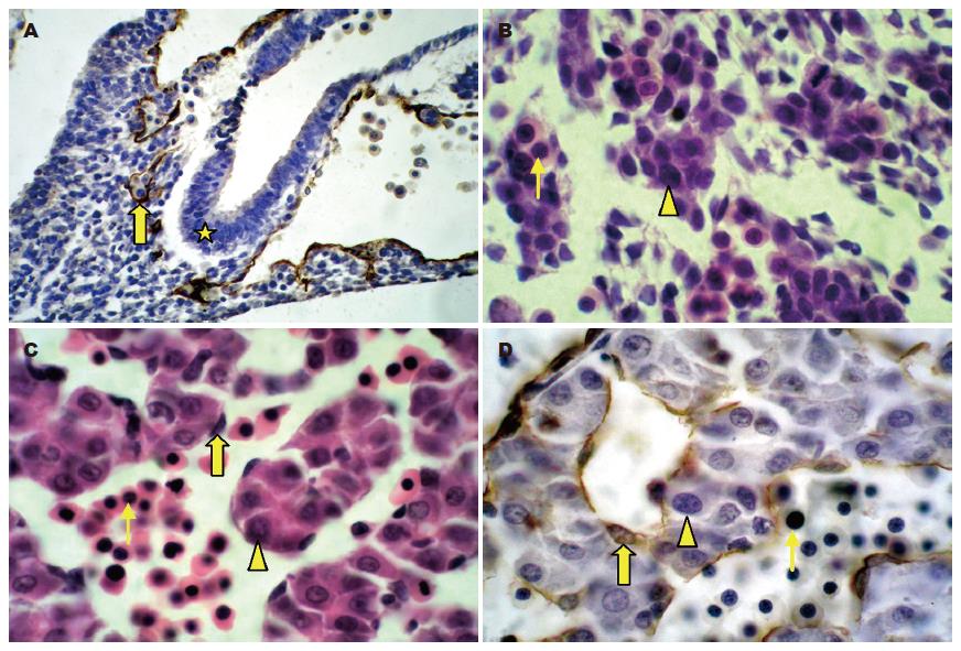
胚胎发育3 wk末, 部分内胚层细胞由立方形变为高柱状, 并聚集形成一个多层的上皮细胞群即肝芽. 此时肝芽内未见血管内皮细胞及造血细胞, 在靠近肝芽的间充质中的血管内皮细胞多, 并可见有的内皮细胞正在形成毛细血管, 而在远离肝芽的间充质中则很少见到内皮细胞及毛细血管(图1A). 4 wk时, 组成肝芽的细胞向横隔间充质迁移, 并在此处分支形成少量的肝细胞索, 肝索处有较多正在形成的毛细血管. 此时的血管内皮细胞扁平、核大、深染, 细胞不连续. 血管内的血细胞有核, 核质比较大(图1B). 5 wk时, 在肝索间出现原始的肝血窦结构, 原始肝血窦内造血细胞的数量较4 wk明显增多, 这些造血细胞和内皮细胞的形态与3-4 wk的相同, 此时肝索和肝血窦的分布无规律(图1C). 4-5 wk时肝内正在形成和已经形成的血管内皮细胞以及游离散在的血管内皮细胞均为CD34强阳性, 阳性反应主要位于内皮细胞的细胞膜(图1D).
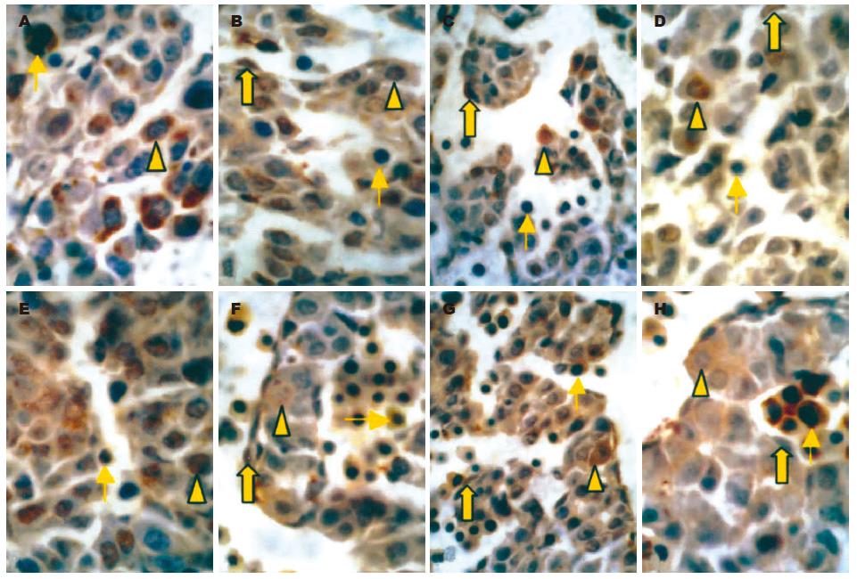
胚胎发育3-5 wk, 部分肝干细胞及造血细胞中出现VEGFA阳性反应, 反应产物积聚在细胞质和胞核; 这些VEGFA阳性的血细胞较大, 核圆形, 核质比大; 血管内皮细胞中无VEGFA阳性反应(图2A). 多数肝干细胞及内皮细胞中均有VEGFC免疫反应, 而造血细胞中无(图2B). 多数肝干细胞和少数血管内皮细胞呈flt-1和flk-1阳性, 虽然flt-1和flk-1免疫组化阳性反应发生的部位相似, 但在肝干细胞和内皮细胞中flt-1的反应较flk-1强, 此时造血细胞呈flt-1、flk-1免疫反应阴性, 而呈flt-4阳性(图2C-E).
发育3-5 wk的胚胎肝血管, 有Ang-1、Ang-2和Tie-2阳性反应的造血细胞, 细胞形态同VEGFA阳性细胞, Tie-2免疫反应的强度超过Ang-1和Ang-2. 与此同时, 内皮细胞表达Ang-1和Ang-2, 而不表达其受体Tie-2. 肝干细胞中Ang-2的阳性反应较Ang-1和Tie-2强(图2F-H).
以往认为内皮细胞的作用主要是运送氧气, 但近年研究发现, 在血液循环形成前, 内皮细胞还可诱导心脏、肝脏、胰腺和牙齿的胚胎发生[9-13]. 在这一过程中, 血管内皮细胞向组织细胞发出信号, 以确定器官生长的位置、形态和分化状态[14]; 另一方面, 受组织细胞的影响, 内皮细胞获得器官特异性的特征, 以适应器官的生长发育[2,15-17]. 本文结果显示, 3 wk时在靠近肝芽的间充质中的血管内皮细胞多, 并可见有的内皮细胞正在形成毛细血管, 而在远离肝芽的横隔间充质中则很少见到内皮细胞及毛细血管, 提示内皮细胞可诱导内胚层细胞向肝细胞的特化. 4 wk时, 组成肝芽的细胞向正在形成毛细血管的间充质处迁移, 并在此形成肝索, 提示横隔间充质中的血管内皮细胞可促进肝内胚层细胞向横隔的迁移. 5 wk时, 组成肝索的细胞增多, 并将穿经此处的卵黄静脉分隔成肝血窦样血管. 以上结果均提示在早期人胚肝发育过程中, 内皮细胞及新生血管可诱导肝细胞的发生. 对VEGF和Angiopoietin家族及其受体的免疫组化研究表明, 发育3-5 wk的肝干细胞表达VEGFA、flt-4、Tie-2等, 而内皮细胞表达flt-1、flk-1、VEGFC、Ang-1、Ang-2, 根据内皮细胞与肝干细胞在发育过程中的时空配布关系和VEGFA、Ang-1、Ang-2及其受体的表达, 我们推测在人胚发育的3-5 wk, 肝干细胞与内皮细胞之间存在相互作用, 其中来自肝干细胞的VEGFA信号可使内皮细胞分化、增殖并形成血管, 而来自内皮细胞的VEGFC、Ang-1、Ang2信号亦可促进胚肝的形态发生和肝索的形成, 两者相互依赖.
胚胎肝是胚内造血器官, 造血干细胞最早出现于卵黄囊, 之后迁移到胚胎肝, 并在此增殖、分化, 说明造血组织与肝脏存在空间上的密切关系. 已有研究表明在造血和成肝过程中有许多共同的特征, 肝干细胞激活时, 能表达几种造血干细胞的标记物(c-kit、CD34和Thy-1)[18,19]; 肝源性有丝分裂源--HGF亦可作为造血干细胞的生长因子[20]. 我们注意到, 3 wk肝芽内尚无造血细胞迁入, 4 wk时胚肝中开始出现少量CD34阳性的造血干细胞, 这些细胞体积大, 圆形, 核圆形, 核/质比大, 5 wk出现造血灶, 并且3-5 wk的肝索细胞几乎全部是肝干细胞[6], 提示胎肝造血与肝干细胞的发育是同步进行的. 4 wk开始, 肝内造血细胞呈VEGFA阳性, 肝干细胞表达VEGFA受体flt-1、flk-1, 这说明造血细胞分泌的VEGFA可作用于肝干细胞; 同时, 肝干细胞表达VEGFC, 多数造血细胞不表达VEGFC而仅表达其受体flt-4, 这提示肝干细胞可通过产生VEGFC经旁分泌途径对造血细胞发挥作用. 肝干细胞强表达Ang-2, 而造血细胞表达其受体Tie-2, 表明肝干细胞可通过产生Ang-2对造血细胞发挥作用.
总之, 本文结果表明, 肝干细胞、造血细胞、肝外和肝内的间充质血管内皮细胞三者的发育存在密切的时间和空间的联系, 这为三者相互作用奠定了基础. 来自肝干细胞的VEGFA信号可使内皮细胞分化、增殖并形成血管; 来自内皮细胞的VEGFC、Angiopoietin信号对3-5 wk肝干细胞的生长和分化起作用; 造血细胞分泌的VEGFA可以通过旁分泌途径调节肝干细胞的发育; 造血细胞的发育依赖于肝干细胞产生的VEGFC和Ang-2.
肝干细胞的发育过程伴随着肝内血管及造血细胞的发育. 近年, 动物研究表明, 肝干细胞与血管内皮细胞之间存在相互诱导、相互作用的关系, 但这些作用通过什么因子和受体介导目前尚不清楚. 在胚胎发育过程中, 造血细胞与肝脏存在着密切关系, 肝干细胞可促进造血细胞的生成. 但肝实质细胞通过产生哪些信号分子来诱导造血细胞的分化不清楚, 造血细胞反过来对肝干细胞有无影响也未见报道.
本文通过免疫组化技术, 原位观察了人胚胎肝干细胞与内皮细胞、造血细胞在肝脏发育过程中的位置关系以及VEGF和Angiopoietin家族及其受体的表达, 发现不同的VEGF和Angiopoietin家族的因子分别介导了肝干细胞与内皮细胞、造血细胞的相互作用.
通过本课题的研究, 可了解肝干细胞增殖、分化的微环境及某些调控因素, 阐明肝干细胞增殖分化与成熟的部分调控机制, 为肝干细胞的体外培养和诱导分化提供试验依据.
本文研究的内容具有一定的新颖性.
编辑: 菅鑫妍 审读: 张海宁 电编: 张敏
| 1. | Matsumoto K, Yoshitomi H, Rossant J, Zaret KS. Liver organogenesis promoted by endothelial cells prior to vascular function. Science. 2001;294:559-563. [PubMed] |
| 2. | Parviz F, Matullo C, Garrison WD, Savatski L, Adamson JW, Ning G, Kaestner KH, Rossi JM, Zaret KS, Duncan SA. Hepatocyte nuclear factor 4alpha controls the development of a hepatic epithelium and liver morphogenesis. Nat Genet. 2003;34:292-296. [PubMed] |
| 3. | Morita M, Qun W, Watanabe H, Doi Y, Mori M, Enomoto K. Analysis of the sinusoidal endothelial cell organization during the developmental stage of the fetal rat liver using a sinusoidal endothelial cell specific antibody, SE-1. Cell Struct Funct. 1998;23:341-348. [PubMed] |
| 4. | Sonoda Y, Sasaki K, Suda M, Itano C. Ultrastruc-tural studies of hepatoblast junctions and liver he-matopoiesis of the mouse embryo. Kaibogaku Zasshi. 2001;76:473-482. [PubMed] |
| 5. | Medlock ES, Haar JL. The liver hemopoietic enviro-nment: I. Developing hepatocytes and their role in fetal hemopoiesis. Anat Rec. 1983;207:31-41. [PubMed] |
| 6. | LeCouter J, Moritz DR, Li B, Phillips GL, Liang XH, Gerber HP, Hillan KJ, Ferrara N. Angiogenesis-independent endothelial protection of liver: role of VEGFR-1. Science. 2003;299:890-893. [PubMed] |
| 7. | Jiang J, Li A, Zhou H, Mei Y, Yang S, Hong H, Song H, Yang H. The characteristics of hepatic stem cells and the expression of growth factor and their receptors in the early embryonic human liver. Shengwu Yixue Gongchengxue Zazhi. 2004;21:995-998. [PubMed] |
| 8. | Zanjani ED, Almeida-Porada G, Livingston AG, Flake AW, Ogawa M. Human bone marrow CD34- cells engraft in vivo and undergo multilineage expression that includes giving rise to CD34+ cells. Exp Hematol. 1998;26:353-360. [PubMed] |
| 9. | Lammert E, Cleaver O, Melton D. Role of endothe-lial cells in early pancreas and liver development. Mech Dev. 2003;120:59-64. [PubMed] |
| 10. | Lammert E, Cleaver O, Melton D. Induction of pan-creatic differentiation by signals from blood vessels. Science. 2001;294:564-567. [PubMed] |
| 11. | Thesleff I, Aberg T. Tooth morphogenesis and the differentiation of ameloblasts. Ciba Found Symp. 1997;205:3-12. [PubMed] |
| 12. | Yoshitomi H, Zaret KS. Endothelial cell interactions initiate dorsal pancreas development by selectively inducing the transcription factor Ptf1a. Development. 2004;131:807-817. [PubMed] |
| 13. | Nikolova G, Lammert E. Interdependent develop-ment of blood vessels and organs. Cell Tissue Res. 2003;314:33-42. [PubMed] |
| 14. | Brutsaert DL. Cardiac endothelial-myocardial signaling: its role in cardiac growth, contractile performance, and rhythmicity. Physiol Rev. 2003;83:59-115. [PubMed] |
| 15. | Wilting J, Christ B. Embryonic angiogenesis: a review. Naturwissenschaften. 1996;83:153-164. [PubMed] |
| 16. | Modis L, Martinez-Hernandez A. Hepatocytes modulate the hepatic microvascular phenotype. Lab Invest. 1991;65:661-670. [PubMed] |
| 17. | Mochida S, Ishikawa K, Toshima K, Inao M, Ikeda H, Matsui A, Shibuya M, Fujiwara K. The mechanisms of hepatic sinusoidal endothelial cell regeneration: a possible communication system associated with vascular endothelial growth factor in liver cells. J Gastroenterol Hepatol. 1998;13:S1-S5. [PubMed] |
| 18. | Grompe M. The role of bone marrow stem cells in liver regeneration. Semin Liver Dis. 2003;23:363-372. [PubMed] |
| 19. | Shu SN, Wei L, Wang JH, Zhan YT, Chen HS, Wang Y. Hepatic differentiation capability of rat bone marrow-derived mesenchymal stem cells and hematopoietic stem cells. World J Gastroenterol. 2004;10:2818-2822. [PubMed] |
| 20. | Kollet O, Shivtiel S, Chen YQ, Suriawinata J, Thung SN, Dabeva MD, Kahn J, Spiegel A, Dar A, Samira S. HGF, SDF-1, and MMP-9 are involved in stress-induced human CD34+ stem cell recruitment to the liver. J Clin Invest. 2003;112:160-169. [PubMed] |


