Gastric Cancer Open Access
Copyright ©The Author(s) 2003. Published by Baishideng Publishing Group Inc. All rights reserved.
World J Gastroenterol. Sep 15, 2003; 9(9): 1915-1919
Published online Sep 15, 2003. doi: 10.3748/wjg.v9.i9.1915
Degradation of retinoid X receptor α by TPA through proteasome pathway in gastric cancer cells
Xiao-Feng Ye, Su Liu, Qiao Wu, Xiao-Feng Lin, Bing Zhang, Jia-Fa Wu, Ming-Qing Zhang, Wen-Jin Su, Key Laboratory of the Ministry of Education for Cell Biology and Tumor Cell Engineering, School of Life Sciences, Xiamen University, Xiamen 361005, Fujian Province, China
Author contributions: All authors contributed equally to the work.
Supported by the National Natural Science Foundation of China, No. 30170477, and the National Natural Science Foundation of Fujian Province, No. 0110004
Correspondence to: Wu Qiao, PhD, Key Laboratory of the Ministry of Education for Cell Biology and Tumor Cell Engineering, School of Life Sciences, Xiamen University, Xiamen 361005, Fujian Province, China. xgwu@xmu.edu.cn
Telephone: +86-592-2187959 Fax: +86-592-2086630
Received: March 3, 2003
Revised: April 23, 2003
Accepted: May 16, 2003
Published online: September 15, 2003

Abstract

AIM: To investigate and determine the mechanism and signal pathway of tetradecanoylphorbol-1, 3-acetate (TPA) in degradation of RXRα.

METHODS: Gastric cancer cell line, BGC-823 was used in the experiments. The expression level of RXRα protein was detected by Western blot. Nuclear and cytoplasmic protein fractions were prepared through lysis of cell and centrifugation. Localization and translocation of RXRα were observed under laser-scanning confocal microscope through labeling specific anti-RXRα antibody and corresponding immunofluorescent antibody as secondary antibody. Different inhibitors were used as required.

RESULTS: In BGC-823 cells, RXRα was expressed in the nucleus. When cells were treated with TPA, expression of RXRα was repressed in a time-dependent and TPA-concentration-dependent manner. Meanwhile, translocation of RXRα from the nucleus to the cytoplasm occurred, also in a time-dependent manner. When cells were pre-incubated with proteasome inhibitor MG132 for 3 hrs, followed by TPA for another 12 hrs, TPA-induced RXRα degradation was inhibited. Further observation of RXRα translocation in the presence of MG132 showed that MG-132 could block TPA-induced RXRα redistribution. Conversely, when RXRα translocation was inhibited by LMB, an inhibitor for blocking protein export from the nucleus, TPA could not repress expression of RXRα.

CONCLUSION: TPA could induce the degradation of RXRα protein in BGC-823 cells, and this degradation is time- and TPA-concentration-dependent. Furthermore, the degradation of RXRα by TPA is via a proteasome pathway and associated with RXRα translocation from the nucleus to the cytoplasm.


INTRODUCTION

Retinoid receptors are nuclear receptors, and belong to members of the steroid and thyroid hormone receptor superfamily[1-4]. As retinoid receptor ligands, retinoids exert their effects on proliferation and differentiation through regulating the transcriptional and post-transcriptional activities of retinoid receptors[5-8]. Besides retinoids, other agents such as tetradecanoylphorbol-1, 3-acetate (TPA), and nerve growth factor (NGF), have an effect on retinoid receptor regulation[9-11]. Although up-regulation of retinoid receptor mRNA by retinoids has been demonstrated among a number of different types of cells[12-15], it is also indicated that retinoid receptors are degraded through a distinct pathway[16]. However, little is known how and where retinoid receptors are degraded, especially when other agents, rather than their ligands, are used to stimulate carcinoma cells.

Eukaryotic cells contain multiple proteolysis systems, which are mainly responsible for degradation of proteins in the extracellular milieu[17]. The multicomplex 26S proteasome contains two 19S regulatory complexes and a 20S catalytic core complex that may be responsible for 80%-90% of protein degradation in the cells[18,19]. It has been reported that proteasomes play an important role in thymocyte apoptosis and inflammatory response[20-23]. Proteasome inhibitors can induce cell apoptosis, accompanied by activation of several Caspases, such as Caspase-3 and Caspase-7[24,25]. In contrast, proteasome inhibitors may prevent cells from apoptosis through promoting resistance of cells at higher temperature and toxicity[26-28].

Retinoid receptors can be divided into two types, retinoic acid receptor (RAR) and retinoid X receptor (RXR)[29-32]. Usually, to regulate retinoid receptor by retinoid is preferential to measure its mRNA level, little has been done on retinoid receptor protein itself. Recently, Tanaka et al[16] pointed out that although retinoic acid caused up-regulation of RARα and RARγ mRNA, it down-regulated the expression of RARα and RARγ protein in breast cancer cell line MCF-7. This evidence suggests that the mechanism of retinoic acid on transcriptional or post-transcriptional activity of retinoid receptor is quite different. In the course of investigating the effect of TPA on RXR protein expression, we found that TPA could down-regulate the expression of RXRα protein effectively in gastric cancer cells. In present study, we focused on the mechanism of TPA in RXRα degradation. The results demonstrated that degradation of RXRα by TPA was proteasome-dependent in gastric cancer cells. More importantly, both degradation and proteasome-dependency of RXRα were associated with translocation of RXRα protein. These findings may provide a novel insight into the cross-talk between degradation and translocation of retinoid receptor and TPA signaling pathway.

MATERIALS AND METHODS
Cancer cell line

Human gastric cancer cell line, BGC-823, was obtained from the Institute of Cell Biology in Shanghai. The cells were maintained in RPMI-1640 medium, supplemented with 10% FCS, 1 mM glutamine, and 100 u/mL penicillin.

Detection of western blot[9,33]

Cells treated by different agents were harvested, and suspended in RIPA buffer (10 mmol/L Tris (pH7.4), 150 mmol/L NaCl, 1% Triton X-100, 1% deoxycholic acid, 0.1% SDS, 5 mmol/L EDTA (pH8.0), 1 mmol/L PMSF). Protein concentration was determined using the Bio-Rad protein assay system according to the manufacturer’s instructions (Bio-Rad Hereules, CA). Total protein (50 μg) was subjected to SDS-PAGE and transferred to nitrocellulose membrane for Western blot analysis. The membrane was subsequently blocked with 5% dry milk in TBS-T and then immunnoblotted with the corresponding antibody. Protein was detected using the ECL kit (Peirce) according to the manufacturer’s directions. Before incubation with antibody, the same membrane was stained by ponceau S (Sigma) to show the amount of protein used in each well. The intensities of bands indicated by Western blot were quantified by a densitometer. To separate the cytoplasmic and nuclear fractions, cells were suspended in 500 μl of 10 mM Tris-Cl (pH7.8), 1% Nonidet P-40, 10 mM mercaptoethanol, 0.5 mM phenylmethylsulfonyl fluoride, 1 mg/L leupeptin and 1 mg/L aprotinin for 2 min at 0 °C, then 500 μl of DDW was added, and the cells were allowed to swell for 2 min. Cells were sheared by 10 passages through a 22 gauge needle. The nuclear and cytoplasmic fraction was obtained by centrifugation at 4000 × g for 5 min. The supernatant was cytoplasmic fraction and pellet was nuclear fraction.

Immunofluorescent labeling and confocal microscopy[9,34]

Cells were cultured on a cover glass overnight, then treated with various agents. After washed with PBS, cells were fixed in 4% paraformaldehyde. To identify RXRα protein, cells were firstly incubated with anti-RXRα IgG antibody (Santa Cruz), and then reacted with corresponding FITC-conjugated anti-IgG (Pharmingen) as secondary antibody. To visualize nuclei, cells were stained with propidium iodine (50 mg/L) containing 100 mg of DNase-free RNase A per liter. Fluorescent images were observed and analyzed under laser-scanning confocal microscope (Bio-Rad MRC-1024ES).

RESULTS
TPA induces degradation of RXRα

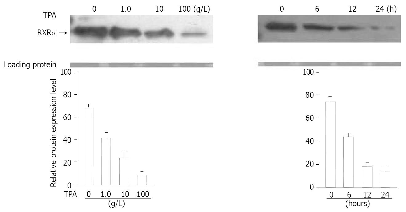
Western blot analysis was carried out to investigate the effect of TPA on the expression level of RXRα protein. When BGC-823 cells were treated at different concentrations of TPA (ranging from 100-1.0 g/L) for 24 hrs, the expression of RXRα protein showed a concentration-dependent decrease (Figure 1A), 87% reduction of RXRα protein expression was observed at the concentration of 100 g/L TPA determined by a densitometer (Figure 1B). Similar repressing tendency was also seen in time-course of TPA treatment. As shown in Figure 1A-B, treatment of cells with TPA for different time periods caused a marked down-regulation of RXRα protein in BGC-823 cells. After 24 hr of TPA treatment, the expression of RXRα protein was almost inhibited. Thus, TPA could induce the degradation of RXRα protein in BGC-823 cells. Moreover, this degradation was time- and TPA concentration-dependent.

Figure 1
Figure 1 Effect of TPA on expression of RXRα protein. Cells were treated at different concentrations of TPA or different time periods. (A) Expression of RXRα was detected by Western blot. The amount of protein used in each lane was indicated by staining with ponceau S. (B) The intensity of each band shown in Figure 1A was quantified by a densitometer.
TPA induces RXRα translocation

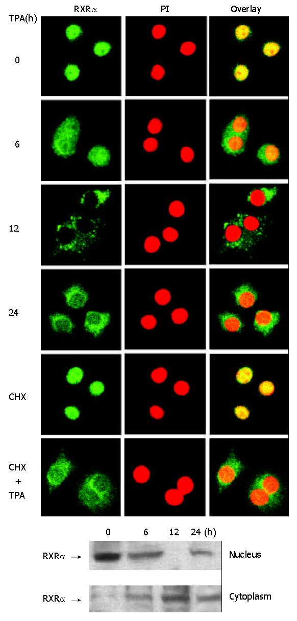
To determine the effect of TPA on the subcellular localization of RXRα, immunofluorescent labeling of RXRα protein was performed and observed under laser-scanning confocal microscope. The result indicated that RXRα was accumulated in the nuclei in intact BGC-823 cells (Figure 2A). Treatment of TPA resulted in redistribution of RXRα protein in a time-dependent manner. When cells were treated with TPA for 6 hrs, RXRα protein started to be translocated from the nucleus to the cytoplasm, and after 12 h of treatment, RXRα protein was completely accumulated in the cytoplasm, and after 24 h of treatment, most of the RXRα protein became localized in the cytoplasm, but little in the nucleus (Figure 2A). To further confirm this translocation, nuclear and cytoplasmic fractions were prepared from BGC-823 cells, and the protein extracts were analyzed by Western blot. It clearly showed that in the intact BGC-823 cells, RXRα was expressed mainly in the nucleus (Figure 2B). TPA treatment resulted in a time-dependent shift of RXRα protein from nuclear portion to cytoplasmic portion. The maximum cytoplasmic RXRα accumulation occurred in the 12 h TPA treatment, and after 24 h TPA treatment, trace RXRα was detected in the nucleus, mostly in the cytoplasm (Figure 2B). Accordingly, this result was in agreement with that of Figure 2A, strongly suggesting that the translocation of RXRα did occur in the course of TPA treatment.

Figure 2
Figure 2 Translocation of RXRα from the nucleus to the cyto-plasm induced by TPA. Cells were treated with TPA for differ-ent time periods or CHX (10 g/L) for 3 hrs as required. (A) Cells were immunostained with anti-RXRα antibody followed by corresponding FITC-conjugated anti-IgG secondary anti-body to show RXRα protein. Simultaneously, cells were stained with PI to display the nucleus. The fluorescent image was ob-served under laser-scanning confocal microscope. (B) Nuclear and cytoplasmic fractions were prepared as described in the Materials and Methods. RXRα was revealed by Western blot.

To verify that cytoplasmic RXRα protein was originated from nuclear RXRα, rather than being synthesized in the cytoplasm, cells were pre-incubated with cycloheximide (CHX) for 3 hrs to specifically prevent new cytoplasmic protein synthesis[35]. Confocal microscopy observation showed that RXRα protein was seen only in the nucleus when cells were treated with CHX alone (Figure 2A), indicating that cytoplasmic protein synthesis was inhibited by CHX effectively. However, when cells were continuously incubated with TPA for another 24 hrs, RXRα protein was still translocated into the cytoplasm (Figure 2A). Therefore, it convincingly demonstrated that cytoplasmic RXRα protein was from the nucleus.

Effect of proteasome inhibitor on RXRα degradation

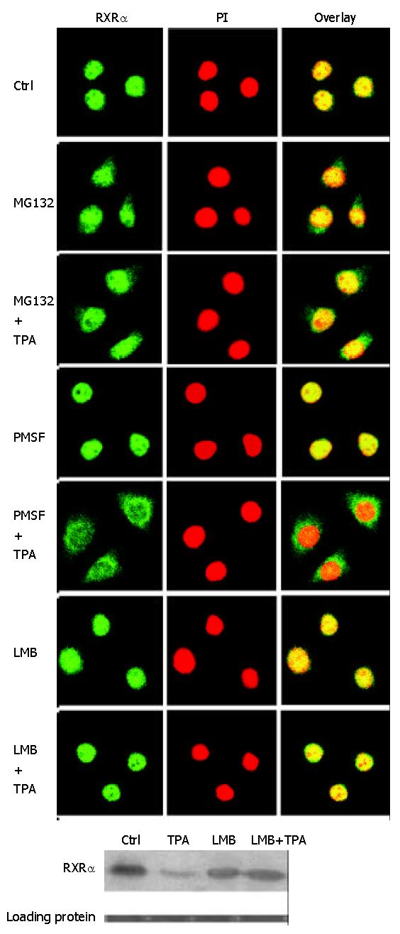
To determine whether the degradation of RXRα by TPA was 26S proteasome-dependent, BGC-823 cells were incubated with proteasome-specific inhibitor MG132[16,18] in the absence or presence of TPA. Western blot analysis showed that MG132 was able to inhibit TPA-induced RXRα degradation (Figure 3). By contrast, when PMSF, a potent inhibitor of protease but not specific-inhibitor of proteasome[36], was used as a positive control, it could not inhibit TPA-induced RXRα degradation, TPA still down-regulated expression of RXRα (Figure 3). Thus, this result suggested that TPA-induced degradation of RXRα occurred through the proteasome pathway.

Figure 3
Figure 3 Effect of different agents, including TPA, MG132 (10 μmol/L) and PMSF (100 mg/L), on expression of RXRα. Cells were pre-treated with MG132 or PMSF for 3 hrs, followed by TPA for another 24 hrs. RXRα expression was analyzed by Western blot.
RXRα translocation by TPA is proteasome-dependent

To determine the relationship between RXRα translocation and proteasome pathway, the behavior of RXRα translocation in the presence of MG132 was investigated. Confocal microscopy observation showed that cells treated with MG132 alone for 3 hrs could hardly induce RXRα protein translocation into the cytoplasm, majority of RXRα protein was localized in the nucleus, but very little in the cytoplasm. After TPA treatment for another 12 hrs, most of RXRα protein remained in the nucleus, similar to MG132 treatment alone (Figure 4A). As a positive control, when PMSF was used to treat cells alone, RXRa was not translocated, still in the nucleus. However, when cells were continuously incubated with TPA, translocation of RXRα was detected (Figure 4A). Thus, it clearly revealed that MG132 and PMSF had different effects on inducing RXRα translocation, MG132 could inhibit TPA-induced translocation of RXRa protein, and RXRα translocation induced by TPA was proteasome-dependent.

Figure 4
Figure 4 Effect of different agents, including TPA, MG132, PMSF and LMB (1 ng/mL), on translocation and expression of RXRα. Cells were pre-treated with different inhibitors for 3 hrs as required, followed by TPA treatment for another 12 hrs. (A) Translocation of RXRα in response to different agents, observed under laser-confocal microscope. (B) Expression of RXRα in-duced by different agents, revealed by Western blot.

In addition, we used another inhibitor LMB, an inhibitor of protein export from the nucleus[37,38], to block RXRα protein nucleocytoplasmic translocation, and then determine whether this blockage could affect TPA-induced RXRα degradation. As shown in Figure 4A, when LMB was incubated with cells alone for 3 hrs, or followed by TPA treatment for another 12 hrs, RXRα was not translocated into the cytoplasm, indicating that LMB did block RXRα translocation no matter TPA existed or not. Under such circumstances, TPA could not repress the expression level of RXRα protein (Figure 4B). These results clearly indicated that TPA-induced RXRα degradation was not only proteasome-dependent, but also translocation-dependent.

DISCUSSION

The redistribution of proteins between nucleus and cytoplasm is an important event for the regulation of their activities and the execution of their functions[39-41]. For example, phosphorylation is needed for redistribution of some proteins. In some cases, phosphorylation of a protein promotes its entry from the cytoplasm to the nucleus[41], in other cases, it causes the protein translocation from the nucleus to the cytoplasm[41,42]. Our study showed that degradation of RXRα protein by TPA was associated with its translocation from the nucleus to the cytoplasm. In gastric cancer BGC-823 cells, TPA could down-regulate the expression of RXRα protein significantly, and this repression was time-dependent and TPA-concentration-dependent (Figure 1A). In the treatment of cells with TPA for 24 hrs, 87% of RXRα protein expression was inhibited (Figure 1B). Moreover, we found that RXRα was translocated from the nucleus to the cytoplasm as degraded by TPA, also in a time-dependent manner (Figure 2A). However, when this translocation was blocked by LMB (Figure 4A), degradation of RXRα by TPA was not detected by Western blot (Figure 4B). Therefore, it clearly demonstrated that TPA-induced RXRα degradation was time-dependent and translocation-dependent.

Although we have detected the degradation of RXRα by TPA during the translocation of RXRα, its molecular mechanism is still largely unknown. More evidences revealed that the proteasome played not only a proteolytic role in protein degradation, but also a non-proteolytic role in transcriptional elongation, nuclear excision repair, and protein trafficking[43-48]. To determine whether degradation of RXRα was responsible for proteasome pathway, we used MG132, a specific proteasome inhibitor, to study the potential linkage among RXRα degradation, translocation and proteasome pathway. The results showed that inhibition of proteasome function by MG132 could repress TPA-induced RXRα degradation (Figure 3), and further block TPA-induced RXRα translocation (Figure 4A), which strongly supported the non-proteolytic role of the proteasome in protein translocation. Accordingly, it is likely that regulation of post-transcriptional activity of RXRα through the proteasome pathway may be involved in multiple mechanisms.

Recently, Tanaka et al[16] found that incubation of breast cancer cells MCF-7 with 10-6 mol/L retinoic acid induced a rapid breakdown of both RARα and RARγ in spite of the accumulation of their mRNA. Eliezer et al[49] also pointed out that retinoic acid-induced degradation of RARs and RXRs could play an important role in the control of the activity of RAR/RXR heterodimer. These reports, at least, provide a crucial clue: retinoid receptor (including RARs and RXRs) activity may be regulated through a distinct mechanism at transcriptional or post-transcriptional level, and formation of heterodimer/or homodimer between these receptors is essential. In our previous studies, we found that RXR could form heterodimer with RAR in the presence of retinoic acid[50]. Moreover, we have indicated that besides RAR, RXR also formed heterodimer with Nur77, an orphan receptor, bound to the RARβ (retinoic acid receptor β) promoter, and then induced RARβ expression, which is critical for inducing apoptosis and inhibiting cell growth in carcinoma cells[15,50]. Therefore, it is likely that degradation and translocation of RXRα by TPA are not a simple event. RXRα might form heterodimer with some proteins, such as Nur77 or RAR, then export from the nucleus to exert its function in the cytoplasm. But, at least, it has been confirmed that the RXRα is degraded through a proteasome pathway in the present study.

Footnotes

Edited by Ma JY

References
1.  Kastner P, Mark M, Chambon P. Nonsteroid nuclear receptors: what are genetic studies telling us about their role in real life. Cell. 1995;83:859-869.  [PubMed]  [DOI]  [Cited in This Article: ]  [Cited by in Crossref: 773]  [Cited by in F6Publishing: 754]  [Article Influence: 26.0]  [Reference Citation Analysis (0)]
2.  Mangelsdorf DJ, Evans RM. The RXR heterodimers and orphan receptors. Cell. 1995;83:841-850.  [PubMed]  [DOI]  [Cited in This Article: ]  [Cited by in Crossref: 2414]  [Cited by in F6Publishing: 2346]  [Article Influence: 80.9]  [Reference Citation Analysis (0)]
3.  Zhang XK, Kahl M. Regulation of retinoid and thyroid hormone action through homodimeric and heterodimeric receptors. Trends Endocrinol Metab. 1993;4:156-162.  [PubMed]  [DOI]  [Cited in This Article: ]  [Cited by in Crossref: 90]  [Cited by in F6Publishing: 64]  [Article Influence: 2.1]  [Reference Citation Analysis (0)]
4.  Xu M, Jin YL, Fu J, Huang H, Chen SZ, Qu P, Tian HM, Liu ZY, Zhang W. The abnormal expression of retinoic acid receptor-beta, p 53 and Ki67 protein in normal, premalignant and malignant esophageal tissues. World J Gastroenterol. 2002;8:200-202.  [PubMed]  [DOI]  [Cited in This Article: ]
5.  Gudas LJ, Sporn MB, Roberts AB. Cellular biology and biochem-istry of the retinoids. In: Sporn MB, Roberts AB, Goodman DS, eds. The retinoids. New York: Raven Press. 1994;443-520.  [PubMed]  [DOI]  [Cited in This Article: ]
6.  Lotan R. Effects of vitamin A and its analogs (retinoids) on normal and neoplastic cells. Biochim Biophys Acta. 1980;605:33-91.  [PubMed]  [DOI]  [Cited in This Article: ]
7.  Roberts AB, Sporn MB. Cellular biology and biochemistry of the retinoids. In: Sporn MB, Roberts AB, Goodman DS, eds. The retinoids. Orlando, Fla: Academic Press. 1984;2:209-286.  [PubMed]  [DOI]  [Cited in This Article: ]
8.  Liu JR, Li BX, Chen BQ, Han XH, Xue YB, Yang YM, Zheng YM, Liu RH. Effect of cis-9, trans-11-conjugated linoleic acid on cell cycle of gastric adenocarcinoma cell line (SGC-7901). World J Gastroenterol. 2002;8:224-229.  [PubMed]  [DOI]  [Cited in This Article: ]
9.  Wu Q, Liu S, Ye XF, Huang ZW, Su WJ. Dual roles of Nur77 in selective regulation of apoptosis and cell cycle by TPA and ATRA in gastric cancer cells. Carcinogenesis. 2002;23:1583-1592.  [PubMed]  [DOI]  [Cited in This Article: ]  [Cited by in Crossref: 88]  [Cited by in F6Publishing: 91]  [Article Influence: 4.1]  [Reference Citation Analysis (0)]
10.  Katagiri Y, Takeda K, Yu ZX, Ferrans VJ, Ozato K, Guroff G. Modulation of retinoid signalling through NGF-induced nuclear export of NGFI-B. Nat Cell Biol. 2000;2:435-440.  [PubMed]  [DOI]  [Cited in This Article: ]  [Cited by in Crossref: 144]  [Cited by in F6Publishing: 149]  [Article Influence: 6.2]  [Reference Citation Analysis (0)]
11.  Xia M, Xue SB, Xu CS. Shedding of TNFR1 in regenerative liver can be induced with TNF alpha and PMA. World J Gastroenterol. 2002;8:1129-1133.  [PubMed]  [DOI]  [Cited in This Article: ]
12.  Liu S, Wu Q, Chen ZM, Su WJ. The effect pathway of retinoic acid through regulation of retinoic acid receptor alpha in gastric cancer cells. World J Gastroenterol. 2001;7:662-666.  [PubMed]  [DOI]  [Cited in This Article: ]
13.  Liu Y, Lee MO, Wang HG, Li Y, Hashimoto Y, Klaus M, Reed JC, Zhang X. Retinoic acid receptor beta mediates the growth-inhibitory effect of retinoic acid by promoting apoptosis in human breast cancer cells. Mol Cell Biol. 1996;16:1138-1149.  [PubMed]  [DOI]  [Cited in This Article: ]  [Cited by in Crossref: 241]  [Cited by in F6Publishing: 252]  [Article Influence: 9.0]  [Reference Citation Analysis (0)]
14.  Li Y, Dawson MI, Agadir A, Lee MO, Jong L, Hobbs PD, Zhang XK. Regulation of RAR beta expression by RAR- and RXR-selective retinoids in human lung cancer cell lines: effect on growth inhibition and apoptosis induction. Int J Cancer. 1998;75:88-95.  [PubMed]  [DOI]  [Cited in This Article: ]  [Cited by in F6Publishing: 2]  [Reference Citation Analysis (0)]
15.  Wu Q, Dawson MI, Zheng Y, Hobbs PD, Agadir A, Jong L, Li Y, Liu R, Lin B, Zhang XK. Inhibition of trans-retinoic acid-resistant human breast cancer cell growth by retinoid X receptor-selective retinoids. Mol Cell Biol. 1997;17:6598-6608.  [PubMed]  [DOI]  [Cited in This Article: ]  [Cited by in Crossref: 67]  [Cited by in F6Publishing: 70]  [Article Influence: 2.6]  [Reference Citation Analysis (0)]
16.  Tanaka T, Rodríguez de la Concepción ML, De Luca LM. Involvement of all-trans-retinoic acid in the breakdown of retinoic acid receptors alpha and gamma through proteasomes in MCF-7 human breast cancer cells. Biochem Pharmacol. 2001;61:1347-1355.  [PubMed]  [DOI]  [Cited in This Article: ]  [Cited by in Crossref: 38]  [Cited by in F6Publishing: 40]  [Article Influence: 1.7]  [Reference Citation Analysis (0)]
17.  Ciechanover A, Schwartz AL. The ubiquitin-mediated proteolytic pathway: mechanisms of recognition of the proteolytic substrate and involvement in the degradation of native cellular proteins. FASEB J. 1994;8:182-191.  [PubMed]  [DOI]  [Cited in This Article: ]
18.  Lin HK, Altuwaijri S, Lin WJ, Kan PY, Collins LL, Chang C. Proteasome activity is required for androgen receptor transcriptional activity via regulation of androgen receptor nuclear translocation and interaction with coregulators in prostate cancer cells. J Biol Chem. 2002;277:36570-36576.  [PubMed]  [DOI]  [Cited in This Article: ]  [Cited by in Crossref: 105]  [Cited by in F6Publishing: 108]  [Article Influence: 4.9]  [Reference Citation Analysis (0)]
19.  Lee DH, Goldberg AL. Proteasome inhibitors: valuable new tools for cell biologists. Trends Cell Biol. 1998;8:397-403.  [PubMed]  [DOI]  [Cited in This Article: ]  [Cited by in Crossref: 1100]  [Cited by in F6Publishing: 1139]  [Article Influence: 43.8]  [Reference Citation Analysis (0)]
20.  Kloetzel PM, Soza A, Stohwasser R. The role of the proteasome system and the proteasome activator PA28 complex in the cellular immune response. Biol Chem. 1999;380:293-297.  [PubMed]  [DOI]  [Cited in This Article: ]  [Cited by in Crossref: 46]  [Cited by in F6Publishing: 47]  [Article Influence: 1.9]  [Reference Citation Analysis (0)]
21.  Hirsch T, Dallaporta B, Zamzami N, Susin SA, Ravagnan L, Marzo I, Brenner C, Kroemer G. Proteasome activation occurs at an early, premitochondrial step of thymocyte apoptosis. J Immunol. 1998;161:35-40.  [PubMed]  [DOI]  [Cited in This Article: ]
22.  Grimm LM, Goldberg AL, Poirier GG, Schwartz LM, Osborne BA. Proteasomes play an essential role in thymocyte apoptosis. EMBO J. 1996;15:3835-3844.  [PubMed]  [DOI]  [Cited in This Article: ]
23.  Schwartz AL, Ciechanover A. The ubiquitin-proteasome pathway and pathogenesis of human diseases. Annu Rev Med. 1999;50:57-74.  [PubMed]  [DOI]  [Cited in This Article: ]  [Cited by in Crossref: 323]  [Cited by in F6Publishing: 327]  [Article Influence: 13.1]  [Reference Citation Analysis (0)]
24.  Wagenknecht B, Hermisson M, Groscurth P, Liston P, Krammer PH, Weller M. Proteasome inhibitor-induced apoptosis of glioma cells involves the processing of multiple caspases and cytochrome c release. J Neurochem. 2000;75:2288-2297.  [PubMed]  [DOI]  [Cited in This Article: ]  [Cited by in Crossref: 69]  [Cited by in F6Publishing: 77]  [Article Influence: 3.2]  [Reference Citation Analysis (0)]
25.  Qiu JH, Asai A, Chi S, Saito N, Hamada H, Kirino T. Proteasome inhibitors induce cytochrome c-caspase-3-like protease-mediated apoptosis in cultured cortical neurons. J Neurosci. 2000;20:259-265.  [PubMed]  [DOI]  [Cited in This Article: ]
26.  Lee DH, Goldberg AL. Proteasome inhibitors cause induction of heat shock proteins and trehalose, which together confer thermotolerance in Saccharomyces cerevisiae. Mol Cell Biol. 1998;18:30-38.  [PubMed]  [DOI]  [Cited in This Article: ]  [Cited by in Crossref: 185]  [Cited by in F6Publishing: 178]  [Article Influence: 6.8]  [Reference Citation Analysis (0)]
27.  Milligan SA, Nopajaroonsri C. Inhibition of NF-kappa B with proteasome inhibitors enhances apoptosis in human lung adenocarcinoma cells in vitro. Anticancer Res. 2001;21:39-44.  [PubMed]  [DOI]  [Cited in This Article: ]
28.  Watanabe K, Kubota M, Hamahata K, Lin Y, Usami I. Prevention of etoposide-induced apoptosis by proteasome inhibitors in a human leukemic cell line but not in fresh acute leukemia blasts. A differential role of NF-kappab activation. Biochem Pharmacol. 2000;60:823-830.  [PubMed]  [DOI]  [Cited in This Article: ]  [Cited by in Crossref: 12]  [Cited by in F6Publishing: 12]  [Article Influence: 0.5]  [Reference Citation Analysis (0)]
29.  Benbrook D, Lernhardt E, Pfahl M. A new retinoic acid receptor identified from a hepatocellular carcinoma. Nature. 1988;333:669-672.  [PubMed]  [DOI]  [Cited in This Article: ]  [Cited by in Crossref: 429]  [Cited by in F6Publishing: 483]  [Article Influence: 13.4]  [Reference Citation Analysis (0)]
30.  Brand N, Petkovich M, Krust A, Chambon P, de Thé H, Marchio A, Tiollais P, Dejean A. Identification of a second human retinoic acid receptor. Nature. 1988;332:850-853.  [PubMed]  [DOI]  [Cited in This Article: ]  [Cited by in Crossref: 719]  [Cited by in F6Publishing: 817]  [Article Influence: 22.7]  [Reference Citation Analysis (0)]
31.  Leid M, Kastner P, Lyons R, Nakshatri H, Saunders M, Zacharewski T, Chen JY, Staub A, Garnier JM, Mader S. Purification, cloning, and RXR identity of the HeLa cell factor with which RAR or TR heterodimerizes to bind target sequences efficiently. Cell. 1992;68:377-395.  [PubMed]  [DOI]  [Cited in This Article: ]  [Cited by in Crossref: 836]  [Cited by in F6Publishing: 975]  [Article Influence: 30.5]  [Reference Citation Analysis (0)]
32.  Mangelsdorf DJ, Ong ES, Dyck JA, Evans RM. Nuclear receptor that identifies a novel retinoic acid response pathway. Nature. 1990;345:224-229.  [PubMed]  [DOI]  [Cited in This Article: ]  [Cited by in Crossref: 1067]  [Cited by in F6Publishing: 1099]  [Article Influence: 32.3]  [Reference Citation Analysis (0)]
33.  Liu S, Wu Q, Ye XF, Cai JH, Huang ZW, Su WJ. Induction of apoptosis by TPA and VP-16 is through translocation of TR3. World J Gastroenterol. 2002;8:446-450.  [PubMed]  [DOI]  [Cited in This Article: ]
34.  Wu Q, Liu S, Ding L, Ye X, Su W. PKCalpha translocation from mitochondria to nucleus is closely related to induction of apoptosis in gastric cancer cells. Sci China C Life Sci. 2002;45:237-244.  [PubMed]  [DOI]  [Cited in This Article: ]  [Cited by in Crossref: 8]  [Cited by in F6Publishing: 8]  [Article Influence: 0.6]  [Reference Citation Analysis (0)]
35.  Jílek F, Hüttelová R, Petr J, Holubová M, Rozinek J. Activation of pig oocytes using calcium ionophore: effect of protein synthesis inhibitor cycloheximide. Anim Reprod Sci. 2000;63:101-111.  [PubMed]  [DOI]  [Cited in This Article: ]  [Cited by in Crossref: 21]  [Cited by in F6Publishing: 18]  [Article Influence: 0.8]  [Reference Citation Analysis (0)]
36.  Eitel K, Wagenknecht B, Weller M. Inhibition of drug-induced DNA fragmentation, but not cell death, of glioma cells by non-caspase protease inhibitors. Cancer Lett. 1999;142:11-16.  [PubMed]  [DOI]  [Cited in This Article: ]  [Cited by in Crossref: 17]  [Cited by in F6Publishing: 17]  [Article Influence: 0.7]  [Reference Citation Analysis (0)]
37.  Yoshida M, Nishikawa M, Nishi K, Abe K, Horinouchi S, Beppu T. Effects of leptomycin B on the cell cycle of fibroblasts and fission yeast cells. Exp Cell Res. 1990;187:150-156.  [PubMed]  [DOI]  [Cited in This Article: ]  [Cited by in Crossref: 97]  [Cited by in F6Publishing: 99]  [Article Influence: 2.9]  [Reference Citation Analysis (0)]
38.  Fornerod M, Ohno M, Yoshida M, Mattaj IW. CRM1 is an export receptor for leucine-rich nuclear export signals. Cell. 1997;90:1051-1060.  [PubMed]  [DOI]  [Cited in This Article: ]  [Cited by in Crossref: 1642]  [Cited by in F6Publishing: 1661]  [Article Influence: 61.5]  [Reference Citation Analysis (0)]
39.  Nigg EA. Nucleocytoplasmic transport: signals, mechanisms and regulation. Nature. 1997;386:779-787.  [PubMed]  [DOI]  [Cited in This Article: ]  [Cited by in Crossref: 791]  [Cited by in F6Publishing: 807]  [Article Influence: 29.9]  [Reference Citation Analysis (0)]
40.  Kaffman A, Rank NM, O'Neill EM, Huang LS, O'Shea EK. The receptor Msn5 exports the phosphorylated transcription factor Pho4 out of the nucleus. Nature. 1998;396:482-486.  [PubMed]  [DOI]  [Cited in This Article: ]  [Cited by in Crossref: 258]  [Cited by in F6Publishing: 261]  [Article Influence: 10.0]  [Reference Citation Analysis (0)]
41.  Ben-Levy R, Hooper S, Wilson R, Paterson HF, Marshall CJ. Nuclear export of the stress-activated protein kinase p38 mediated by its substrate MAPKAP kinase-2. Curr Biol. 1998;8:1049-1057.  [PubMed]  [DOI]  [Cited in This Article: ]  [Cited by in Crossref: 269]  [Cited by in F6Publishing: 277]  [Article Influence: 10.7]  [Reference Citation Analysis (0)]
42.  Jans DA, Hübner S. Regulation of protein transport to the nucleus: central role of phosphorylation. Physiol Rev. 1996;76:651-685.  [PubMed]  [DOI]  [Cited in This Article: ]
43.  Pickart CM. Ubiquitin enters the new millennium. Mol Cell. 2001;8:499-504.  [PubMed]  [DOI]  [Cited in This Article: ]  [Cited by in Crossref: 305]  [Cited by in F6Publishing: 319]  [Article Influence: 13.9]  [Reference Citation Analysis (0)]
44.  Shenoy SK, McDonald PH, Kohout TA, Lefkowitz RJ. Regulation of receptor fate by ubiquitination of activated beta 2-adrenergic receptor and beta-arrestin. Science. 2001;294:1307-1313.  [PubMed]  [DOI]  [Cited in This Article: ]  [Cited by in Crossref: 661]  [Cited by in F6Publishing: 656]  [Article Influence: 28.5]  [Reference Citation Analysis (0)]
45.  Yu A, Malek TR. The proteasome regulates receptor-mediated endocytosis of interleukin-2. J Biol Chem. 2001;276:381-385.  [PubMed]  [DOI]  [Cited in This Article: ]  [Cited by in Crossref: 88]  [Cited by in F6Publishing: 90]  [Article Influence: 3.9]  [Reference Citation Analysis (0)]
46.  Ferdous A, Gonzalez F, Sun L, Kodadek T, Johnston SA. The 19S regulatory particle of the proteasome is required for efficient transcription elongation by RNA polymerase II. Mol Cell. 2001;7:981-991.  [PubMed]  [DOI]  [Cited in This Article: ]  [Cited by in Crossref: 214]  [Cited by in F6Publishing: 220]  [Article Influence: 9.6]  [Reference Citation Analysis (0)]
47.  Gillette TG, Huang W, Russell SJ, Reed SH, Johnston SA, Friedberg EC. The 19S complex of the proteasome regulates nucleotide excision repair in yeast. Genes Dev. 2001;15:1528-1539.  [PubMed]  [DOI]  [Cited in This Article: ]  [Cited by in Crossref: 80]  [Cited by in F6Publishing: 83]  [Article Influence: 3.6]  [Reference Citation Analysis (0)]
48.  Lommel L, Chen L, Madura K, Sweder K. The 26S proteasome negatively regulates the level of overall genomic nucleotide excision repair. Nucleic Acids Res. 2000;28:4839-4845.  [PubMed]  [DOI]  [Cited in This Article: ]  [Cited by in Crossref: 33]  [Cited by in F6Publishing: 36]  [Article Influence: 1.5]  [Reference Citation Analysis (0)]
49.  Kopf E, Plassat JL, Vivat V, de Thé H, Chambon P, Rochette-Egly C. Dimerization with retinoid X receptors and phosphorylation modulate the retinoic acid-induced degradation of retinoic acid receptors alpha and gamma through the ubiquitin-proteasome pathway. J Biol Chem. 2000;275:33280-33288.  [PubMed]  [DOI]  [Cited in This Article: ]  [Cited by in Crossref: 114]  [Cited by in F6Publishing: 118]  [Article Influence: 4.9]  [Reference Citation Analysis (0)]
50.  Wu Q, Li Y, Liu R, Agadir A, Lee MO, Liu Y, Zhang X. Modulation of retinoic acid sensitivity in lung cancer cells through dynamic balance of orphan receptors nur77 and COUP-TF and their heterodimerization. EMBO J. 1997;16:1656-1669.  [PubMed]  [DOI]  [Cited in This Article: ]  [Cited by in Crossref: 124]  [Cited by in F6Publishing: 115]  [Article Influence: 4.3]  [Reference Citation Analysis (0)]