Liver Cancer Open Access
Copyright ©The Author(s) 2003. Published by Baishideng Publishing Group Inc. All rights reserved.
World J Gastroenterol. Jul 15, 2003; 9(7): 1460-1464
Published online Jul 15, 2003. doi: 10.3748/wjg.v9.i7.1460
Characteristics and application of established luciferase hepatoma cell line that responds to dioxin-like chemicals
Zhi-Ren Zhang, Shun-Qing Xu, Xi Sun, Yong-Jun Xu, Xiao-Kun Cai, Zhi-Wei Liu, Xiang-Lin Tan, Yi-Kai Zhou, Jun-Yue Zhang, Hong Yan, Institute of Environmental Medicine, Tongji Medical College, Huazhong University of Science and Technology, Wuhan, 430030, Hubei Province, China
Author contributions: All authors contributed equally to the work.
Supported by the National Natural Science Foundation of China, No. 29877020 and 20107002
Correspondence to: Dr. Shun-Qing Xu, Institute of Environmental Medicine, Tongji Medical College, Huazhong University of Science and Technology, Wuhan, 430030, Hubei Province, China. shunqing@public.wh.hb.cn
Telephone: +86-27-83693417 Fax: +86-27-83657705
Received: January 28, 2002
Revised: March 4, 2002
Accepted: May 11, 2002
Published online: July 15, 2003

Abstract

AIM: To establish a luciferase reporter cell line that responds dioxin-like chemicals (DLCs) and on this basis to evaluate its characteristics and application in the determination of DLCs.

METHODS: A recombinant luciferase reporter plasmid was constructed by inserting dioxin-responsive element (DREs) and MMTV promoter segments into the pGL3-promoter plasmid immediately upstream of the luciferase gene, which was structurally demonstrated by fragment mapping analysis in gel electrophoresis and transfected into the human hepatoma cell line HepG2, both transiently and stably, to identify the inducible expression of luciferase by 2, 3, 7, 8-tetrachlorodibenzo-p-dioxin (TCDD). The time course, responsive period, sensitivity, structure-inducibility and dose-effect relationships of inducible luciferase expression to DLCs was dynamically observed in HepG2 cells stably transfected by the recombinant vector (HepG2-Luc) and compared with that assayed by ethoxyresorufin-O-deethylase (EROD) in non-transfected HepG2 cells (HepG2-wt).

RESULTS: The inducible luciferase expression of HepG2-Luc cells was noted in a time-, dose-, and AhR-dependent manner, which peaked at 4 h and then decreased to a stable level at 14 h after TCDD treatment. The responsiveness of HepG2-Luc cells to TCDD induction was decreased with culture time and became undetectable at 10th month of HepG2-Luc cell formation. The fact that luciferase activity induced by 3, 3', 4, 4'-PCB in HepG2-Luc cells was much less than that induced by TCDD suggests a structure-inducibility relationship existing among DLCs. Within the concentrations from 3.5 × 10⁻¹² to 5 × 10⁻⁹ mol/L, significant correlations between TCDD doses and EROD activities were observed in both HepG2-luc and HepG2-wt cells. The correlation between TCDD doses from 1.1 × 10⁻¹³ to 1 × 10⁻⁸ mol/L and luciferase activities was also found to be significant in HepG2-luc cells (r = 0.997, P < 0.001), but not in their HepG2-wt counterparts. For the comparison of the enzyme responsiveness between cell lines to TCDD, the luciferase sensitivity and reproducibility in HepG2-luc cells were both better than that of EROD in HepG2-wt cells, the former was at 1.1 × 10⁻¹³ mol/L and 3.5 × 10⁻¹² mol/L, and the coefficients of variation (CV) of the latter was 15%-30% and 22%-38%, respectively.

CONCLUSION: The luciferase expression of HepG2-luc cells established in the present study could sensitively respond to the DLCs stimulation and might be a prospective tool for the determination of DLCs.


INTRODUCTION

It has been well known in recent years that dioxin-like chemicals (DLCs) such as 2, 3, 7, 8- tetrachlorodibenzo-p-dioxin (TCDD) can produce a wide variety of species- and tissue- specific toxic and biological effects, such as epidermal lesion, wasting syndrome, birth defect, hepatotoxicity, lethality, alteration in endocrine homeostasis, tumor promotion, myelotoxicity, immunotoxicity and induction of numerous enzymes (e.g. cytochrome P4501A1)[1-4]. Many of these responses are mediated by the cytosolic aryl hydrocarbon receptor (AhR) and modulated by the interaction of DLCs: AhR complex with its DNA recognition sequence [the dioxin-responsive element (DRE)][5-9]. Generally, the combination of DLCs with cytoplasmic AhR is the initial step of DLCs-triggered cell signaling pathway. After that, two molecules of hsp90 dissociate from DLCs: AhR complex, and the latter enters the nucleus to form a new complex with AhR nuclear translocator protein (ARNT), which further binds to the DRE, leading to the transcriptional activation of adjacent genes and toxic effects[10,11]. Therefore, AhR and its related signal pathway have been usually taken as the useful targets to be investigated for the determination of DLCs in different situations. Till now, the DLCs-responsive receptor assays have been established in mammalian cell lines[12] and in mice[13].

However, not all of these assays have been considered to be satisfactory yet. For example, the ethoxyresorufin-O-deethylase (EROD) induced as a common and rapid response to DLCs under DREs control has been widely used to evaluate the relatively toxicological potency of a complex mixture containing DLCs[14-16] except that higher concentrations appear when the enzyme activity is inhibited. Some detecting methods are time-consuming, expensive and inadequate to be used for screening and diagnosing dioxin and dioxin-like compounds in large numbers of samples[17-19]. Even if the chemical-activated luciferase expression (CALUX) bioassay method based on the pGL2 vector[20] is to a certain extent lack of sensitivity and needs to be improved.

We therefore conducted the present study in an attempt to establish a more effective luciferase reporter cell line that responds to the alteration of DLCs concentrations and to evaluate its characteristics and applications in measurement of DLCs.

MATERIALS AND METHODS
Materials

Plasmids pHAV and pMcat were generously provided by Dr. James P. Whitlock Jr., Stanford University. Restriction endonuclease and other reagents used for molecular cloning were from Huamei Ltd. (China, Shanghai) or Promega Corporation. 2, 3, 7, 8-TCDD and 3', 4, 4'- polychlorinated biphenyl (PCB) were purchased from Accustanfards Inc. (New Haven, CT). Dimethylsulphoxide (DMSO) was from BIB Corporation. Luciferase assay reagents came from Promega Corporation.

Construction of inducible luciferase expression vector

Plasmid pHAV containing DREs was cleaved with HindIII. Following purification by agarose gel electrophoresis, the smallerHindIII fragment was further digested by BamHI and EcoRV to produce a 630-bp segment containing DREs, which was then ligased with BglII linkers and cleaved with BglII to create cohesive ends. To get the MMTV promoter, plasmid pMCat was cleaved with HindIII and the smaller HindIII fragment was then cleaved with BglII to produce cohesive 5' BglII and 3' HindIII termini. The two segments containing DREs and MMTV promoter were connected with T4 DNA ligase to form a 1020 bp fragment with cohesive 5' BglII and 3' HindIII termini, which was further subcloned into the BglII- HindIII site immediately upstream of the luciferase gene in the pGL3-promoter plasmid (Promega). As such, the luciferase's expression was under the control of DREs. The recombinant vector was identified structurally with restriction endonuclease analysis.

Inducible expression of recombinant vector

HepG2 cells were seeded in a 60 mm culture dish at a density of 3 × 10⁵ cells in 5 mL of RPMI1640 supplemented with 10% heat-inactivated fetal calf serum at 37 °C with saturated humidity and a 5% (v/v) CO2 atmosphere[21]. After cultured for 24 h, the cells were transiently or stably transfected by 15 μg of recombinant vector with calcium phosphate mediated method[22]. For transient transfection, the cells were allowed to grow for 48 h, followed by adding 0.5% DMSO or 0.1-1 nmol/L TCDD dissolved in DMSO. After cultivated for another 24 h[22], cells were harvested to determine luciferase expression.

For the establishment of stable transfection, HepG2 cells were cotransfected with the selective plasmid PTK-Hyg and the recombinant vector simultaneously. Following 24 h of cultivation in nonselective medium, the transfected cells were transferred into a selective medium containing hygromycin and cultured for 4 wk when TCDD-induced luciferase expression was conducted. The clone with the largest ratio of inducible to constitutive expression of luciferase was selected for further study[23]. To identify the time course of inducible luciferase expression, the stably transfected cells (HepG2-Luc) were cultured with exposure to 0.5% DMSO or 1 nmol/L TCDD and the induced luciferase activities were determined at every 2 h up to 28 h post-exposure. For determination of the responsive period of luciferase induction, the HepG2-Luc cells were treated with the same kinds of inducers for 24 h and their luciferase activity was assayed, which was performed at every month up to 12 mo. The structure-inducibility and dose-effect relationships of different inducers that might represent their ability to bind AhR were also analyzed in HepG2-Luc cells with the indicated concentrations of 3, 3', 4, 4'-PCB, another member of DLCs, and TCDD.

Luciferase assay was performed by the routine procedures. Briefly, after removal of the culture medium, the incubated cells were washed twice with phosphate buffered saline (PBS) and lysed using luciferase lysis reagent for 15 min at room temperature. Following centrifugation, 20 μL of supernatant was added into 100 μL luciferase assay reagent, the resulting bioluminescence was quantified immediately with Lumate LB 9570 luminometer over 3 s. The concentration of the sample protein was determined with Bio-Rad method[24]. The luciferase activity was finally expressed as relative light unit (RLU) per microgram protein[21,25].

Comparative study with EROD determination

EROD activity was employed as a variable to evaluate the effectiveness of HepG2-luc cells in response to TCDD and to compare with luciferase. HepG2 (HepG2-wt) and HepG2-luc cells were seeded in 6-well dishes at a density of 2 × 10⁵ cells in 3 mL of medium and cultivated for 24 h, then they were exposed to the indicated concentrations of TCDD in DMSO and further incubated for 72 h. Following removal of the culture medium, the cells were washed twice with PBS and stored at -80 °C. EROD activity was assayed using fluorescent method. The concentrations of sample protein were determined with Bio-Rad method[24]. EROD activity was finally expressed as pmol resorufin production per microgram protein per minute[26,27].

Statistical analysis

Data values in the present study were presented as the mean for each independent determination of four replicates. The detection limit was expressed as the average value plus three times of standard deviations (SD). Coefficients of variation (CV) were calculated by SD/mean × 100%. Correlation coefficients (r) were obtained using least-squares linear regression analysis.

RESULTS
Identification of inducible expression of recombinant vector

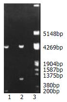
Structurally, segment analysis by restriction endonuclease digestion confirmed that the inserted fragment sequences containing DREs and MMTV promoter in the constructed plasmids were completely consistent with that of the theoretical calculations as shown in Figure 1.

Figure 1
Figure 1 Fragment mapping analysis of recombinant plasmid digested with BglII and HindIII in gel electrophoresis. Lane 1: pGL3 plasmid was cleaved into two fragments, which showed about 5000 bp and 200 bp respectively. Lane 2: Recombinant plasmid was cut into a fragment of about 5000 bp and the one of about 1000 bp. Lane 3: DNA marker.

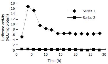
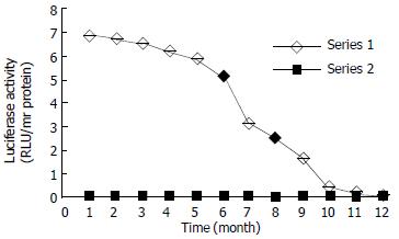
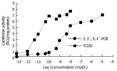
Inducible expression of luciferase by the recombinant plasmid was identified in transiently transfected HepG2 cells, which produced a significantly higher induction of luciferase activity (80-fold, data not shown) compared with that of their non-transfected counterparts when exposed to TCDD. Dynamically, TCDD-induced luciferase activity in HepG2-Luc cells peaked at 4 h and then decreased to a stable level at about 14 h after TCDD treatment (Figure 2). Responsiveness of HepG2-Luc cells to the TCDD induction was decreased with culture time and became undetectable at 10th month of HepG2-Luc cell formation (Figure 3), which indicats that the recombinant plasmid could not integrate stably with HepG2 chromosome. The structure-inducibility relationship of different DLCs inducers was demonstrated by the fact that the luciferase activity induced by 3, 3', 4, 4'-PCB was much less than that induced by TCDD and a dose-dependent increase of luciferase activity was evoked by both of the DLCs inducers as shown in Figure 4.

Figure 2
Figure 2 The time course of luciferase activities induced by TCDD and DMSO in stably transfected cells. The TCDD-induced luciferase activity in HepG2-Luc cells peaked at about 4 h, and then decreased to a stable level at about 14 h after TCDD treatment.
Figure 3
Figure 3 Responsive period of HepG2-Luc cells to TCDD induction. The responsiveness of HepG2-Luc cells to TCDD induction was decreased with culture time and became undetectable at about 10th month.
Figure 4
Figure 4 Comparison of luciferase activity induced by TCDD and 3, 3', 4, 4'-PCB in HepG2-Luc cells. The luciferase activity induced by indicated concentrations of 3, 3', 4, 4'-PCB was much less than that induced by TCDD.
Comparative study

Within concentrations from 3.5 × 10⁻¹² to 5 × 10⁻⁹ mol/L, significant correlations between TCDD doses and EROD activities were observed in both HepG2-luc and HepG2-wt cells. The correlation between TCDD doses from 1.1 × 10⁻¹³ to 1 × 10⁻⁸ mol/L and luciferase activities was also found to be significant in HepG2-luc cells (r = 0.997, P < 0.001), but not in their HepG2-wt counterparts. For comparison of the enzyme responsiveness between cell lines to TCDD, the luciferase sensitivity and reproducibility in HepG2-luc cells were better than those of EROD in HepG2-wt cells, which were 1.1 × 10⁻¹³ mol/L and 3.5 × 10⁻¹² mol/L, whose CV was 15%-30% and 22%-38% (data not shown), respectively.

DISCUSSION

Few environmental compounds have generated much interest within the scientific community and in the lay public as polychlorinated dibenzo-p-dioxins (PCDDs) and polychlorinated dibenzofurans (PCDFs). Their ubiquitous presence in the environment and the risk of accidental exposure have raised concern over a possible threat of PCDDs or PCDFs to human health. The most extensively studied potent isomer is TCDD or dioxin, which is known to induce a wide range of toxic and biochemical responses in laboratory animals and humans[8,11]. Therefore, monitoring the levels of DLCs in environmental pollutants is important for assessing and maintaining the safety of food and the health of environment. Exploring new biodetectors such as bioassays, biomarkers, enzyme immunoassays (EIAs), or other bioanalytical tools has been a continuously growing area in recent years[15]; in which, however, establishing luciferase reporter cell lines for bioassays has been considered as the rapid, sensitive and inexpensive methods for the screening and diagnosis of dioxin and dioxin-like compounds[20,28].

In the present study, we constructed a recombinant expression plasmid that contained the luciferase gene under TCDD-inducible control of several DREs and responded to DLCs with the induction of firefly luciferase, which has been identified by both transient and stable transfection of this vector into HepG2 cells. Our results indicate that this established HepG2-luc cell bioassay reporter system can respond sensitively to DLCs with the induction of luciferase in a time-, dose-, and AhR-dependent manner and harbors lots of better features. Firstly, it bears higher level of sensitivity and can detect TCDD within the linear range from 1.1 × 10⁻¹³ to 1 × 10⁻⁸ mol/L, which is 10-fold more sensitive than that of previously similar studies and 30-fold more sensitive than the EROD assay. This is partially because the vector plasmid employed in the study is pGL3, whose expression efficiency of luciferase is 10-100 times more than that of pGL2. In addition, no post- transcriptional procedures are needed to regulate the expression of prokaryotic luciferase, thus its linear relationship is better than that of endogenous gene. Secondly, its pronounced accuracy has been demonstrated by the significant correlation between inducible luciferase activities and TCDD doses (r = 0.997, P < 0.001), and by the satisfactory reproducibility of TCDD determination with HepG2-luc cells (CV = 15%-30%). Finally, the simplicity and facility of HepG2-luc cells in analytical methodology make it possible to perform a rapid screening and semi-quantitation of DLCs[29,30] and to deal with lots of samples in a short period of time.

Besides the detection of DLCs described above, HepG2-luc cells can also be applied to many other DLCs-related research fields. Since ARNT is required to exert biological effects by some TCDD and related ligands, interactions between AhR and hypoxia signaling pathways can be studied by using luciferase transfected cell line[31]. In accordance with the fact that the toxic factor of 3, 3', 4, 4'-PCB equivalent to TCDD is 0.01[32], our results showed that the ability of 3, 3', 4, 4'-PCB to induce luciferase expression was much less than that of TCDD, suggesting that the structure-inducibility relationship existing in some DLCs could be reflected by HepG2-luc cells, and the latter can be used alone or with chromatographer to evaluate the relative toxicity of DLCs[19,33,34]. In addition, this kind of recombinant cell lines can also be used for the detection and relative quantitation of AhR agonists/antagonists in complex mixtures of environmental and biological samples, for identification and characterization of novel AhR agonists, and for examination of species differences in DLCs responsiveness[12].

One limitation of the present study is the relatively short duration of responsiveness to DLCs by established HepG2-luc cells, although it is in accordance with that reported by most of the other studies. Up to the present, many established luciferase reporter cell lines such as MLE/BV, H4IIE, GPC16 and HGL1.1c3 responding to DLCs maintain their stable TCDD responsiveness for 6-12 mo, except for Hepa1 which maintains its sensitivity for over 3 years[27]. Therefore, our further study has been designed aiming at improving and maintaining the sensitivity of HepG2-luc cells to DLCs for a longer time.

ACKNOWLDGEMENTS

We thank Dr. James P. Whitlock Jr. (Stanford University) for his kind gift of plasmids pHAV and pMcat.

Footnotes

Edited by Zhu L

References
1.  Couture LA, Abbott BD, Birnbaum LS. A critical review of the developmental toxicity and teratogenicity of 2, 3, 7, 8-tetrachlorodibenzo-p-dioxin: recent advances toward understanding the mechanism. Teratology. 1990;42:619-627.  [PubMed]  [DOI]  [Cited in This Article: ]  [Cited by in Crossref: 181]  [Cited by in F6Publishing: 192]  [Article Influence: 5.6]  [Reference Citation Analysis (0)]
2.  Martinez JM, Afshari CA, Bushel PR, Masuda A, Takahashi T, Walker NJ. Differential toxicogenomic responses to 2, 3, 7, 8-tetrachlorodibenzo-p-dioxin in malignant and nonmalignant human airway epithelial cells. Toxicol Sci. 2002;69:409-423.  [PubMed]  [DOI]  [Cited in This Article: ]  [Cited by in Crossref: 78]  [Cited by in F6Publishing: 75]  [Article Influence: 3.4]  [Reference Citation Analysis (0)]
3.  Dong W, Teraoka H, Yamazaki K, Tsukiyama S, Imani S, Imagawa T, Stegeman JJ, Peterson RE, Hiraga T. 2, 3, 7, 8-tetrachlorodibenzo-p-dioxin toxicity in the zebrafish embryo: local circulation failure in the dorsal midbrain is associated with increased apoptosis. Toxicol Sci. 2002;69:191-201.  [PubMed]  [DOI]  [Cited in This Article: ]  [Cited by in Crossref: 136]  [Cited by in F6Publishing: 139]  [Article Influence: 6.3]  [Reference Citation Analysis (0)]
4.  Kerkvliet NI. Recent advances in understanding the mechanisms of TCDD immunotoxicity. Int Immunopharmacol. 2002;2:277-291.  [PubMed]  [DOI]  [Cited in This Article: ]  [Cited by in Crossref: 166]  [Cited by in F6Publishing: 167]  [Article Influence: 7.6]  [Reference Citation Analysis (0)]
5.  Lindström G, Hooper K, Petreas M, Stephens R, Gilman A. Workshop on perinatal exposure to dioxin-like compounds. I. Summary. Environ Health Perspect. 1995;103 Suppl 2:135-142.  [PubMed]  [DOI]  [Cited in This Article: ]
6.  Legare ME, Hanneman WH, Barhoumi R, Burghardt RC, Tiffany-Castiglioni E. 2, 3, 7, 8-Tetrachlorodibenzo-p-dioxin alters hippocampal astroglia-neuronal gap junctional communication. Neurotoxicology. 2000;21:1109-1116.  [PubMed]  [DOI]  [Cited in This Article: ]
7.  Hankinson O. The aryl hydrocarbon receptor complex. Annu Rev Pharmacol Toxicol. 1995;35:307-340.  [PubMed]  [DOI]  [Cited in This Article: ]  [Cited by in Crossref: 1227]  [Cited by in F6Publishing: 1171]  [Article Influence: 40.4]  [Reference Citation Analysis (0)]
8.  Wilson CL, Safe S. Mechanisms of ligand-induced aryl hydrocarbon receptor-mediated biochemical and toxic responses. Toxicol Pathol. 1998;26:657-671.  [PubMed]  [DOI]  [Cited in This Article: ]
9.  Unkila M, Pohjanvirta R, Tuomisto J. Biochemical effects of 2, 3, 7, 8-tetrachlorodibenzo-p-dioxin (TCDD) and related compounds on the central nervous system. Int J Biochem Cell Biol. 1995;27:443-455.  [PubMed]  [DOI]  [Cited in This Article: ]  [Cited by in Crossref: 33]  [Cited by in F6Publishing: 34]  [Article Influence: 1.2]  [Reference Citation Analysis (0)]
10.  Dragan YP, Schrenk D. Animal studies addressing the carcinogenicity of TCDD (or related compounds) with an emphasis on tumour promotion. Food Addit Contam. 2000;17:289-302.  [PubMed]  [DOI]  [Cited in This Article: ]  [Cited by in Crossref: 58]  [Cited by in F6Publishing: 59]  [Article Influence: 2.5]  [Reference Citation Analysis (0)]
11.  Vanden Heuvel JP, Lucier G. Environmental toxicology of polychlorinated dibenzo-p-dioxins and polychlorinated dibenzofurans. Environ Health Perspect. 1993;100:189-200.  [PubMed]  [DOI]  [Cited in This Article: ]  [Cited by in Crossref: 42]  [Cited by in F6Publishing: 34]  [Article Influence: 1.1]  [Reference Citation Analysis (0)]
12.  Garrison PM, Tullis K, Aarts JM, Brouwer A, Giesy JP, Denison MS. Species-specific recombinant cell lines as bioassay systems for the detection of 2, 3, 7, 8-tetrachlorodibenzo-p-dioxin-like chemicals. Fundam Appl Toxicol. 1996;30:194-203.  [PubMed]  [DOI]  [Cited in This Article: ]  [Cited by in Crossref: 303]  [Cited by in F6Publishing: 282]  [Article Influence: 10.1]  [Reference Citation Analysis (0)]
13.  Willey JJ, Stripp BR, Baggs RB, Gasiewicz TA. Aryl hydrocarbon receptor activation in genital tubercle, palate, and other embryonic tissues in 2, 3, 7, 8-tetrachlorodibenzo-p-dioxin-responsive lacZ mice. Toxicol Appl Pharmacol. 1998;151:33-44.  [PubMed]  [DOI]  [Cited in This Article: ]  [Cited by in Crossref: 36]  [Cited by in F6Publishing: 36]  [Article Influence: 1.4]  [Reference Citation Analysis (0)]
14.  Whyte JJ, Jung RE, Schmitt CJ, Tillitt DE. Ethoxyresorufin-O-deethylase (EROD) activity in fish as a biomarker of chemical exposure. Crit Rev Toxicol. 2000;30:347-570.  [PubMed]  [DOI]  [Cited in This Article: ]  [Cited by in Crossref: 552]  [Cited by in F6Publishing: 465]  [Article Influence: 19.4]  [Reference Citation Analysis (0)]
15.  Behnisch PA, Hosoe K, Sakai S. Bioanalytical screening methods for dioxins and dioxin-like compounds a review of bioassay/biomarker technology. Environ Int. 2001;27:413-439.  [PubMed]  [DOI]  [Cited in This Article: ]  [Cited by in Crossref: 185]  [Cited by in F6Publishing: 95]  [Article Influence: 4.1]  [Reference Citation Analysis (0)]
16.  Whitlock JP. Induction of cytochrome P4501A1. Annu Rev Pharmacol Toxicol. 1999;39:103-125.  [PubMed]  [DOI]  [Cited in This Article: ]  [Cited by in Crossref: 856]  [Cited by in F6Publishing: 872]  [Article Influence: 37.9]  [Reference Citation Analysis (0)]
17.  Diehl-Jones WL, Bols NC. Use of response biomarkers in milk for assessing exposure to environmental contaminants: the case for dioxin-like compounds. J Toxicol Environ Health B Crit Rev. 2000;3:79-107.  [PubMed]  [DOI]  [Cited in This Article: ]  [Cited by in Crossref: 12]  [Cited by in F6Publishing: 5]  [Article Influence: 0.2]  [Reference Citation Analysis (0)]
18.  Schwirzer SM, Hofmaier AM, Kettrup A, Nerdinger PE, Schramm KW, Thoma H, Wegenke M, Wiebel FJ. Establishment of a simple cleanup procedure and bioassay for determining 2, 3, 7, 8-tetrachlorodibenzo-p-dioxin toxicity equivalents of environmental samples. Ecotoxicol Environ Saf. 1998;41:77-82.  [PubMed]  [DOI]  [Cited in This Article: ]  [Cited by in Crossref: 39]  [Cited by in F6Publishing: 18]  [Article Influence: 0.7]  [Reference Citation Analysis (0)]
19.  Behnisch PA, Hosoe K, Brouwer A, Sakai S. Screening of dioxin-like toxicity equivalents for various matrices with wildtype and recombinant rat hepatoma H4IIE cells. Toxicol Sci. 2002;69:125-130.  [PubMed]  [DOI]  [Cited in This Article: ]  [Cited by in Crossref: 45]  [Cited by in F6Publishing: 45]  [Article Influence: 2.0]  [Reference Citation Analysis (0)]
20.  Sanderson JT, Aarts JM, Brouwer A, Froese KL, Denison MS, Giesy JP. Comparison of Ah receptor-mediated luciferase and ethoxyresorufin-O-deethylase induction in H4IIE cells: implications for their use as bioanalytical tools for the detection of polyhalogenated aromatic hydrocarbons. Toxicol Appl Pharmacol. 1996;137:316-325.  [PubMed]  [DOI]  [Cited in This Article: ]  [Cited by in Crossref: 185]  [Cited by in F6Publishing: 187]  [Article Influence: 6.7]  [Reference Citation Analysis (0)]
21.  Chen CA, Okayama H. Calcium phosphate-mediated gene transfer: a highly efficient transfection system for stably transforming cells with plasmid DNA. Biotechniques. 1988;6:632-638.  [PubMed]  [DOI]  [Cited in This Article: ]
22.  Zhang ZR, Xu SQ, Zhou YK, Ren S, Lü B. [Construction of luciferase reporter plasmid which is under the control of dioxin-responsive enhancers]. Shengwu Gongcheng Xuebao. 2001;17:170-174.  [PubMed]  [DOI]  [Cited in This Article: ]
23.  Chen C, Okayama H. High-efficiency transformation of mammalian cells by plasmid DNA. Mol Cell Biol. 1987;7:2745-2752.  [PubMed]  [DOI]  [Cited in This Article: ]
24.  Bradford MM. A rapid and sensitive method for the quantitation of microgram quantities of protein utilizing the principle of protein-dye binding. Anal Biochem. 1976;72:248-254.  [PubMed]  [DOI]  [Cited in This Article: ]  [Cited by in Crossref: 173357]  [Cited by in F6Publishing: 153368]  [Article Influence: 3195.2]  [Reference Citation Analysis (0)]
25.  Murk AJ, Legler J, Denison MS, Giesy JP, van de Guchte C, Brouwer A. Chemical-activated luciferase gene expression (CALUX): a novel in vitro bioassay for Ah receptor active compounds in sediments and pore water. Fundam Appl Toxicol. 1996;33:149-160.  [PubMed]  [DOI]  [Cited in This Article: ]  [Cited by in Crossref: 223]  [Cited by in F6Publishing: 186]  [Article Influence: 6.6]  [Reference Citation Analysis (0)]
26.  Li W, Wu WZ, Xu Y, Li L, Schramm KW, Kettrup A. Measuring TCDD equivalents in environmental samples with the micro-EROD assay: comparison with HRGC/HRMS data. Bull Environ Contam Toxicol. 2002;68:111-117.  [PubMed]  [DOI]  [Cited in This Article: ]  [Cited by in Crossref: 8]  [Cited by in F6Publishing: 4]  [Article Influence: 0.2]  [Reference Citation Analysis (0)]
27.  Postlind H, Vu TP, Tukey RH, Quattrochi LC. Response of human CYP1-luciferase plasmids to 2, 3, 7, 8-tetrachlorodibenzo-p-dioxin and polycyclic aromatic hydrocarbons. Toxicol Appl Pharmacol. 1993;118:255-262.  [PubMed]  [DOI]  [Cited in This Article: ]  [Cited by in Crossref: 115]  [Cited by in F6Publishing: 116]  [Article Influence: 3.7]  [Reference Citation Analysis (0)]
28.  Hahn ME. Biomarkers and bioassays for detecting dioxin-like compounds in the marine environment. Sci Total Environ. 2002;289:49-69.  [PubMed]  [DOI]  [Cited in This Article: ]  [Cited by in Crossref: 81]  [Cited by in F6Publishing: 38]  [Article Influence: 1.7]  [Reference Citation Analysis (0)]
29.  Allen SW, Mueller L, Williams SN, Quattrochi LC, Raucy J. The use of a high-volume screening procedure to assess the effects of dietary flavonoids on human cyp1a1 expression. Drug Metab Dispos. 2001;29:1074-1079.  [PubMed]  [DOI]  [Cited in This Article: ]
30.  Khim JS, Lee KT, Villeneuve DL, Kannan K, Giesy JP, Koh CH. In vitro bioassay determination of dioxin-like and estrogenic activity in sediment and water from Ulsan Bay and its vicinity, Korea. Arch Environ Contam Toxicol. 2001;40:151-160.  [PubMed]  [DOI]  [Cited in This Article: ]  [Cited by in Crossref: 35]  [Cited by in F6Publishing: 18]  [Article Influence: 0.8]  [Reference Citation Analysis (0)]
31.  Nie M, Blankenship AL, Giesy JP. Interactions between aryl hydrocarbon receptor (AhR) and hypoxia signaling pathways. Environ Toxicol Pharmacol. 2001;10:17-27.  [PubMed]  [DOI]  [Cited in This Article: ]  [Cited by in Crossref: 76]  [Cited by in F6Publishing: 52]  [Article Influence: 2.3]  [Reference Citation Analysis (0)]
32.  Safe SH. Polychlorinated biphenyls (PCBs): environmental impact, biochemical and toxic responses, and implications for risk assessment. Crit Rev Toxicol. 1994;24:87-149.  [PubMed]  [DOI]  [Cited in This Article: ]  [Cited by in Crossref: 1222]  [Cited by in F6Publishing: 1002]  [Article Influence: 33.4]  [Reference Citation Analysis (0)]
33.  Machala M, Vondrácek J, Bláha L, Ciganek M, Neca JV. Aryl hydrocarbon receptor-mediated activity of mutagenic polycyclic aromatic hydrocarbons determined using in vitro reporter gene assay. Mutat Res. 2001;497:49-62.  [PubMed]  [DOI]  [Cited in This Article: ]  [Cited by in Crossref: 233]  [Cited by in F6Publishing: 225]  [Article Influence: 9.8]  [Reference Citation Analysis (0)]
34.  Hamers T, van Schaardenburg EC, Murk AJ, Koeman JH. The application of reporter gene assays for the determination of the toxic potency of diffuse air pollution. Sci Total Environ. 2000;262:159-174.  [PubMed]  [DOI]  [Cited in This Article: ]  [Cited by in Crossref: 48]  [Cited by in F6Publishing: 41]  [Article Influence: 1.7]  [Reference Citation Analysis (0)]