Liver Cancer Open Access
Copyright ©The Author(s) 2003. Published by Baishideng Publishing Group Inc. All rights reserved.
World J Gastroenterol. May 15, 2003; 9(5): 946-950
Published online May 15, 2003. doi: 10.3748/wjg.v9.i5.946
Signal transduction of gap junctional genes, connexin32, connexin43 in human hepatocarcinogenesis
Xiang-Dong Ma, Department of Obstetrics & Gynecology, Xijing Hospital, Fourth Military Medical University, 17 Changle Xilu, Xi’an 710033, Shaanxi Province, China
Xing Ma, Department of Orthopaedics, Xijing Hospital, Fourth Military Medical University, 17 Changle Xilu, Xi’an 710033, Shaanxi Province, China
Yan-Fang Sui, Weng-Liang Wang, Department of Pathology, Fourth Military Medical University, 17 Changle Xilu, Xi’an 710033, Shaanxi Province, China
Chun-Mei Wang, Department of Electronic Microscopy, Fourth Military Medical University, 17 Changle Xilu, Xi’an 710033, Shaanxi Province, China
Author contributions: All authors contributed equally to the work.
Correspondence to: Dr. Xiang-Dong Ma, Department of Obstetrics & Gynecology, Xijing Hospital, Fourth Military Medical University, 17 Changle Xilu, Xi’an 710033, Shaanxi Province, China. maping@fmmu.edu.cn
Telephone: +86-29-3373569
Received: June 1, 2002
Revised: June 23, 2002
Accepted: July 15, 2002
Published online: May 15, 2003

Abstract

AIM: To investigate gap junctional intercellular communication (GJIC) in hepatocellular carcinoma cell lines, and signal transduction mechanism of gap junction genes connexin32(CX32), connexin43(CX43) in human hepatocarcinogenesis.

METHODS: Scarped loading and dye transfer (SLDT) was employed with Lucifer Yellow (LY) to detect GJIC function in hepatocellular carcinoma cell lines HHCC, SMMC-7721 and normal control liver cell line QZG. After Fluo-3AM loading, laser scanning confocal microscope (LSCM) was used to measure concentrations of intracellular calcium [Ca2+]i in the cells. The phosphorylation on tyrosine of connexin proteins was examined by immunoblot.

RESULTS: SLDT showed that ability of GJIC function was higher in QZG cell than that in HHCC and SMMC-7721 cell lines. By laser scanning confocal microscopy, concentrations of intracellular free calcium [Ca2+]i was much higher in QZG cell line (108.37 nmol/L) than those in HHCC (35.13 nmol/L) and SMMC-7721 (47.08 nmol/L) cells. Western blot suggested that only QZG cells had unphosphorylated tyrosine in CX32 protein of 32 ku and CX43 protein of 43 ku; SMMC-7721 cells showed phosphorylated tyrosine CX43 protein.

CONCLUSION: The results indicated that carcinogenesis and development of human hepatocellular carcinoma related with the abnormal expression of CX genes and disorder of its signal transduction pathway, such as decrease of [Ca2+]i, post-translation phosphorylation on tyrosine of CX proteins which led to a dramatic disruption of GJIC.


INTRODUCTION

Hepatocellular carcinoma has been one of the most common malignant threatening human health[1-9], and much higher incidences of human hepatocellular carcinoma (HCC) are observed recently[10-18]. Gap junctional intercellular communication (GJIC) is performed by intercellular hemichannels[19], which are formed by six basic protein subunits named connexin (CX) expressed in neighboring cells[20]. Six connexins form around a central pore[21], through which adjacent cells can exchange low weight molecules of information and energy directly[22]. At least 16 members of connexin super gene family have been identified, which are divided into two groups according to amino acid sequence. GJIC mediated via gap junctions plays important roles in tissue homeostasis, embryonic development as well as carcinogenensis[23-25]. The mechanisms of carcinogenesis may be related with the block of GJIC of homologous and heterologous communication between tumor cells and surrounding normal cells[26].

Connexin32 (CX32) and connexin43 (CX43) are the major kinds of gap junction proteins expressing in hepatocytes[18]. Our works showed that expressions of CX32, CX43 proteins decreased in hepatocellular carcinoma tissues and cell lines whereas increased in normal tissues and cell lines[19-21], but CX32 mRNA and CX43 mRNA kept a high level in both hepatocellular carcinoma and normal liver cells lines described previously[22,23]. It suggested that the mechanism involved in abnormal processes in post-translation or signal transduction is worthy of discussing[24].

MATERIALS AND METHODS
Cell culture

Hepatocellular carcinoma cell lines HHCC, SMMC-7721 and normal hepatocyte cell line QZG, were kindly provided by professor Chen in 863 Project Group in Fourth Military Medical University. The cells were cultured in RPMI 1640 medium (Gibco BRL, USA), supplemented with 10 mL/L fetal bovine serum (Gibco BRL, USA), incubated in a humidified atmosphere which contains 950 mL/L air and 5 mL/L CO2 at 37 °C.

Scrape loading dye transfer (SLDT)

Functions of gap junctional intercellular communication (GJIC) in adjacent cells were assessed by scrape loading dye transfer (SLDT) with fluorescent dye Lucifer Yellow (LY, Sigma, USA). The cultures were rinsed three times with serum free RPMI 1640 medium, several scrapes were made on the monolayer using a surgical scalpel, cells were exposed to 100 g/L LY in 0.33 mol/L LiCl, incubated for 3 min, and rinsed by serum free RPMI 1640 medium for several times to remove any background fluorescence. The GJIC function in adjacent cells revealed by LY dye transfer could be detected under fluorescent microscope (Nikon UFX-II, Japan).

Concentration of intracellular calcium ([Ca2+]i) measured by laser scanning confocal microscopy (LSCM)

Digested by trypsin and collected, HHCC, SMMC-7721 and QZG cells were loaded by 100 nmol/L special fluorescent dye indicator of calcium Fluo-3 AM (Sigma, USA) in serum free RPMI-1640 medium, 37 °C for 90 min, rinsed by Ca2+ free D-Hank’s for three times. MRC-1024 laser scanning confocal microscope imaging system (Bio-Rad, USA) was used to obtain the images determining the location and intensity of intracellular Ca2+ in various cell lines.

MRC-1024 laser scanning confocal microscope imaging system consists of a laser scanning system intermediate optics and a detector controlled by lasership software (Bio-Rad, USA). Images were obtained by Zesis 100 microscope and images of interest were saved to a 512 × 512 pixel, Zoom was 4.0. Besides analysis of location of Ca2+, quantities of intracellular concentration [Ca2+]i were counted with formation below: [Ca2+]i = kd × (F - Fmin)/(Fmax - F), kd = 400 nmol/L.

Immunoblot analysis of phosphorylation on tyrosine

Western blot was performed to analyses the phosphorylation on tyrosine. Cells were rinsed with cold 0.01 mol/L PBS containing 5 mmol/L EDTA, 10 mmol/L NaF and 1 mmol/L PMSF on ice for 20 min, harvested by scraping in the same solution, and centrifuged at 13000 r/min at 4 °C for 30 min. Pellets were rinsed dissolved in 20 g/L SDS. Concentrations of proteins were measured by the Bio-Rad DC protein assay (Bio-Rad, USA).

Twenty ug protein of various cells in equal amounts of 2 × SDS denatured at 100 °C for 4 min. 100 g/L SDS-PAGE vertical electrophoretically Min-PROTEAN II (Bio-Rad, USA) was used at 80V for 30 min and 120V for 60 min. Mini Trans-Blot transferred proteins form gel to NC membranes (Hybond, USA) by transferring electrophoretically (Bio-Rad, USA) at 100V 350 mA 4 °C for 2 h.

Normal serum blocked NC membranes at room temperature for 2 h. CX32 and CX43 protein bands were detected using a mouse monoclonal anti-CX32 antibody (1:2000, Zymed, USA), a mouse monoclonal anti-CX43 antibody(1:2000, Zymed, USA). Phosphorylations on tyrosine were detected by special anti-phosphorylation tyrosine mAb 4G10 (1:2000, CA, USA) at room temperature for 2 h. NC membranes were washed by TBST for 10 min for several times, incubated with a biotin-streptavidin alkaline phosphatase conjugated secondary IgG (1:32, Johnson, USA) at room temperature for 2 h and washed by TBST for 10 min for several times again. The enhanced chemilunimescence (ECL) detection kit (Amersham, UK) was employed to detect immunoblot signal.

RESULTS
Gap junctional intercellular communication of various cells

After stained with special fluorescent dye Lucifer Yellow (LY), function of gap junctional intercellular communication were detected by scrape loading and dye transfer (SLDT). Molecular weight of LY is below 1000 u that can pass via gap junction smoothly. Normal liver cell line QZG showed stronger intensity and amount yellow fluorescent among neighboring cells, whereas hepatocellular carcinoma cells HHCC and SMMC-7721 showed much slight fluorescent staining limited among adjacent cells.

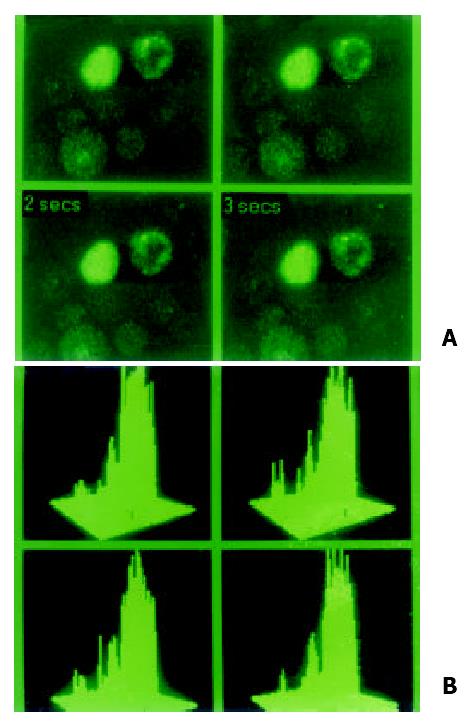
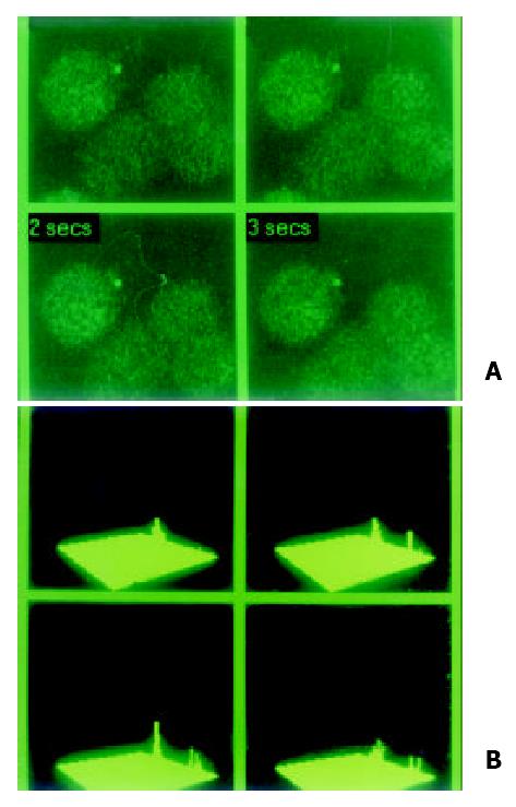
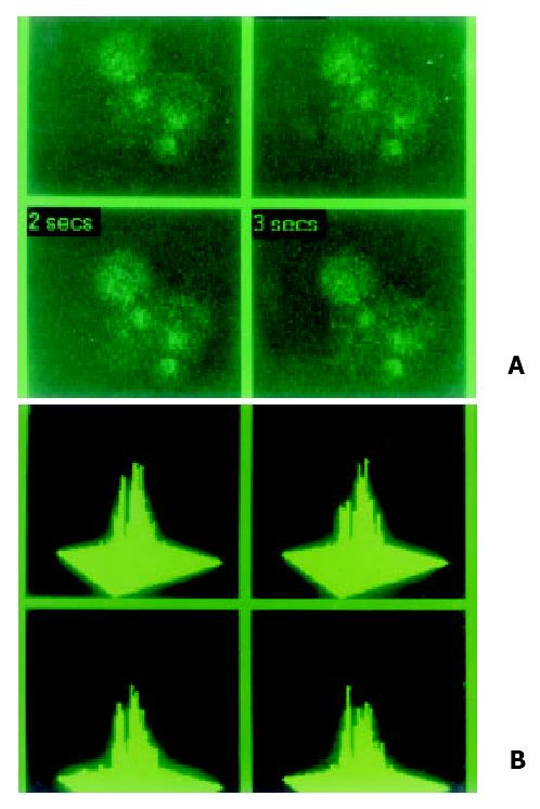
Analysis of concentrations and location of intracellular Ca2+ ([Ca2+]i) LSCM was used to determine intracellular Ca2+ in cultured cells after being loaded with special indicator of Ca2+ Fluo-3 AM. The results showed that intensity of Ca2+ was much higher with a very bright and higher peak signal of Ca2+ in QZG cells. Intracellular concentration of Ca2+ in QZG cells was 108.37 nmol/L. In HHCC and SMMC-7721 cells peak signals of Ca2+ were lower. [Ca2+]i in HHCC cell decreased to 35.13 nmol/L, SMMC-7721 cells also had lower peak Ca2+ and its [Ca2+]i decreased to 47.08 nmol/L as shown in Figure 1, Figure 2, Figure 3.

Figure 1
Figure 1 Special indicator Fluo-3 AM was loading at 37 °C for 90 min in hepatocarcinoma cell lines in order to examine localization and concentration of intracellular calcium in MRC-1024 laser scanning confocal microscope imaging system. (A). In normal hepatocyte cell line QZG, stronger signal of calcium with lower peak released. (B). Concentration of intracellular calcium in HHCC was 108.37 nmol/L.
Figure 2
Figure 2 Special indicator Fluo-3 AM was loading at 37 °C for 90 min in hepatocarcinoma cell lines in order to examine localization and concentration of intracellular calcium in MRC-1024 laser scanning confocal microscope imaging system. (A). In hepatocarcinoma cell line HHCC, slight signal of calcium with lower peak was observed. (B). Concentration of intracellular calcium in HHCC was 35.13 nmol/L.
Figure 3
Figure 3 Special indicator Fluo-3 AM was loading at 37 °C for 90 min in hepatocarcinoma cell lines in order to examine localizatio and concentration of intracellular calcium in MRC-1024 laser scanning confocal microscope imaging system. (A). Loading by Fluo-3 AM, dark signal of calcium and lower peak was also absent in hepatocarcinoma cell line SMMC-7721. (B). Concentration of intracellular calcium was 47.08 nmol/L.
Phosphorylation on tyrosine of CX32, CX43 proteins

The expressions of CX32, CX43 proteins were assessed using western blot analysis with anti-CX32 mAb, anti-CX43 mAb. We also verified the phosphorylation on tyrosine using western blot analysis with anti-phosphorylated tyrosine mAb 4G10.

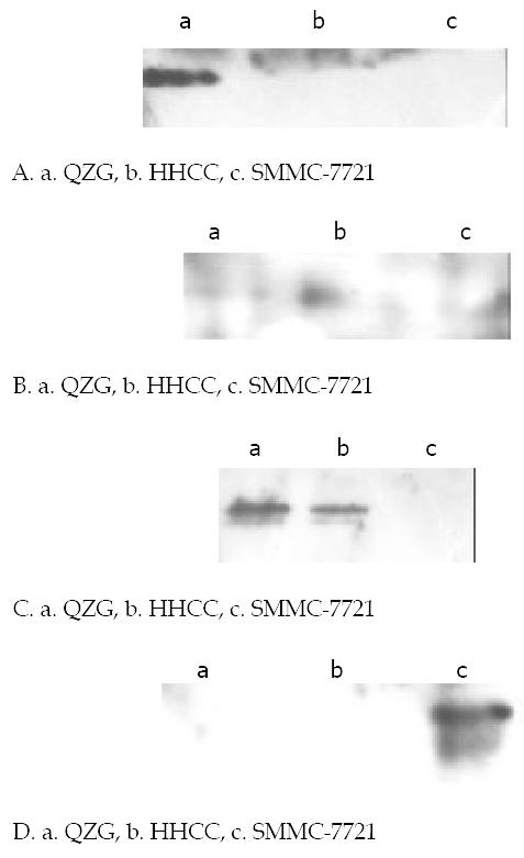
Western blot showed that major bands at 32 ku, 44 ku in a blot is unphosphorylated tyrosine in QZG cells. It recognized a band at 32 ku phosphorylated tyrosine CX32 protein and phosphorylated tyrosine CX43 protein of 43 ku in SMMC-7721 cells. No evidence of phosphorylated tyrosine at 32 ku of CX32 and 44 ku of CX43 proteins were detected in HHCC cells.

These results conform that the mouse monoclonal antibody selectively binds phosphorylated tyrosine CX43 proteins was corresponding decrease of concentrations of calcium and block of gap junctional intercellular communication in same hepatocellular carcinoma cell lines as shown in Figure 4A-D.

Figure 4
Figure 4 Western blot analysis of phosphorylation on tyrosine of CX32, CX43 proteins in various cells lines. Cells were harvested and lysed, equal amount of cell lystates were resolved of SDS-PAGE, transferred to NC membranes, and then probed with anti-phophorylation tyrosine mAb 4G10. (A). Expressions of CX32 proteins in cell lines with special anti-CX32 mAb. CX32 showed high immunoblot signal only in normal hepatocyte cell line QZG with very slight signal in hepatocarcinoma cell lines HHCC, SMMC-7721. (B). Expressions of CX43 proteins in cell lines with special anti-CX43 mAb. CX43 apperared in both QZG and SMMC-7721 cells but no in HHCC. (C). Phosphorylation on tyrosine of CX43 with special anti-phosphrylation tyrosine 4G10, unphosphorylation appeared in QZG cells even they showed high level expression of CX32, CX43. (D). Phosphorylated tyrosine of CX43 protein was detected in SMMC-7721 cells.
DISCUSSION

Gap junctions are channels between neighboring cells, which allow small molecules, and ions (mass molecules are below 1000 u) to pass through directly. Normal hepatocytes express high quantities of connexin32, connexin43 as the major gap junction forming proteins[20,27]. As described previously, immunohistochemical analysis showed gap junctional proteins CX32, CX43 frequently decreased in hepatocellular carcinoma tissues and cell lines[28-31]. But by in situ hybridization (ISH), CX32 mRNA, CX43 mRNA in hepatocellular carcinoma tissues and cell lines showed same strong positive rates and density without significant difference[32,33]. The aberrant location of CX32 and CX43 proteins might be responsible for hepatocarcinogenesis[34], the mechanism might be defect of CX genes in post-translational processing[35].

In various neoplasm, including hepatocellular carcinoma cells often express less connexin, but the mechanisms are unknown. It suggested that decrease of expression and lacking of function of CX32, CX43 might keep close relationship with liver hepatocarcinogenesis. Disruption of GJIC activity could contribute to the multi-step, multi-mechanism process of carcinogenesis[36]. Most tumors demonstrate a reduction between either homologous and heterologous gap junctional intercellular communication (GJIC). GJIC was assessed by transfer of the fluorescent dye Lucifer Yellow (LY) after scrape loading dye transfer (SLDT) in hepatocellular carcinoma cell lines HHCC, SMMC-7721 and normal liver cell line QZG. Molecular mass of LY is below 1000 u, which can pass via gap junction smoothly[37]. Our results showed that QZG had high ability of GJIC function than that in HHCC and SMMC-7721 cell lines, which were almost blocked.

Presumably down-regulation of GJIC activity would lead to the removal of growth inhibitory signaling. It was hypothesized that an initiated cell would be growth suppressed by being coupled by gap junctions to surrounding normal cells. Disturbance of GJIC facilitate cells to escape from intercellular signals involved regulations on proliferation, differentiation and apoptosis. Many factors, tumor-promoting chemicals[38] and oncogenes influence signal transduction in the initiated and surrounding normal cells, which would cause the down-regulation of GJIC. Many kinds of tumor promoting agents have been shown to decrease expressions of gap junction proteins or block GJIC associated with hepatocarcinogenesis[39]. H-ras, v-myc also cooperate malignancy[40]. Connexin genes might belong to the group of tumor suppressor genes.

Our results suggested that multiple mechanisms likely contributed to block of GJIC, including decrease of expressions of gap junction proteins and abnormal pathway of signal transduction including deceased level of intracellular Ca2+, phosphorylation on tyrosine and other potential mechanisms. In different tissues molecules smaller than approximately 1000 u may pass through gap junctions which implies that the most common second messengers cAMP, inositol-1,4,5-trisphosphate (IP3) and intracellular free calcium ions Ca2+ can be transferred. The mechanisms of intracellular calcium signaling are currently being investigated. Mechanisms during hepatocarcinogenesis are related to multiple pathophysiological processes including decreased in intracellular Ca2+. Calcium signals might be an important mechanism for controlling and synchronizing physiology. With intact gap junctional coupling, calcium in connected hepatocytes can communicate directly through gap junctions.

Whereas, gap junctional permeability and conductance affected by the calcium concentrations spikes in hepatocytes, a reduction of junctional conductance by calcium concentrations has been observed in hepatoma cells[42,43]. In our study, after special indicator of calcium Fluo-3AM loading, LSCM was used to measure concentrations of intracellular calcium [Ca2+]i in hepatocellular carcinoma cell lines HHCC, SMMC-7721 and normal liver cell line QZG. Concentrations of intracellular calcium [Ca2+]i was much higher in QZG (108.37 nmol/L) cell line than that in HHCC (35.13 nmol/L) and SMMC-7721(47.08 nmol/L) cell lines. QZG cells showed much higher calcium peaks than HHCC, SMMC-7721 cells which was associated with disruption of GJIC in these cell lines. Decreased level of intracellular Ca2+ contributed to conduction abnormalities of GJIC which immediate loss of calcium fluxes and influence the calcium dynamics in hepatocellular carcinoma cell lines.

Gap junctions are modulated by a multi-step molecular components and signaling pathway. Our observations also suggested that disruption of GJIC in hepatoma cells was associated with multiple distinct steps such as phosphorylation on tyrosine of CX32, CX43 proteins[43]. CX contains phosphorylations sites for several protein kinases including protein kinase C (PKC) and mitogen-activated protein kinase (MAPK) which are involved in CX phophorylation leading to the closure of GJIC[44,45]. Disruption of GJIC requires CX43 phosphorylation and interaction with other cellular factors. Phosphorylation of CX proteins has been postulated to be a critical regulatory element for gap junctional communication[46]. In our study, western blot suggested that only QZG cells had unphosphorylated tyrosine in CX32 protein at 32ku and CX43 protein at 43ku; SMMC-7721 cells had phosphorylated tyrosine CX43 proteins. Change in the amount and distribution of phosphorylated and nonphosphorylated isoforms of CX32 and CX43 were measured by immunoblot using isoform-secific antibodies anti-CX32 mAb, anti-CX43 mAb. Dramatic decreases in gap junctinal communication and loss of phosphorylation on tyrosine[47]. It indicated post-translation phosphorylation on tyrosine of CX proteins, which associated with a dramatic disruption of GJIC[48]. Functional recovery was associated with increased levels of unphosphorylated CX43.

As above all, disruption of GJIC in hepatocellular carcinoma cells could also be achieved by multi-mechanisms. We could make conclusions that the establishment and permanent of functional gap junctions were regulated by transcriptions as well as post-translational controls including reduce of concentrations of intracellular calcium and phosphorylation on tyrosine of connexin proteins even the biological co nseq uences o f sp ecific chang es in CX p ro tein s phosphorylation are not understood in detail. The decrease of CX proteins or the disorder of signal transduction of CX genes might keep close relationships with hepatocellular carcinoma.

Footnotes

Edited by Yuan HT

References
1.  Wang Y, Liu H, Zhou Q, Li X. Analysis of point mutation in site 1896 of HBV precore and its detection in the tissues and serum of HCC patients. World J Gastroenterol. 2000;6:395-397.  [PubMed]  [DOI]  [Cited in This Article: ]
2.  Mei MH, Xu J, Shi QF, Yang JH, Chen Q, Qin LL. Clinical significance of serum intercellular adhesion molecule-1 detection in patients with hepatocellular carcinoma. World J Gastroenterol. 2000;6:408-410.  [PubMed]  [DOI]  [Cited in This Article: ]
3.  Qin Y, Li B, Tan YS, Sun ZL, Zuo FQ, Sun ZF. Polymorphism of p16INK4a gene and rare mutation of p15INK4b gene exon2 in primary hepatocarcinoma. World J Gastroenterol. 2000;6:411-414.  [PubMed]  [DOI]  [Cited in This Article: ]
4.  Wang XZ, Jiang XR, Chen XC, Chen ZX, Li D, Lin JY, Tao QM. Seek protein which can interact with hepatitis B virus X protein from human liver cDNA library by yeast two-hybrid system. World J Gastroenterol. 2002;8:95-98.  [PubMed]  [DOI]  [Cited in This Article: ]
5.  Yin ZZ, Jin HL, Yin XZ, Li TZ, Quan JS, Jin ZN. Effect of Boschniakia rossica on expression of GST-P, p53 and p21(ras)proteins in early stage of chemical hepatocarcinogenesis and its anti-inflammatory activities in rats. World J Gastroenterol. 2000;6:812-818.  [PubMed]  [DOI]  [Cited in This Article: ]
6.  Lin GY, Chen ZL, Lu CM, Li Y, Ping XJ, Huang R. Immunohistochemical study on p53, H-rasp21, c-erbB-2 protein and PCNA expression in HCC tissues of Han and minority ethnic patients. World J Gastroenterol. 2000;6:234-238.  [PubMed]  [DOI]  [Cited in This Article: ]
7.  Liu L, Jiang Z, Teng GJ, Song JZ, Zhang DS, Guo QM, Fang W, He SC, Guo JH. Clinical and experimental study on regional administration of phosphorus 32 glass microspheres in treating hepatic carcinoma. World J Gastroenterol. 1999;5:492-505.  [PubMed]  [DOI]  [Cited in This Article: ]
8.  Liu LX, Liu ZH, Jiang HC, Qu X, Zhang WH, Wu LF, Zhu AL, Wang XQ, Wu M. Profiling of differentially expressed genes in human gastric carcinoma by cDNA expression array. World J Gastroenterol. 2002;8:580-585.  [PubMed]  [DOI]  [Cited in This Article: ]
9.  Lin NF, Tang J, Ismael HS. Study on environmental etiology of high incidence areas of liver cancer in China. World J Gastroenterol. 2000;6:572-576.  [PubMed]  [DOI]  [Cited in This Article: ]
10.  Tian DY, Yang DF, Xia NS, Zhang ZG, Lei HB, Huang YC. The serological prevalence and risk factor analysis of hepatitis G virus infection in Hubei Province of China. World J Gastroenterol. 2000;6:585-587.  [PubMed]  [DOI]  [Cited in This Article: ]
11.  Zhong DR, Ji XL. Hepatic angiomyolipoma-misdiagnosis as hepatocellular carcinoma: A report of 14 cases. World J Gastroenterol. 2000;6:608-612.  [PubMed]  [DOI]  [Cited in This Article: ]
12.  Riordan SM, Williams R. Transplantation of primary and reversibly immortalized human liver cells and other gene therapies in acute liver failure and decompensated chronic liver disease. World J Gastroenterol. 2000;6:636-642.  [PubMed]  [DOI]  [Cited in This Article: ]
13.  Li Y, Su JJ, Qin LL, Yang C, Luo D, Ban KC, Kensler T, Roebuck B. Chemopreventive effect of oltipraz on AFB(1)-induced hepatocarcinogenesis in tree shrew model. World J Gastroenterol. 2000;6:647-650.  [PubMed]  [DOI]  [Cited in This Article: ]
14.  Xu HY, Yang YL, Gao YY, Wu QL, Gao GQ. Effect of arsenic trioxide on human hepatoma cell line BEL-7402 cultured in vitro. World J Gastroenterol. 2000;6:681-687.  [PubMed]  [DOI]  [Cited in This Article: ]
15.  Zang GQ, Zhou XQ, Yu H, Xie Q, Zhao GM, Wang B, Guo Q, Xiang YQ, Liao D. Effect of hepatocyte apoptosis induced by TNF-alpha on acute severe hepatitis in mouse models. World J Gastroenterol. 2000;6:688-692.  [PubMed]  [DOI]  [Cited in This Article: ]
16.  Huang XF, Wang CM, Dai XW, Li ZJ, Pan BR, Yu LB, Qian B, Fang L. Expressions of chromogranin A and cathepsin D in human primary hepatocellular carcinoma. World J Gastroenterol. 2000;6:693-698.  [PubMed]  [DOI]  [Cited in This Article: ]
17.  Xu HY, Yang YL, Guan XL, Song G, Jiang AM, Shi LJ. Expression of regulating apoptosis gene and apoptosis index in primary liver cancer. World J Gastroenterol. 2000;6:721-724.  [PubMed]  [DOI]  [Cited in This Article: ]
18.  Chen YP, Liang WF, Zhang L, He HT, Luo KX. Transfusion transmitted virus infection in general populations and patients with various liver diseases in south China. World J Gastroenterol. 2000;6:738-741.  [PubMed]  [DOI]  [Cited in This Article: ]
19.  Falk MM. Biosynthesis and structural composition of gap junction intercellular membrane channels. Eur J Cell Biol. 2000;79:564-574.  [PubMed]  [DOI]  [Cited in This Article: ]  [Cited by in Crossref: 58]  [Cited by in F6Publishing: 55]  [Article Influence: 2.3]  [Reference Citation Analysis (0)]
20.  Kumar NM, Gilula NB. The gap junction communication channel. Cell. 1996;84:381-388.  [PubMed]  [DOI]  [Cited in This Article: ]  [Cited by in Crossref: 1372]  [Cited by in F6Publishing: 1339]  [Article Influence: 47.8]  [Reference Citation Analysis (0)]
21.  Windoffer R, Beile B, Leibold A, Thomas S, Wilhelm U, Leube RE. Visualization of gap junction mobility in living cells. Cell Tissue Res. 2000;299:347-362.  [PubMed]  [DOI]  [Cited in This Article: ]  [Cited by in Crossref: 26]  [Cited by in F6Publishing: 16]  [Article Influence: 0.7]  [Reference Citation Analysis (0)]
22.  Verselis VK, Trexler EB, Bukauskas FF. Connexin hemichannels and cell-cell channels: comparison of properties. Braz J Med Biol Res. 2000;33:379-389.  [PubMed]  [DOI]  [Cited in This Article: ]  [Cited by in Crossref: 38]  [Cited by in F6Publishing: 39]  [Article Influence: 1.6]  [Reference Citation Analysis (0)]
23.  Yamasaki H, Naus CC. Role of connexin genes in growth control. Carcinogenesis. 1996;17:1199-1213.  [PubMed]  [DOI]  [Cited in This Article: ]  [Cited by in Crossref: 341]  [Cited by in F6Publishing: 340]  [Article Influence: 12.1]  [Reference Citation Analysis (0)]
24.  Yamasaki H, Krutovskikh V, Mesnil M, Tanaka T, Zaidan-Dagli ML, Omori Y. Role of connexin (gap junction) genes in cell growth control and carcinogenesis. C R Acad Sci III. 1999;322:151-159.  [PubMed]  [DOI]  [Cited in This Article: ]  [Cited by in Crossref: 102]  [Cited by in F6Publishing: 107]  [Article Influence: 4.3]  [Reference Citation Analysis (0)]
25.  Krutovskikh V, Yamasaki H. The role of gap junctional intercellular communication (GJIC) disorders in experimental and human carcinogenesis. Histol Histopathol. 1997;12:761-768.  [PubMed]  [DOI]  [Cited in This Article: ]
26.  Iwai M, Harada Y, Muramatsu A, Tanaka S, Mori T, Okanoue T, Katoh F, Ohkusa T, Kashima K. Development of gap junctional channels and intercellular communication in rat liver during ontogenesis. J Hepatol. 2000;32:11-18.  [PubMed]  [DOI]  [Cited in This Article: ]  [Cited by in Crossref: 26]  [Cited by in F6Publishing: 53]  [Article Influence: 2.2]  [Reference Citation Analysis (0)]
27.  Steinberg TH. Gap junction function: the messenger and the message. Am J Pathol. 1998;152:851-854.  [PubMed]  [DOI]  [Cited in This Article: ]
28.  Ma XD, Sui YF, Wang WL. Expression of gap junction proteins CX32, CX43 in human hepatocarcinoma cell lines and normal liver cell line: Study with laser scanning confocal microscope. Zhongguo Aizheng Zazhi. 2000;10:1-2.  [PubMed]  [DOI]  [Cited in This Article: ]
29.  Ma XD, Sui YF, Wang WL, Xiu LQ. A flow cytometric study of gap junction CX32, CX43 in human hepatocellular carcinoma cell lines. Fenzi Yu Xibao Mianyixue Zazhi. 1999;15:51-54.  [PubMed]  [DOI]  [Cited in This Article: ]
30.  Ma XD, Sui YF, Wang WL. The expression of gap junction protein connexin32 in human hepatocellular carcinoma, cirrhotic and viral hepatitide liver tissues. Zhongguo Aizheng Zazhi. 1999;18:133-135.  [PubMed]  [DOI]  [Cited in This Article: ]
31.  Ma XD, Sui YF, Wang WL. Expression of gap junction genes connexin32, connexin43 and their proteins in human hepatocellular carcinoma cell lines and normal liver cell line. Shiyong Aizheng Zazhi. 1999;14:161-163.  [PubMed]  [DOI]  [Cited in This Article: ]
32.  Ma XD, Sui YF, Wang WL. Expression of gap junction genes connexin 32, connexin 43 and their proteins in hepatocellular carcinoma and normal liver tissues. World J Gastroenterol. 2000;6:66-69.  [PubMed]  [DOI]  [Cited in This Article: ]
33.  Ma XD, Ma X, Sui YF, Wang WL. Expression of gap junction genes connexin32 and connexin43 mRNAs and proteins, and their role in hepatocarcinogenesis. World J Gastroenterol. 2002;8:64-68.  [PubMed]  [DOI]  [Cited in This Article: ]
34.  Neveu MJ, Hully JR, Babcock KL, Hertzberg EL, Nicholson BJ, Paul DL, Pitot HC. Multiple mechanisms are responsible for altered expression of gap junction genes during oncogenesis in rat liver. J Cell Sci. 1994;107:83-95.  [PubMed]  [DOI]  [Cited in This Article: ]
35.  Piechocki MP, Burk RD, Ruch RJ. Regulation of connexin32 and connexin43 gene expression by DNA methylation in rat liver cells. Carcinogenesis. 1999;20:401-406.  [PubMed]  [DOI]  [Cited in This Article: ]  [Cited by in Crossref: 52]  [Cited by in F6Publishing: 54]  [Article Influence: 2.2]  [Reference Citation Analysis (0)]
36.  Niessen H, Willecke K. Strongly decreased gap junctional permeability to inositol 1,4, 5-trisphosphate in connexin32 deficient hepatocytes. FEBS Lett. 2000;466:112-114.  [PubMed]  [DOI]  [Cited in This Article: ]  [Cited by in Crossref: 43]  [Cited by in F6Publishing: 42]  [Article Influence: 1.8]  [Reference Citation Analysis (0)]
37.  Juul MH, Rivedal E, Stokke T, Sanner T. Quantitative determination of gap junction intercellular communication using flow cytometric measurement of fluorescent dye transfer. Cell Adhes Commun. 2000;7:501-512.  [PubMed]  [DOI]  [Cited in This Article: ]  [Cited by in Crossref: 14]  [Cited by in F6Publishing: 14]  [Article Influence: 0.6]  [Reference Citation Analysis (0)]
38.  Ren P, Ruch RJ. Inhibition of gap junctional intercellular communication by barbiturates in long-term primary cultured rat hepatocytes is correlated with liver tumour promoting activity. Carcinogenesis. 1996;17:2119-2124.  [PubMed]  [DOI]  [Cited in This Article: ]  [Cited by in Crossref: 14]  [Cited by in F6Publishing: 15]  [Article Influence: 0.5]  [Reference Citation Analysis (0)]
39.  Moennikes O, Buchmann A, Romualdi A, Ott T, Werringloer J, Willecke K, Schwarz M. Lack of phenobarbital-mediated promotion of hepatocarcinogenesis in connexin32-null mice. Cancer Res. 2000;60:5087-5091.  [PubMed]  [DOI]  [Cited in This Article: ]
40.  DeoCampo ND, Wilson MR, Trosko JE. Cooperation of bcl-2 and myc in the neoplastic transformation of normal rat liver epithelial cells is related to the down-regulation of gap junction-mediated intercellular communication. Carcinogenesis. 2000;21:1501-1506.  [PubMed]  [DOI]  [Cited in This Article: ]  [Cited by in Crossref: 8]  [Cited by in F6Publishing: 7]  [Article Influence: 0.3]  [Reference Citation Analysis (0)]
41.  Røttingen JA, Camerer E, Mathiesen I, Prydz H, Iversen JG. Synchronized Ca2+ oscillations induced in Madin Darby canine kidney cells by bradykinin and thrombin but not by ATP. Cell Calcium. 1997;21:195-211.  [PubMed]  [DOI]  [Cited in This Article: ]  [Cited by in Crossref: 21]  [Cited by in F6Publishing: 21]  [Article Influence: 0.8]  [Reference Citation Analysis (0)]
42.  Peracchia C, Wang XG, Peracchia LL. Slow gating of gap junction channels and calmodulin. J Membr Biol. 2000;178:55-70.  [PubMed]  [DOI]  [Cited in This Article: ]  [Cited by in Crossref: 31]  [Cited by in F6Publishing: 31]  [Article Influence: 1.3]  [Reference Citation Analysis (0)]
43.  Matesic DF, Rupp HL, Bonney WJ, Ruch RJ, Trosko JE. Changes in gap-junction permeability, phosphorylation, and number mediated by phorbol ester and non-phorbol-ester tumor promoters in rat liver epithelial cells. Mol Carcinog. 1994;10:226-236.  [PubMed]  [DOI]  [Cited in This Article: ]  [Cited by in Crossref: 128]  [Cited by in F6Publishing: 123]  [Article Influence: 4.1]  [Reference Citation Analysis (0)]
44.  Elcock FJ, Deag E, Roberts RA, Chipman JK. Nafenopin causes protein kinase C-mediated serine phosphorylation and loss of function of connexin 32 protein in rat hepatocytes without aberrant expression or localization. Toxicol Sci. 2000;56:86-94.  [PubMed]  [DOI]  [Cited in This Article: ]  [Cited by in Crossref: 13]  [Cited by in F6Publishing: 15]  [Article Influence: 0.6]  [Reference Citation Analysis (0)]
45.  Hossain MZ, Jagdale AB, Ao P, Boynton AL. Mitogen-activated protein kinase and phosphorylation of connexin43 are not sufficient for the disruption of gap junctional communication by platelet-derived growth factor and tetradecanoylphorbol acetate. J Cell Physiol. 1999;179:87-96.  [PubMed]  [DOI]  [Cited in This Article: ]  [Cited by in F6Publishing: 1]  [Reference Citation Analysis (0)]
46.  Kwak BR, Hermans MM, De Jonge HR, Lohmann SM, Jongsma HJ, Chanson M. Differential regulation of distinct types of gap junction channels by similar phosphorylating conditions. Mol Biol Cell. 1995;6:1707-1719.  [PubMed]  [DOI]  [Cited in This Article: ]  [Cited by in Crossref: 120]  [Cited by in F6Publishing: 126]  [Article Influence: 4.3]  [Reference Citation Analysis (0)]
47.  Omori Y, Krutovskikh V, Mironov N, Tsuda H, Yamasaki H. CX32 gene mutation in a chemically induced rat liver tumour. Carcinogenesis. 1996;17:2077-2080.  [PubMed]  [DOI]  [Cited in This Article: ]  [Cited by in Crossref: 41]  [Cited by in F6Publishing: 42]  [Article Influence: 1.5]  [Reference Citation Analysis (0)]
48.  Hossain MZ, Ao P, Boynton AL. Rapid disruption of gap junctional communication and phosphorylation of connexin43 by platelet-derived growth factor in T51B rat liver epithelial cells expressing platelet-derived growth factor receptor. J Cell Physiol. 1998;174:66-77.  [PubMed]  [DOI]  [Cited in This Article: ]  [Cited by in F6Publishing: 1]  [Reference Citation Analysis (0)]