Published online Dec 15, 2003. doi: 10.3748/wjg.v9.i12.2870
Revised: September 10, 2003
Accepted: October 23, 2003
Published online: December 15, 2003
Carcinoid tumors generally appear as yellow/gray or tan submucosal nodules. We experienced a case of pedunculated rectal carcinoid showing a mushroom-like appearance. The case was a forty years old woman who was admitted to our hospital due to rectal bleeding. Colonoscopy revealed a pedunculated polyp presenting a mushroom-shaped appearance measuring 13 mm in diameter in the rectum. The histological diagnosis of specimens obtained by biopsy was adenocarcinoma and transanal ultrasonography revealed the tumor localization within the submucosal layer in the rectum. Endoscopic mucosal resection (EMR) was performed. Histopathological examination established the diagnosis of carcinoid tumor in the rectum. Frequencies of the pedunculated type in rectal carcinoids were reported to be 2.4% to 7.1% in the literature. Because of its rarity, pedunculated configuration may confuse the endoscopic diagnosis of carcinoids. Treatment for carcinoids of 1 to 1.5 cm in size remains controversial. Although such tumors are technically respectable by EMR, careful attention must be paid in dealing with these tumors because there may be unexpected behaviors of the tumors.
- Citation: Hamada H, Shikuwa S, Wen CY, Isomoto H, Nakao K, Miyashita K, Daikoku M, Yano K, Ito M, Mizuta Y, Chen LD, Xu ZM, Murata I, Kohno S. Pedunculated rectal carcinoid removed by endoscopic mucosal resection: a case report . World J Gastroenterol 2003; 9(12): 2870-2872
- URL: https://www.wjgnet.com/1007-9327/full/v9/i12/2870.htm
- DOI: https://dx.doi.org/10.3748/wjg.v9.i12.2870
Carcinoid tumors characteristically appear as yellow/gray or tan submucosal nodules, but they are occasionally polypoid or sessile. However, there have been few reports describing a pedunculated type of carcinoid. We presented here an extremely rare case with a pedunculated rectal carcinoid showing a mushroom-like appearance, and referred to the diagnosis and treatment of this rare tumor.
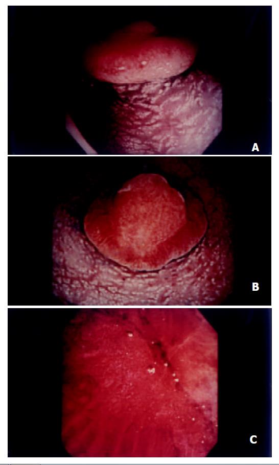
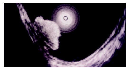
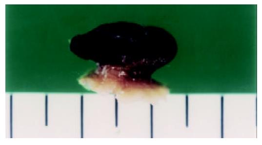
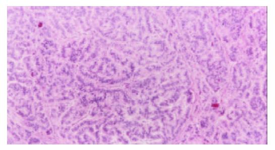
A 40 years old woman was admitted to our hospital due to rectal bleeding. Physical examination and laboratory data including serum tumor markers and hormones such as urinary 5-hydroxyindoleacetic acid (5-HIAA) were normal. Barium enema contrast examination showed a fungiform polyp in the rectum. Colonoscopy revealed a pedunculated polyp presenting a mushroom-shaped appearance in the rectum (Figure 1A, B). There was a hemispherical protrusion with a shallow central erosion in the top, surrounded by a marked mucosal bulge of the edge. Magnifying endoscopy (OLYMPUS CF-type XQ240ZI, Olympus, Tokyo, Japan) revealed no absence of the pit pattern in the center and enlarged pits at the edge, corresponding to the non-structure type of the pit pattern classification proposed by Kudo et al[1] (Figure 1C). Endoscopic ultrasonography (EUS), using a miniature probe (12-20 Hz) with a water-filling method, demonstrated a homogeneous hypoechoic mass, but the structure deeper than the third layer of the rectal wall was unclear (Figure 2). Therefore, we employed a soft-balloon technique using an ultrasonic probe with a balloon filled with deaerated water, which provided a clear ultrasonographic picture of the deeper part of the rectum. Judging from findings on EUS, abdominal computed tomography and chest roentgenography, there were no signs of metastasis in the regional lymph nodes or distant organs. Histology of the biopsy specimens suggested an adenocarcinoma, yielding a diagnosis of polypoid type of early rectal cancer. The depth of mural invasion was estimated to be limited to the submucosa by magnifying endoscopy and EUS. After an injection of saline in the submucosa, the lesion was excised by EMR. Macroscopically, the lesion was located in the submucosal layer. The mass was white-yellowish and solid, measuring 13 mm in diameter (Figure 3). Microscopically, the tumor was composed of small uniform cells, arranged in small nests and cords and with an anastomosing ribbon-like pattern in the submucosal layer (Figure 4). There were no atypical histopathologic features such as mitosis or nuclear atypism. Histochemically, the tumor cells possessed an argyrophil but not an argentaffin nature. Immunohistologically, the tumor cells were positive for neuron specific enolase (NSE) and chromogranin A, but were negative for p53 and Ki67. These findings established the diagnosis of carcinoid tumor in the rectum.
Carcinoid tumors are enigmatic slow growing malignancies and controversy remains as to their origin[2]. Gastrointestinal carcinoid is regarded as a tumor arising from subepithelial neuroendocrine cells or the totipotential crypt cells in the deep mucosa, usually presenting as a submucosal tumor. Therefore, endoscopic diagnosis is difficult unless biopsy provides a correct histological diagnosis. Macroscopically, a gastrointestinal carcinoid tumor appears as a yellow-gray tan nodule beneath the mucosa and develops a round or oval, sessile polyp as it grows[3]. A pedunculated polyp of carcinoid tumor is extremely rare in the rectum. To our knowledge, only nine cases of rectal carcinoid, including the present case, have been reported[4-9]. Frequencies of the pedunculated type in rectal carcinoids were reported to be 2.4% to 7.1% in the literature[5,6]. Because of its rarity, pedunculated configuration may confuse the endoscopic diagnosis of carcinoids.
Recently, Kudo et al[1] reported the usefulness of the pit pattern observation using magnifying endoscopy and stereomicroscopy in the diagnosis of epithelial neoplasms of the large intestine. They classified the pit patterns into five types based on fine morphology of the surface, histology and size. Type V pit pattern showed an irregular or nonstructural surface, which was frequently seen in carcinomas[1]. In this case, we misdiagnosed this tumor as carcinoma because it showed the V pit pattern and pathological diagnosis of biopsy specimens was not correct. It was suggested that the carcinoid in the present case showed non-structural pit patterns because overlying mucosa of the tumor disappeared. It appears inappropriate, therefore, to use pit pattern analysis for evaluation of nature of the tumor in this case.
The size of carcinoid has been accepted as an important factor predicting its prognosis. Several surgical studies have shown that tumors less than 1 cm seldom metastasize, whereas tumors greater than 2 cm have a high incidence of metastasis[2,3,10]. As for the risk of metastasis from rectal carcinoid, Bates et al[11] reported an incidence of 1.7% for tumors less than 9 mm in size, 10% for tumor of 10 to 19 mm and 82% for tumors larger than 20 mm. It is uncertain whether gross appearance of the carcinoids is a predicting factor, although Haraguchi et al[12] reported that no metastasis was observed in pedunculated carcinoid in a review of 496 cases of rectal carcinoid.
These reports indicated that the most effective therapy for rectal carcinoid was a complete surgical excision, particularly in tumors greater than 2 cm[3,13]. Recently, endoscopic treatment has been applied for gastrointestinal tumors including carcinoids. Ishikawa et al[14] suggested the following indications of conventional endoscopic polypectomy for rectal carcinoid tumors. They were tumors less than 15 mm in diameter, flat tumors with normal or yellow color, tumors consisting of nodular nests or trabecular or ribbon-like structures in histology. This guideline of endoscopic treatment for rectal carcinoid may remain controversial. We do not recommend a conventional endoscopic polypectomy for common rectal carcinoid because there was a high incidence of tumor residue after treatment[15]. Recently, endoscopic mucosal resection (EMR) has been generally accepted as a choice of treatment for rectal carcinoid tumors less than 1 cm[9,14,16,17]. Treatment for carcinoids of 1 to 1.5 cm in size remains controversial. Although such tumors have been technically respectable by endoscopic aspiration mucosectomy using the hood technique or a ligating device[18], careful attention must be paid to these tumors because there may be unexpected behaviors of the tumors.
The carcinoid in the present case was negative for p53 and Ki-67. Hasegawa et al[4] reported that carcinoid tumors expressing p53 and Ki-67 had a high malignant potential and metastatic activity. In future, the induction of molecular biology may be helpful in predicting the prognosis of carcinoid tumors[19,20].
Edited by Wang XL
| 1. | Kudo S, Rubio CA, Teixeira CR, Kashida H, Kogure E. Pit pattern in colorectal neoplasia: endoscopic magnifying view. Endoscopy. 2001;33:367-373. [RCA] [PubMed] [DOI] [Full Text] [Cited by in Crossref: 317] [Cited by in RCA: 309] [Article Influence: 12.9] [Reference Citation Analysis (0)] |
| 2. | Koura AN, Giacco GG, Curley SA, Skibber JM, Feig BW, Ellis LM. Carcinoid tumors of the rectum: effect of size, histopathology, and surgical treatment on metastasis free survival. Cancer. 1997;79:1294-1298. [RCA] [PubMed] [DOI] [Full Text] [Cited by in RCA: 3] [Reference Citation Analysis (0)] |
| 3. | Läuffer JM, Zhang T, Modlin IM. Review article: current status of gastrointestinal carcinoids. Aliment Pharmacol Ther. 1999;13:271-287. [RCA] [PubMed] [DOI] [Full Text] [Cited by in Crossref: 72] [Cited by in RCA: 58] [Article Influence: 2.2] [Reference Citation Analysis (0)] |
| 4. | Hasegawa O, Iwashita A, Futami T, Kitamura K, Arima S. Patho-logical study of rectal carcinoid. J the Japan society of colon Proctol-ogy. 1997;50:163-176. |
| 5. | Pon JL, Walke L. Carcinoid tumors of the rectum. Dis Colon Rectum. 1971;14:46-56. [RCA] [PubMed] [DOI] [Full Text] [Cited by in Crossref: 5] [Cited by in RCA: 5] [Article Influence: 0.1] [Reference Citation Analysis (0)] |
| 6. | QUAN SH, BADER G, BERG JW. CARCINOID TUMORS OF THE RECTUM. Dis Colon Rectum. 1964;7:197-206. [RCA] [PubMed] [DOI] [Full Text] [Cited by in Crossref: 40] [Cited by in RCA: 41] [Article Influence: 0.7] [Reference Citation Analysis (0)] |
| 7. | Masumori S, Nogaki M, Ozeki T, Ktsuragi T, Koshimura Y, Higaki A, Hosoda S. The rectal carcinoid- clinicopathologic Study of five cases.. Jap J Cancer Clin. 1975;21:1181-1188. |
| 8. | Kira J, Fuchigami T, Murakami M, Koga A, Iwashita A. A case report of rectal carcinoid and an analysis of rectal carcinoids re-ported in Japan. I to Cho(Stomach and Intestine). 1980;15:1105-1110. |
| 9. | Kobayashi K, Katsumata T, Otani Y, Atari E. [Diagnosis and treatment of rectal carcinoid]. Nihon Rinsho. 1991;49:233-237. [PubMed] |
| 10. | Soga J. Carcinoids of the rectum: an evaluation of 1271 reported cases. Surg Today. 1997;27:112-119. [RCA] [PubMed] [DOI] [Full Text] [Cited by in Crossref: 129] [Cited by in RCA: 113] [Article Influence: 4.0] [Reference Citation Analysis (0)] |
| 11. | Bates HR. Carcinoid tumors of the rectum: a statistical review. Dis Colon Rectum. 1966;9:90. [RCA] [PubMed] [DOI] [Full Text] [Cited by in Crossref: 27] [Cited by in RCA: 28] [Article Influence: 1.2] [Reference Citation Analysis (0)] |
| 12. | Haraguchi M, Makiyama K, Yamakawa M, Yamasaki K, Iwanaga S, Mizuta Y, Ide T, Komori M, Tanaka T, Osabe M. Six cases of rectal carcinoid treated by endo-scopic polypectomy. Gastroenterol Endosc. 1998;30:2612-2620. |
| 13. | Kulke MH, Mayer RJ. Carcinoid tumors. N Engl J Med. 1999;18:358-368. [RCA] [DOI] [Full Text] [Cited by in Crossref: 647] [Cited by in RCA: 549] [Article Influence: 21.1] [Reference Citation Analysis (0)] |
| 14. | Ishikawa H, Imanishi K, Otani T, Okuda S, Tatsuta M, Ishiguro S. Effectiveness of endoscopic treatment of carcinoid tumors of the rectum. Endoscopy. 1989;21:133-135. [RCA] [PubMed] [DOI] [Full Text] [Cited by in Crossref: 33] [Cited by in RCA: 30] [Article Influence: 0.8] [Reference Citation Analysis (0)] |
| 15. | Okamoto T, Higaki K, Kawabata K. [Autopsy case of malignant carcinoid tumor of the ascending colon]. Gan No Rinsho. 1983;29:1361-1365. [PubMed] |
| 16. | Fujimura Y, Mizuno M, Takeda M, Sato I, Hoshika K, Uchida J, Kihara T, Mure T, Sano K, Moriya T. A carcinoid tumor of the rectum removed by strip biopsy. Endoscopy. 1993;25:428-430. [RCA] [PubMed] [DOI] [Full Text] [Cited by in Crossref: 14] [Cited by in RCA: 15] [Article Influence: 0.5] [Reference Citation Analysis (0)] |
| 17. | Higaki S, Nishiaki M, Mitani N, Yanai H, Tada M, Okita K. Effectiveness of local endoscopic resection of rectal carcinoid tumors. Endoscopy. 1997;29:171-175. [RCA] [PubMed] [DOI] [Full Text] [Cited by in Crossref: 40] [Cited by in RCA: 43] [Article Influence: 1.5] [Reference Citation Analysis (0)] |
| 18. | Shikuwa S, Matsunaga K, Osabe M, Ofukuji M, Omagari K, Mizuta Y, Takeshima F, Murase K, Otani H, Ito M. Esophageal granular cell tumor treated by endoscopic mucosal resection using a ligating device. Gastrointest Endosc. 1998;47:529-532. [RCA] [PubMed] [DOI] [Full Text] [Cited by in Crossref: 17] [Cited by in RCA: 15] [Article Influence: 0.6] [Reference Citation Analysis (0)] |
| 19. | Lundqvist M, Wilander E. Subepithelial neuroendocrine cells and carcinoid tumours of the human small intestine and appendix. A comparative immunohistochemical study with regard to serotonin, neuron-specific enolase and S-100 protein reactivity. J Pathol. 1986;148:141-147. [RCA] [PubMed] [DOI] [Full Text] [Cited by in Crossref: 43] [Cited by in RCA: 40] [Article Influence: 1.0] [Reference Citation Analysis (0)] |
| 20. | Moyana TN, Satkunam N. Crypt cell proliferative micronests in rectal carcinoids. An immunohistochemical study. Am J Surg Pathol. 1993;17:350-356. [RCA] [PubMed] [DOI] [Full Text] [Cited by in Crossref: 5] [Cited by in RCA: 5] [Article Influence: 0.2] [Reference Citation Analysis (0)] |


