INTRODUCTION
Bile leakage is one of the most common and serious complications after hepatobiliary surgery. In recent years, the treatment strategy of bile leakage has generated fundamental changes due to the excellent effect of non-surgical therapy and its minimally invasive intervention[1-6]. Non-surgical treatment has been regarded as the first choice in the management of postoperative bile leakage in many cases. Only when there is no essential condition or no effect by non-surgery, will operation be considered[1,2]. Although there are many nonoperative approaches that could be used to cure bile leakage, most literatures only reported one of these methods[7-53], being deficient in systematization and comprehensiveness. Thus, surgeons could not often apply them synthetically and reasonably. Over the past decade from 1991 to 2000, we treated 57 patients with postoperative bile leakage non-surgically in our two hospitals. In this article, we summarized the methods and indications of the non-surgical treatment for postoperative bile leakage.
MATERIALS AND METHODS
Patients
From January 1991 to December 2000, a total of 57 patients with postoperative bile leakage, including 23 males and 34 females, were treated non-surgically in the Department of Surgery of the Second Affiliated Hospital, Medical School of Zhejiang University and the Affiliated Yijishan Hospital of Wannan Medical College. The mean age of the patients was 46 years, with a range from 25 to 73 years. All of them belonged to postoperative bile leakage. The original operations were performed with simple open cholecystectomy (OC) in 21 cases, OC plus choledochotomy with T tube drainage in 25, simple hepatectomy in 7, hepatectomy plus Roux-en-Y cholangiojejunostomy in 3, and repair of bile duct injury plus choledochoduodenostomy in 1, respectively. The clinical presentations were diverse. Most patients were presented with sudden or gradual abdominal pain after operation, and T tube removal/slippage or U tube exchange. Almost half of the cases had much bile-like liquid outflowing from the original drainage tubes or the fistulous tracts of T tube. Twelve patients had light to moderate fever, four hyperpyrexia, six jaundice, and two had nausea and vomiting.
Diagnosis
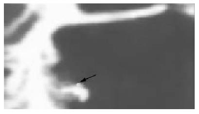
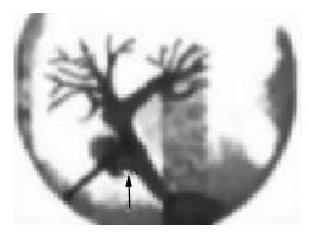
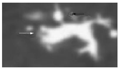
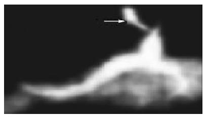
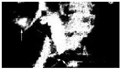
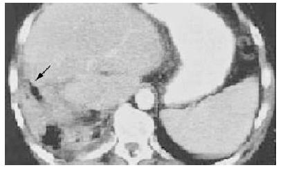
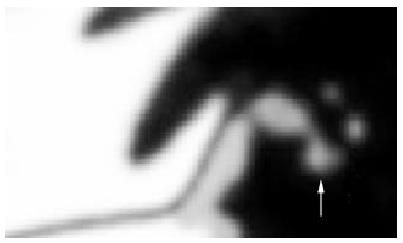
The diagnoses were made according to the operational history, clinical situation, abdominal paracentesis (21 person times), ultrasonography (17), ERCP (8), PTC (5), MRI/MRCP (3), gastroscopy (3), and percutaneous fistulography (2). The site of the leakage was the cystic duct in 3 cases, the fossae of gallbladder in 14 (Figure 1), the disrupted or damaged fistulous tracts of T tube in 25 (Figure 2 and Figure 3), the damaged bile duct in 3 (Figure 4 and Figure 5), the cut surface of liver in 7 (Figure 6), the stomas of bilioenteric anastomosis in 3 and it was undetectable in the other 2 cases. Besides bile leakage, the wrong ligation of bile ducts beyond the leakages was also found in 3 patients with common bile duct injury. Of the 25 patients with bile leakage following T tube removal, residual stones of the distal bile duct were also found in 5 cases (Figure 3 and Figure 7) and benign papillary strictures were detected in 3 (Figure 5). Bile collections in 2 patients developed biloma, which oppressed pylorus, resulting in pyloric obstruction. Bile collections at two different sites, including hepatorenal recess and hepatogastric interspace with only a slender and bent tract (diameter < 0.5 cm) between them, were confirmed in one patient undergoing hepatectomy, whose original subphrenic drainage was removed owing to occlusion before the bile leakage was found. Although the drainage on the anterior abdominal wall was still retained, it only drained bile from hepatogastric interspace.
Figure 1 ERCP showed a bile leak from the fossae of gallbladder (↑).
Figure 2 Cholangiography through T tube showed a leak from common bile duct (↑).
Figure 3 ERCP showed a leak from the fistulous tract of T tube (↑) and stones at distal duct (Δ).
Figure 4 ERCP showed a leak from right hepatic duct (Δ).
Figure 5 MRCP showed stricture at distal duct (↑) and liquid collection in the right subhepatic region (Δ).
Figure 6 CT showed bile accumulating in the cut surface of right liver (↑).
Figure 7 The distal duct stones was removed with choledochoscope (Δ).
Management
When patients revealed the symptoms of bile leakage or biliary peritonitis, some essential disposal measures were immediately used such as right lateral decubitus position, semi-reclining position, gastrointestinal decompression, complementing water-electrolyte, antibiotic and appropriate nutritional support[1,2]. After that, specific non-surgical methods were chosen according to the volume, site of bile leakage and patient's condition. The original drainage in 19 patients undergoing OC or hepatectomy were kept unobstructed because they still produced some effect of drainage. The cannulae were changed in 5 cases of OC or hepatectomy due to their occlusion. In addition, washing with antibiotic was performed through the cannulae. In all the 25 patients with bile leakage following T tube removal or slippage, a new drainage was once again placed into the fistulous tract of T tube. Repeated abdominal cavity suction and drainage with a percutaneous catheter were performed by percutaneous abdominal paracentesis in 3 of 25 patients while percutaneous transhepatic cholangial drainage (PTCD) or percutaneous transhepatic biliary drainage (PTBD) were performed in 2 patients whose intrahepatic bile ducts were dilated. The residual ductal stones in 5 patients were eliminated by choledochoscope (Figure 7), endoscopic sphincterotomy (EST) and the traditional Chinese medicine, respectively. The traditional Chinese medicines included bitter orange, costusroot, mongolian milkvetch, rhubarb, christina loosestrife herb, etc. In order to maintain the pressure of bile duct and facilitate stones passage, it was necessary to obstruct the T tube at intervals when the Chinese medicines were administered. The bile duct stricture in 3 cases was relieved by choledochoscope and EST, respectively. Percutaneous catheter drainages guided by ultrasound were used in one patient with biloma and 3 patients with common bile leakage. As to the patient with subhepatic fluid accumulations at two different sites detected by ultrasonography, MRI and percutaneous fistulography, it was inappropriate to reoperate on him or her due to the postoperative weak health, and it was very dangerous to apply abdominal paracentesis because the hepatorenal recess was just near the right thoracic cavity. A new drain tube was first placed into hepatogastric interspace and afterwards into the bent tract and hepatorenal recess through the fistulous tract on the anterior abdominal wall. The whole process was guided by gastroscope and X-ray. Flushing with antibiotic was performed everyday.
RESULTS
In the early stage of our study, one patient with stoma leakage died of infectious shock, and one with post-hepatectomy bile leakage died of multiple organ failure (MOF). After 1996, no patient died of bile leakage. Five patients were converted to operation due to ineffective non-surgical management, presentation of jaundice or concomitant wrong ligation of bile duct. All the 5 patients were cured by late reoperation. Of the 5 cases, one with biloma belonged to the early stage of our study. Owing to the absence of experience in the non-surgical treatment at that time (1993), he only received some essential disposals except percutaneous catheter drainage at the beginning of bile leakage. He underwent exploratory laparotomy plus drainage a week later because nausea and vomiting were not relieved. Two patients with bile leakage from cystic duct stump underwent exploratory laparotomy plus re-ligation of cystic duct after 2-3 d drainage. The other two patients all underwent hepatojejunostomy 2-3 mo later due to concomitant wrong ligation of bile duct. The other 50 patients were directly cured by non-surgical treatment without correlated complications. The mean closure time of bile leakage was 9 d, with a range from 5 d to 3 mo. The cure rate of non-surgical treatment was 82.5% (50/57).
DISCUSSION
When bile leakage occurs, it is necessary to adopt some essential disposals such as gastrointestinal decompression, fluid replacement, antibiotic therapy, etc. However, it is important to choose operation or nonoperation according to the volume of bile leakage and patient's condition. In recent years, the treatment strategy of bile leakage has generated fundamental changes due to the invention or finding of some new non-surgical methods, which have further improved the therapeutic effect of bile leakage with minimal invasive intervention or no trauma. Non-surgical treatment has been regarded as the first choice in the management of postoperative bile leakage in many cases, and it is acceptable by such patients. Only when there is no essential condition for non-surgery, or no effect, will operation be considered[1-6].
Non-surgical methods
In our study, the 57 patients with postoperative bile leakage were all managed by non-surgical treatment at the beginning of bile leakage. Fifty patients were cured except that two died and five were converted to reoperation later. The cure rate of non-surgical treatment was 82.5% (50/57). The total therapeutic efficacy is satisfactory. Based on our data, the following non-surgical methods could be used in the treatment of bile leakage. (1) Keep the original drain tube unobstructed or chang it[1,2,5]. The original drainage should be maintained when it still drains some bile, and should be changed for a new drain tube when obstructed. In addition, we think it is equally of importance to wash with antibiotic through the cannulae[9], because it could increase the local concentration of antibiotic and improve anti-infection effect. It was difficult for the bile leakage to heal in patients of older age, malnutrition, complicated diabetes and using hormone. Besides drainage, some related therapy should be continued in these cases, the hormone should be cancelled, and the time of drainage be properly prolonged. (2) Insert a new catheter through the fistulous tract of T tube[1,5,16,17]. It is necessary to place a new catheter immediately through the fistulous tract of T tube if the bile leakage occurs after a T tube removal or slippage. The procedure should be performed as early as possible to prevent the occlusion of fistulous tract. Suction with negative pressure could be additionally used when necessary[1,16,18]. (3) Percutaneous abdominal paracentesis[1,5,6,8,10-13]. As an important nonoperative method, abdominal paracentesis can be applied because it does not need special device except an injection syringe. If the site of leakage was deep or near important organs, abdominal paracentesis should be guided by ultrasound. It is helpful to not only the diagnosis of bile leakage but also the abdominal cavity suctions or percutaneous catheter drainage. In this group, abdominal paracentesis was performed in five patients (4 with bile leakage after T tube removal and 1 with post-cholecystectomy biloma). Among them, the biloma was punctured under the guidance of ultrasound[1]. All 5 were cured. (4) PTCD/PTBD or placement of a percutaneous transhepatic stent[1,14,15]. Owing to some dangers (including hemorrhage, new bile leakage and so on), it should be generally performed under the guidance of ultrasound in patients with intrahepatic cholangiectasis[54]. PTCD is helpful to the healing of bile leakage because it may reduce the volume of leakage. In our study, PTCDs were used in 2 cases because both abdominal drainages were insufficient. Two weeks later, both patients were cured. (5) Endoscopic management[1,6,8,24-53]. As a new progression in the nonoperative treatment of bile leakage, endoscopic management generally means EST, endoscopic nasobiliary tube drainage (ENBD) and endoscopic biliary stent (EBS)[32,33,55], which are all finished by duodenoscope. It is mainly applied to the patients with bile duct stone, stricture or biloma[32,33,55]. These endoscopic methods could effectively remove the residual ductal stones, relieve the stricture of bile duct and facilitate the healing of leakage. However, endoscopes include choledochoscope and gastroscope besides duodenoscope. Because choledochoscope may not only gets rid of the residual stones but also dilate the narrow bile duct, it should also play an important role in the nonoperative treatment of bile leakage, and should not be neglected. In our study, choledochoscopic treatments were given to 4 patients (2 with residual stones, 2 with bile duct stenosis). All were cured. Gastroscope may occasionally be used in the nonoperative treatment of bile leakage. In this group, the new drain tube in the patient with bile accumulations at two different sites was placed under the guidance of gastroscope and X-ray. The leakage closed 3 mo later. Thus, we should not confine our sights to the duodenoscope when using endoscopic treatment. Besides duodenoscope, gastroscope and choledochoscope may also be applied. (6) Traditional Chinese medicines[1]. It is mainly applied to the patients with bile duct stone. Some Chinese medicines may faciliate the passage of residual stones, resulting in accelerated healing of leakage. In this group, after Chinese medicines were administered to 2 patients for 5-7 d, the residual stones were all precluded that was confirmed by ultrasound and contrast examination. Both leakages closed within 2 wk. In addition, there are some other non-surgical methods including laparoscopy[19-22], ethanol ablation[23] and so on.
Indications
Non-surgical treatment therapy plays an important role in the management of bile leakage due to its many advantages. First, the cure rate is high[33,39]. Binmoeller et al[33] reported that the total cure rate of endoscopic management was 86%. In Fuji's study, all 8 postoperative bile leakages were cured by endoscopic treatment[39]. In our study, 57 patients were treated with various non-surgical methods, 50 were directly cured, the rate being 82.5%. Next, non-surgical treatment is convenient, safe[1,10,40,41], atraumatic or micro-traumatic[43]. If only it is properly used, reoperation could be avoided in most patients with postoperative bile leakage, and the pain and costs of patients could be reduced. Thus, it is acceptable by patients, and fits the general trend and direction of modern surgery. The non-surgical therapy is especially suitable for the elderly patients who are physically weak, poor in common state, complicated by other serious diseases, unbearable or unwilling to accept reoperation in a short time. In this group, more than half of the patients belonged to such cases. Owing to correct choice of treatment, they not only avoided reoperation, but also safely passed the dangerous period. As for the patients who need surgery but could not be operated on at the early stage of bile leakage due to their conditions, non-surgical treatment can not only serves as interim measure before operation but also wins precious time for preoperative preparation. It was quite obvious that such condition was present in the 5 patients who were converted to late reoperation. When the patients with bile leakage were treated non-surgically, its indications should be strictly controlled. Based on our data, we think that nonoperative therapy could be first attempted in the following cases[1]. (1) Patients with early bile leakage[2] (within 4-6 h), relatively minor leakage, rather light or local peritonitis and no infectious shock or following shock by estimation; (2) Postoperative early patients who are poor in general state, complicated with other serious diseases, unbearable or unwilling to accept reoperation in a short time; (3) Patients whose original subhepatic drainages were still reserved and unobstructed; (4) Patients who suffer from bile leakages after T tubes removal or slippage and can be implanted with new drain tubes through the fistulous tracts of T tubes; (5) Patients who can be treated with percutaneous abdominal paracentesis or drainage; (6) Patients with intrahepatic bile duct dilataltion who can be managed by PTCD/PTBD and (7) Patients whose residual bile duct stones or strictures can be cured by endoscopic management or traditional Chinese Medicine. Of course, it is essential to pay close attention to the change of patient's condition and complications during nonoperative treatment, and it also necessary to be simultaneously ready to reoperate at any time. Surgery should be considered as soon as possible if the patient's condition has not improved even aggravated such as abdominal pain degree worsening, scope expanding, temperature apparently rising and so on[1,10]. Reoperation should also be considered if only the patient's condition goes beyond the limit of nonoperative therapy such as relatively major bile leakage, serious peritonitis[1,2], persistent leakage or concomitant wrong ligation of bile duct. In our study, 5 patients were converted to reoperation later due to inefficiency of non-surgical management, occurrence of jaundice or wrong ligation of bile duct.


