Published online Jun 15, 2000. doi: 10.3748/wjg.v6.i3.424
Revised: February 3, 2000
Accepted: March 2, 2000
Published online: June 15, 2000
- Citation: Pei WF, Xu GS, Sun Y, Zhu SL, Zhang DQ. Protective effect of electroacupuncture and moxibustion on gastric mucosal damage and its relation with nitric oxide in rats. World J Gastroenterol 2000; 6(3): 424-427
- URL: https://www.wjgnet.com/1007-9327/full/v6/i3/424.htm
- DOI: https://dx.doi.org/10.3748/wjg.v6.i3.424
Gastric mucosal injury is one of the common disorders, there are many reports subjected to its pathogenesis treatment and prevention[1-4]. We investigated the protective effect of electroacupuncture and moxibustion of Zusanli point on gastric mucosal damage and its relation with nitric oxide (NO) on the animal model with acute gastric mucosal damage induced by ethanol. The detected indexes include the content of NO, gastric mucosal blood flow (GMBF), gastric mucosal lesion index (LI) and transmucosal potential difference (PD).
Drugs L-arginine (L-arg) the precursor of NO (purchased from Huamei Biological Engineering Corporation); Natrii Nitroprussidum (SNP) the donor of NO (provided by Experimental Drug Plant of the Beijing Institute of Pharmaceutical Industry), diluted with distilled water prior to use, Nw-nitro-L-arginine (L-NNA), an inhibitor of NOS (produced by Sigma), and diluted with 0.2 mol·L-1 PBS; 200 g·L-1 pentobarbital sodium was prepared with distilled water immediately before use.
Wistar rats weighing 180-250 g (provided by the Institute of Acupuncture and Moxibustion, Anhui College of Traditional Chinese Medicine) were fasted for 12 h and allowed free access to water. The animals were divided into 16 groups randomly, including: control group (c), model (m), m + electroacu puncture (EA), m + moxibustion (M), L-arg + m, SNP + m, L-NNA + m, L-NNA + L-arg + m, L-arg + m + EA, L-arg + m + M, SNP + m + EA, SNP + m + M, L-NNA + m + EA, L-NNA + m + M, L-NNA + L-arg + m + EA, L-NNA + L-arg + m + M.
The rats fasted for 12 h were injected with pentobarbital intraperitoneally at a dose of (30-40) mg·Kg-1. The middle line incision below xiphoid process after anesthesia was performed, then stomach was exposed and injected with 2 mL 700 mL/L ethanol, and the abdomen closed, 30 min later the preparation of model was fulfilled.
Different procedures were used for different groups. In groups injected drugs or equivalent amount of physiological saline, these were injected respectively into blood flow slowly through the great saphenous vein of the anesthetized rats 15 min before the preparation of the model. In groups which required electroacupuncture or moxibustion, were treated with electroacupuncture or moxibustion on bilateral Zusanli points after the model was successfully prepared. The electroacupuncture was performed with PCE2 electroacupuncture therapy device on the condition of frequency 20 Hz, voltage 5-8 v for 30 min (produced by the Institute of Acupuncture and Moxibustion of Anhui College of Traditional Chinese Medicine). During moxibustion, the lighted pure moxacigar was maintained 1 cm from the bilateral Zusanli points. The GMBF and PD values were measured for all rats. Then the blood was obtained through decephalization or from retro-ocular vassel for the NO assay. Subsequently the rat stomach was resected for the analysis of LI, and the mucosa from gastric antrum and the body for assaying NO contents.
GMBF was assayed by analyzing hydrogen gas clearance curves[6]; PD was assayed with direct Ag-AgCl. Electrode measurement technique[7] with few alterations, i.e. the efficient electrode was put on the mucosa while the reference one on the serosa. The electric potential difference between two membranes was recorded. For the detection of LI, the stomach was resected and incised along the greater curvature, washed with physiological saline. Guth index assessment[8] (slightly modefied) was used to compute the damage, on a scale of grades 1-5 as follows: Grade 1, the petechia or ecchymosis, Grade 2, 3, 4 and 5, pathological focus of 1 mm; 1-2 mm, 2-4 mm, and over 4 mm, respectively. The NO content in blood and mucosa of gastric antrum and body were assayed based on the method from Green et al[9]. The Kit for NO assay was provided by the Department of Biochemistry, Institute of Radiology, Academy of Medical Science of PLA. The analysis was based on the protocol, but the mucosa were homogenized before the assay.
All data were expressed as -x±s, and the Student's t test was used for the comparison between groups.
| Group | n | LI | GMBF (mL·min-1·100 g-1) | PD (mV) | NO | |
| Blood (μmol·L-1) | Antrum (ng·mg-1) | |||||
| C | 5 | 0 | 138.20 ± 4.37 | 20.82 ± 0.99 | 21.12 ± 1.89 | 53.30 ± 2.65 |
| m | 5 | 45.6 ± 3.2a | 60.60 ± 6.09a | 11.85 ± 0.82a | 12.84 ± 1.54a | 35.17 ± 1.57a |
| m + EA | 5 | 31.4 ± 3.3c | 92.55 ± 6.35c | 15.84 ± 0.48c | 18.65 ± 0.69c | 48.51 ± 2.12c |
| m + M | 5 | 33.8 ± 2.4c | 95.91 ± 5.59c | 15.96 ± 0.34c | 18.53 ± 1.04c | 48.49 ± 2.39c |
| L-arg + m | 5 | 32.4 ± 3.2 | 106.22 ± 5.30 | 17.73 ± 0.22 | 18.93 ± 1.30 | 47.57 ± 2.86 |
| SNP + m | 5 | 30.0 ± 3.1 | 104.03 ± 7.57 | 17.89 ± 0.39 | 19.07 ± 1.29 | 47.18 ± 2.17 |
| L-NNL + m | 5 | 55.8 ± 2.8b | 56.85 ± 5.96d | 6.97 ± 0.32c | 8.37 ± 0.27b | 27.29 ± 1.71b |
| L-NNA + L-arg + m | 5 | 46.8 ± 2.6 | 78.90 ± 5.10 | 10.93 ± 0.39 | 12.10 ± 1.23 | 33.76 ± 2.44 |
| L-arg + m + EA | 5 | 21.0 ± 0.9e | 121.50 ± 3.55d | 18.14 ± 0.44 | 24.70 ± 0.75d | 50.63 ± 2.34 |
| L-arg + m + M | 5 | 27.1 ± 2.7d | 97.46 ± 4.90 | 16.08 ± 0.45 | 21.06 ± 1.17 | 44.26 ± 2.01 |
| SNP + m + EA | 5 | 20.9 ± 0.8e | 118.50 ± 4.21d | 17.79 ± 0.59 | 24.80 ± 0.50d | 45.17 ± 2.01 |
| SNP + m + M | 5 | 26.8 ± 2.2d | 101.11 ± 5.23 | 14.73 ± 0.61 | 23.14 ± 1.52d | 43.33 ± 1.97 |
| L-NNA + m + EA | 6 | 41.8 ± 2.1e | 73.57 ± 5.43d | 11.87 ± 0.41d | 12.37 ± 1.78e | 39.25 ± 1.66d |
| L-NNA + m + M | 6 | 40.5 ± 3.0d | 66.42 ± 5.42e | 10.25 ± 0.72e | 11.43 ± 1.03e | 32.17 ± 2.35e |
| L-NNA + L-arg + m + EA | 5 | 31.0 ± 1.2g | 95.00 ± 3.78f | 16.15 ± 0.74g | 19.24 ± 0.54g | 51.51 ± 2.14f |
| L-NNA + L-arg + m + M | 5 | 31.5 ± 2.1g | 70.76 ± 4.78 | 11.81 ± 0.53 | 12.88 ± 0.68 | 36.37 ± 2.29 |
The mean of GMBF, PD in group m showed statistically significant difference compared with the control group (P < 0.01), demonstrating marked decrease of GMBF and PD after gastric mucosal damage, the integrity of gastric mucosa was depended on adequate blood flow. GMBF and PD in group m + M was obviously increased (P < 0.01) compared with group m, suggesting the electroacupunctur e and moxibustion of Zusanli point may relieve and cure the gastric mucosal damage induced by ethanol.
From Table 1 we can find, NO contents in group m showed significant difference compared with normal control (P < 0.01); LI in group m + EA and group m + M was markedly lower than that in group m (P < 0.01). The results showed that the gastric mucosal damage aggravated as NO content decreased while NO content increased and the gastric mucosal damage relieved, which suggests that NO content is associated with the integrity of gastric mucosa.
The L-Arg (150 mg·Kg-1), SNP(200 μg·Kg-1) and L-NNA (3 mg·Kg-1) were administered through the great saphenous vein respectively 15 min before ig administration of 700 mL/L ethanol in different groups. After that the electroacupuncture and moxibustion of Zusanli point were performed for 30 min. Subsequently, the GMBF, PD and LI as well as NO content in blood and gastric mucosa were assayed and detected respectively in each group. It was found that there was little difference in the indexes when group L-arg + m and group SNP + m were compared with group m + EA or group m + M (P < 0.05), indicating the function of electroacupuncture and moxibustion on gastric mucosal damage was similar to that of L-arg or SNP. Comparing group L-Arg + m + EA (or M) and group SNP + m + EA (or M) with group m + EA (or M), and group SNP + m + EA (or M) with group m + EA (or M), NO content, GMBF and PD values were markedly increased, and LI value was obviously decreased (P < 0.01 or P < 0.05), which suggested that electroacupuncture and moxibustion of Zusanli point had the effect of strengthening NO pathway and protecting gastric mucosa.
In comparison of group L-NNA + m with group m., NO content, GMBF and PD value decreased, and LI value increased (P < 0.01 or P < 0.05), showing that L-NNA aggravated ethanol induced gastric mucosal damage. When we compared group L-NNA + m + EA (or M) with group m + EA (or M), NO content, GMBF and PD value also decreased, but LI value increased (P < 0.01 or P < 0.05), which suggests L-NNA decreased the protective action of electroacupunctureand moxibustion on gastric damage. While we compared group L-NNA + L-arg + m + EA (or M) with group L-NNA + m + EA (or M), NO contents, GMBF and PD value increased and LI value decreased (P < 0.01 or P < 0.05), demonst rating L-arg may reverse the inhibitory function of L-NNA. In conclusion, the above mentioned results further suggest that the protective action on gastric mucosa by the electroacupuncture and moxibustion of Zusanli point is NO mediated.
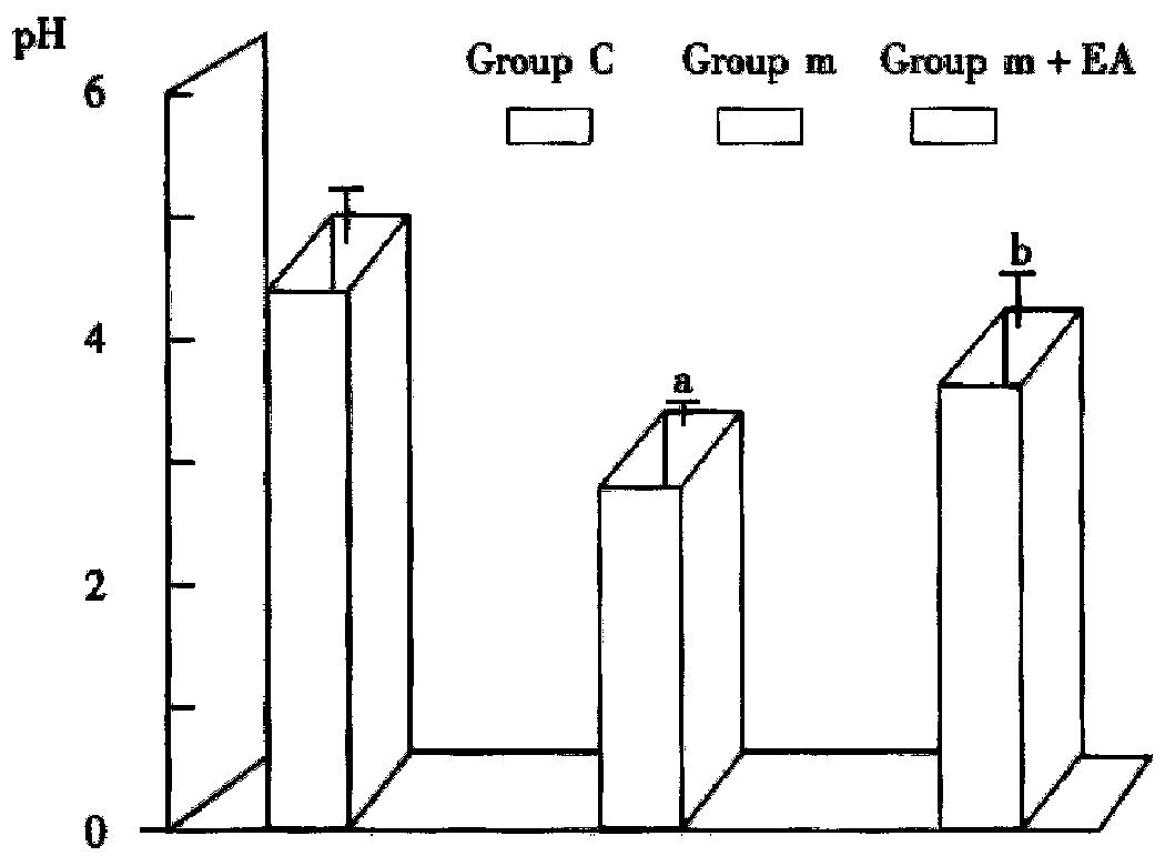
pH in group c was 4.33 ± 0.40 and that in Group m 2.92 ± 0.37, decreasing significantly in the latter group (P < 0.01). The pH value in Group m + EA, 3.84 ± 0.69 was much higher than that in Group m indicating 700 mL/L ethanol promotes the stomach to secrete acids while electroacupuncture inhibits the secretion.
Acupuncture and moxibustion may treat and prevent gastrointestinal disorders, the mechanism of which is being further investigated. The acupuncture and moxibustion on Zusanli points show the two-way regulation of gastrointestinal function. In recent years, it is frequently reported that they may relieve symptom an d promote the ulcer healing to certain extent[1-4]. There is relative point specificity, the first choice is the Zusanli point in stomach channel of foot yangming. Our study investigated the therapeutic and protective effect of the acupuncture and moxibustion on acute gastric mucosal damage induced by 700 mL/L ethanol. It is found that GMBF and PD value as well as NO content, pH value in rat gastric mucosa of model group with acupuncture and moxibustion increased more markedly than those without any therapy (P < 0.01), while the gastric mucosal damage alleviated (P < 0.01) which denotes the therapeutic and protective effect of acupuncture and moxibustion on the damage and suggests that the protective action is related with the level of NO content.
NO is a small molecular gas produced from the procursor L-Arg under the NOS catalysis. The process is called as L-Arg-NO pathway, NOS activity plays a key role in NO synthesis. NOS is existed in many kinds of tissues including vascular endothelial cell, thrombocyte, brain cell, renal epithelial cell, macropha gocyte, neutrophil, hepatocyte, etc. Some factors can induce the expression of NOS and increase the NO synthesis. As a special bioinformational molecule, the neurotransmitter or the humoral factor, NO involves in the functional regulation and pathophysiology of many diseases. Our study investigates the protective effect of acupuncture and moxibustion on acute gastric mucosal damage and in the meantime measures NO content in blood and gastric mucosa and finds that NO content is negatively correlated with the severity of gastric mucosal damage, which proves that the protective action of acupuncture and moxibustion of Zusanli point is NO mediated; and in other words NO involves in the regulative effect of acupuncture and moxibustion on gastrointestinal function.
The good balance between NO and ET keeps the endothelium intact, If the unbalance is developed, the gastric mucosal damage will be formed and the gastric dysfunction presented. Our study showed[1] that when NO content decreased ET content increased and gastric mucosal damage aggravated, and on the contrary, the damage alleviated. Our results have verified it. When L-arg or SNP was administered 15 min before establishing the model with 700 mL/L ethanol; NO content was higher than that in the model group and concurrently GMBF and PD value increased, and LI value decreased; if NOS inhibitor-L-NNA was given, NO content, GMPF and PD value were lower than those in the model group while LI value increased, which indicates that NO plays an important role in maintaining the intact gastric mucosa. GMBF decrease may prominently respond to the gastric mucosal damage after ethanol induction. It is thought that NO is an EDRF, which is capable of relaxing the vascular smooth muscle obviously, Our research further confirmed that the vascular relaxing action of NO can protect gastric mucosa from damage.
NO initiates the protective effect of acupuncture and moxibustion on gastric mucosa, which has been proved in our work. During experiment, in the model rats, the NO content in their blood and gastric mucosa increased after acupuncture and moxibustion were performed, and GMBF and PD values were also increased while LI value decreased. If L-NNA was preliminarily given, the above effect of acupuncture and moxibustion of Zusanli point disappeared, but concurrently given L-ary, the action of L-NNA could be reversed. In addition, if L-arg or SNP was preliminarily administered and then the model established, NO content in the group with the acupuncture and moxibustion of Zusanli point increased more than that in the corresponding groups without acupuncture and moxibustion, moreover, GMBF and PD values increased and LI decreased correspondingly, showing the synergetic action between them. The above phenomena further confirm that the acupuncture and moxibustion of Zusanli points alliviate gastric mucosal damage, which is NO mediated, and this illustrates that the acupuncture an d moxibustion of Zusanli point can activate endogenous NOS.
As far as the possible mechanism why Zusanli point can initiate NO system is concerned, we consider that the main nerves regulating gastric function are vagus nerves, the dorsal vagus nucleus (DMV) being the major motor nucleus. The efficient stimulus transmitted to DMV by the somatic sensory nerves through the spinal cord. DMV also receives afferent information from gastro intestinal tract and integrates other information from central nervous system (CNS), and transfers to the stomach by efferent vagus nerves, resulting in the increase of NO synthesis and release.
The possible mechanism about how NO can protect gastric mucosa from damage is as follows: NO acts as one of endothelial diastolic factors which can cause the dilatation of vascular smooth muscles, resulting in GMBF increase, improvement of blood supply of gastric mucosa, maintaining the integrity of gastric mucosal epithelium, protecting the gastric mucosa from the stimulation of gastric contents and from damage, preventing from H+ invasion and keeping normal ionic concentration gradient across gastric mucosa as well as maintaining appropriate PD value, while acupuncture and moxibustion may activating NOS to increase the synthesis and release of NO. Further research remains to be done as how does the acupuncture and moxibustion activate NOS.
Dr. Wen-Fen Pei, graduated from the Department of Medicine, Anhui Medical College in 1976. Associate professor, engaged in study of digestive physiology, having more than 10 papers published.
Edited by You DY
proofread by Sun SM
| 1. | Xu GS, Wang ZJ, Zhu SL, Chen QZ, Jiao J, Zhang DQ. Nitric Oxide participates in protective effects of acupuncture on gastric mucosal damages in rats. Anhui Zhongyi Xueyuan Xuebao. 1996;15:36-38. |
| 2. | Ma TF, Yang Z. Therapeutic effect and mechanism of acupuncture on digestive tract disease. Gastrointestinal physiology. Peking: Science press 1991; 755-772. |
| 3. | Qiao X, Yin K. [Therapeutic effect of moxibustion on experimental gastric ulcer of rats and its mechanism]. Zhenci Yanjiu. 1992;17:270-273. [PubMed] |
| 4. | Xu G. [Effect of moxibustion on gastrointestinal electric activity in rabbits through zusanli (ST36) and its mechanism]. Zhenci Yanjiu. 1992;17:274-276. [PubMed] |
| 5. | Masuda E, Kawano S, Nagano K, Tsuji S, Takei Y, Tsujii M, Oshita M, Michida T, Kobayashi I, Nakama A. Endogenous nitric oxide modulates ethanol-induced gastric mucosal injury in rats. Gastroenterology. 1995;108:58-64. [RCA] [PubMed] [DOI] [Full Text] [Cited by in Crossref: 63] [Cited by in RCA: 66] [Article Influence: 2.2] [Reference Citation Analysis (0)] |
| 6. | Livingston EH, Reedy T, Leung FW, Guth PH. Computerized curve fitting in the analysis of hydrogen gas clearance curves. Am J Physiol. 1989;257:G668-G675. [PubMed] |
| 7. | Xu GS, Sun Y, Wang ZJ, Zhang DQ, Gu XJ. Effects of electroacupuncture on gastric mucosal blood flow and transmucosal potential difference in stress rats. Huaren Xiaohua Zazhi. 1998;6:4-6. |
| 8. | Guth PH, Aures D, Paulsen G. Topical aspirin plus HCl gastric lesions in the rat. Cytoprotective effect of prostaglandin, cimetidine, and probanthine. Gastroenterology. 1979;76:88-93. [PubMed] |


