Published online Apr 15, 2000. doi: 10.3748/wjg.v6.i2.248
Revised: June 26, 1999
Accepted: July 1, 1999
Published online: April 15, 2000
AIM: To further study the properties of bile liquid crystals, and probe into the relationship between bile liquid crystals and gallbladder stone formation, and provide evidence for the prevention and treatment of cholecystolithiasis.
METHODS: The optic properties of bile liquid crystals in human body were determined by the method of crystal optics under polarizing microscope with plane polarized light and perpendicular polarized light.
RESULTS: Under a polarizing microscope with plane polarized light, bile liquid crystals scattered in bile appeared round, oval or irregularly round. The color of bile liquid crystals was a little lighter than that of the bile around. When the stage was turned round, the color of bile liquid crystals or the darkness and lightness of the color did not change obviously. On the border between bile liquid crystals and the bile around, brighter Becke-Line could be observed. When the microscope tube is lifted, Becke-Line moved inward, and when lowered, Becke-Line moved outward. Under a perpendicular polarized light, bile liquid crystals showd some special interference patterns, called Malta cross. When the stage was turning round at an angle of 360°, the Malta cross showed four times of extinction. In the vibrating direction of 45° angle of relative to upper and lower polarizing plate, gypsum test-board with optical path difference of 530 nm was inserted, the first and the third quadrants of Malt a cross appeared to be blue, and the second and the fourth quadrants appeared orange. When mica test-board with optical path difference of 147 nm was inserted, the first and the third quadrants of Malta cross appeared yellow, and the second and the fourth quadrants appeared dark grey.
CONCLUSION: The bile liquid crystals were distributed in bile in the form of global grains. Their polychroism and absorption were slight, but the edge and Becke*Line were very clear. Its refractive index was larger than that of the bile. These liquid crystals were uniaxial positive crystals. The interference colors were the first order grey-white. The double refractive index of the liquid crystals was Δn = 0.011-0.015.
- Citation: Yang HM, Wu J, Li JY, Zhou JL, He LJ, Xu XF. Optic properties of bile liquid crystals in human body. World J Gastroenterol 2000; 6(2): 248-251
- URL: https://www.wjgnet.com/1007-9327/full/v6/i2/248.htm
- DOI: https://dx.doi.org/10.3748/wjg.v6.i2.248
Researches of recent years show that there are liquid crystals in the bile of the patients with gallbladder stone[1-4]. Some scholars inferred the function of bile liquid crystals[2]. In order to find out the properties of bile liquid crystals, probe into the relationship between bile liuqid crystals and the formation of gallbladder stone and provide evidence for the prevention and treatment of cholecystolithiasis, we used the method of crystal optics, and determined the optic properties of bile liquid crystals in human under polarizing microscope with plane polarized light and perendicular polarized light. No similar reports have been found so far.
The bile was collected from the gallbladder extirpated surgically in the Second Affiliated Hospital of Kunming Medical College and kept at 4.0 °C in the refrigerator for use.
Extraction of bile liquid crystals Firstly, the bile was centrifuged at 4000 r/min for 30 min with type 800 centrifuge sedimentor made in China. Secondly, the supernatant was centrifuged at 30000 r/min for 40 min of equivalent time of centrifugation through 50 g/L-150 g/L linear concentration gradients of sucrose solution with Hitachi RPS 50-2 swing- out rotor and model SCP85H Hitachi automatic preparative ultracentrifuge at 25 °C. After centrifugalization, the bile was taken out in different colors and put into different sample cups. After examination under a polarizing microscope, liquid crystals were selected for microslide preparation.
Microslide preparation Bile (6.0 μL) was taken with China-made W-104 micropipet (0.2 μL), and dropped to a slide and covered with a glass. It was stretched into a slice naturally for observation and measurement.
Measurement of bile refractive index With China-made 2WA871430 Abbe refractometer (0.0005), bile refractive index was measured after gradient centrifugation of sucrose density.
Measurement of bile liquid crystals thickness With China-made 0 mm - 25 mm micrometer screw gauge (0.01 mm) the area of cover glass was measured and the thickness of liquid crystals was worked out according to the volume taken out from the micropipet.
Polarizing microscopic observation Under a Japan-made BHSP Olympus advanced polarizing microscope, observations were made on the outside character of bile liquid crystals, the optical nature involved with light wave absorption and optical nature related with refraction rate. Under a perpendiculer polarizing microscope, interference patterns of bile liquid crystals, disappearing phenomenon and mobility were examined. The size of Malta cross was measured with micrometer eyepiece (0.1 mm) and micrometer objective (0.01 mm). In the vibration direction at 45° angle relative to upper and lower polarizing plate, gypsum test-board with optical path difference of 530 nm and the mica test-board with optical path difference of 147 nm were inserted to measure the optical symbol, interference color level and double refraction rate of the bile liquid crystals.
Under a polarizing and a perpendicular polarized light microscope, 100 pictures of different bile liquid crystals from 17 cases of cholecystolithiasis were observed and measured.
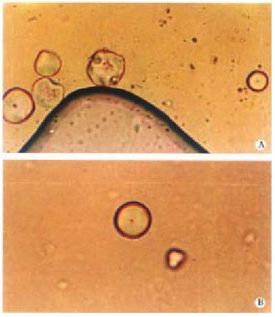
Under a polarizing microscope, bile liquid crystals were found scattered in the bile, appearing round, oval or irregularly round in shape (Figure 1A).
Under a polarizing microscope, the color of bile liquid crystals was a little lighter than that of the bile being around. When the stage was turned round, no obvious changes occurred in the color of bile liquid crystals or the darkness and lightness of the color.
Under a polarizing microscope, on the border between bile liquid crystals and bile being around, darker edge can be observed with a brighter line nearby, i.e., Becke-Line (Figure 1B). When the microscope tube was lifted, Becke-Line moved inward (to the liquid crystals), and when lowered, Becke-Line moved outwardly (to the bile). The refractive index of bile liquid crystals was bigger than that of the bile being around (1.3408-1.3494) (Table 1).
| No. | Refractive index | No. | Refractive index | No. | Refractive index |
| 001 | 1.3470 | 007 | 1.3441 | 013 | 1.3446 |
| 002 | 1.3494 | 008 | 1.3418 | 014 | 1.3443 |
| 003 | 1.3485 | 009 | 1.3408 | 015 | 1.3451 |
| 004 | 1.3486 | 010 | 1.3416 | 016 | 1.3454 |
| 005 | 1.3479 | 011 | 1.3435 | 017 | 1.3456 |
| 006 | 1.3473 | 012 | 1.3445 | 018 | 1.3457 |
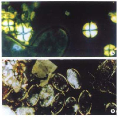
Under a perpendicular polarized light, bile liquid crystals in human body presented some special interference patterns, called Malta cross. These Malta crosses had various kinds of shapes, which can be divided generally into three types. The first one was round in shape, with four symmetry, equal and broad bright double refraction areas, and the middle part was a narrow, dark cross; the second one was irregularly round in shape, with four symmetry bright double-refraction areas but not equal each other, in the middle, the narrow dark cross was lean towards different directions; and the third one shaped oval with four narrow bright double-refraction areas, in the middle there was a bigger dark cross (Figure 2A, B).
Brightness or darkness of Malta cross was changed regularly when the stage was rotated, which changed once for each 45° turning. If there was a turning of 360°, four times of brightness and darkness appeared, i.e., four extinction and four interference color phenomena occurred.
Little distilled water was added to the space between a slide and a cover glass containing bile liquid crystals. Under a perpendicular polarized light, Malta cross was observed from bile liquid crystals, flowing swiftly accompanying with the mobile of the water. In the process of flowing, the shape of Malta cross changed incessantly with the flowing of liquid crystals. When it flew to the edge, Malta cross was changed into crystal grains of double refraction after drying.
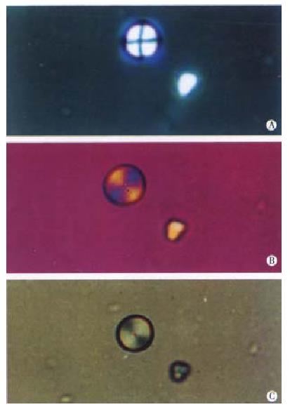
Under a perpendicular polarized light, in the relative direction of 45° angle of viration of upper and lower polarizing plate, a plaster test-board with optical path difference of 530 nm was inserted, the first and the third quadrants of Malta cross was found present in bile liquid crystals being blue in color, and the second and the fourth quadrants being orange, and the black cross being purplish. When a mica test-board with optical path difference of 147 nm was inserted, the first and the third quadrants of Malta cross appeared light yellow, and the second and the fourth quadrants appeared dark grey (Figure 3A, B, C). It is suggested that optical symbol of bile liquid crystals is uniaxial positive and interference color level is the first level grey[5].
Under a perpendicular polarized light microscope, the diameter (or long axis) of Malta cross was measured to be between 6.2 × 10⁻³ mm and 2.9 × 10⁻² mm.
It was measured that the area of bile was 328.3 mm2± 0.7 mm2, the thickness of liquid crystals d = (1.8 ± 0.1) × 10-2 mm, the interference color level was the first level grey-white, and optical path difference was 200 nm-270 nm[6]. According to the formula of that optical path difference: R = d (Ne-No) = d·Än, the double refractive index of bile liquid crystals was calculated as Än = 0.011-0.015.
Under plane polarized light, the outside character of bile liquid crystals was observed, which were not steric pattern, but a section contour of a certain direction formed by bile liquid crystals crossed with cover glass. Because the direction of section varies, different shapes can be observed, based on which its outside character can be determined. Under a polarizing microscope, the section of bile liquid crystals appeared totally round, oval or irregularly round, which only occurred in the section of different directions of a ball, indicating that bile liquid crystals existed in the bile in the form of ball-shaped grains.
Under a polarizing microscope, when the microscope stage is turned round, changes take place in the color of many crystals and the darkness or lightness of the color. This phenomenon which makes the color of crystals change by different vibrating directions of the light waves of the crystals is called polychromatophilia. The phenomenon which makes the darkness or lightness of the color change is called absorptivity[5]. Under a polarizing microscope, when the stage was turned around, changes did not take place distinctively in the color of bile liquid crystals and the darkness or lightness of the color, indicating that polychromatophilia and absorptivity of bile liquid crystals were not prominent.
In the view of kinetic theory, in the surface layer of liquid, the direction of the absorptive force among the liquid elements points to the interior part of the liquid. If a slice of liquid is not acted upon by the outer force, it is bound to be in the shape of a ball under the action of element pressure, in fact, the little liquid drop is therefore, in the shape of a total ball[7]. Liquid crystals in the bile existed in the bile in the form of ball grains, which confirmed that bile liquid crystals did have the character of liquid. Under the microscope with perpendicular polarized light, when little distilled water was added between a slide and a cover glass containing bile liquid crystals, the bile liquid crystals were found flowing swiftly along with water flow. In the process of flowing, the interference pattern shown by the bile liquid crystals-the shape of Malta cross, was changing continuously along with the flowing of bile liquid crystals, indicating that mobility of bile liquid crystals was very good.
Crystals are anisotropic substance, which is also called optical anisotropic substance. Its optical nature is different in directions. When light waves enter anisotropic substance, double refraction occurs except the direction of optical axis, and they are separated into two kinds of polarizing light whose vibrating directions are vertical one another, with different travelling speeds, and unequal refractive indexes. When anisotropic substance is observed under a polarizing microscope with perpendicular polarized light, distructive interference pattern and four times extinction can be observed. Under a polarizing microscope with plane polarized light, on the boundary plane of two kinds of crystals, because of the reflection and refraction of light, darker edge and brighter Becke-Line could be observed[5]. Under a polarizing microscope with perpendicular polarized light, bile liquid crystals in human body appeared in special interference pattern and four times extinction. The optic symbol was uniaxial positive. Under a polarizing microscope with plane polarized light, darker edge and brighter Becke-Line could be seen on the edge of bile liquid crystals contacting the bile around. It is shown that bile liquid crystals did have the character of crystals and the optical nature was similar to uniaxial positive crystals.
Bile in human body is a solution composed of water, bile pigment, bile salt, cholesterol, fatty acid, lecithin and inorganic salt. Under the physiological condition, the scale of bile salt, cholesterol, bile pigment lecithin, etc. in the bile was maintained in a proper scope, and the bile was in a liquid state, but when their scale was out of order, the cholesterol might produce crystals in the bile to form the concretion[8]. In 1983, B.H. Brown et al[2], an American scholar, for the first time put forward that bile liquid crystals were the preform of gallstone. Under polarizing microscope with plane polarized light and perpendicular polarized light, we have observed that bile liquid crystals had the character of both crystals and liquid. Malta cross shown by bile liquid crystals after drying turned into grains of double refraction. The results support the views mentioned above, which are of important value of reference to the research in the form of gallstone and the prevention and treatment of cholelithiasis.
Edited by Ma JY
| 1. | Olszewski MF, Holzbach RT, Saupe A, Brown GH. Liquid crystals in human bile. Nature. 1973;242:336-337. [RCA] [PubMed] [DOI] [Full Text] [Cited by in Crossref: 44] [Cited by in RCA: 29] [Article Influence: 0.6] [Reference Citation Analysis (0)] |
| 2. | Brown GH, Wolken JJ. Liquid crystals and biological structures. New York: Academic Press 1979; 172-173. |
| 3. | Hou MS, Li BL, Zheng GF, Hu DX. Study on liquid crystals of bile. Shengwu Wuli Xuebao. 1991;7:281-283. |
| 4. | Fu AA, Xie DB, Chen Y, Huang BB, Zhu Y. The first report on the studies of liquid crystals in the bile of pig, chicken and human being. Jiangxi Nongye Daxue Xuebao. 1990;12:90-93. |
| 5. | Wang HS. Crystal Optics. 1st editor. Shenyang: Dongbei Industry Institute Press 1988; 36-68, 75-90. |
| 6. | Geography Department of Peking University. Double refractive index-interference colours table. Beijing: Peking University Press 1976; . |
| 7. | Liang BH. Physics. 1st editor. Beijing: People′s Education Publishing House 1954; 311-316. |
| 8. | Zhou YJ, Zhan JR. Physiology. 3rd editor. Beijing: People′s Health Publishing House 1991; 257. |


