Published online Feb 15, 1999. doi: 10.3748/wjg.v5.i1.90
Revised: November 10, 1998
Accepted: December 11, 1998
Published online: February 15, 1999
- Citation: Liu HF, Liu WW, Fang DC, Liu FX, He GY. Clinical significance of Fas antigen expression in gastric carcinoma. World J Gastroenterol 1999; 5(1): 90-91
- URL: https://www.wjgnet.com/1007-9327/full/v5/i1/90.htm
- DOI: https://dx.doi.org/10.3748/wjg.v5.i1.90
In order to determine whether Fas antigen plays a role in the gastric carcinogenic sequence, an im-munohistochemical study of Fas antigen expression in gastric carcinoma and its relation to clinical status, pathomorphological parameters and prognosis was carried out and reported below.
Fifty-nine cases of surgically resected gastric carci-nomas (male 37, female 22; mean age 55.6 years) were selected from the files of the Department of Pathol ogy of our hospital. All blocks were fixed in 10% formalin and embedded in paraffin. Serial sections were cut from each block in 4 μm, HE stained and confirmed pathologically. All patients under-went curative resection, and followed up for 2.7 to 52 months.
Immunohistochemical staining for Fas antigen was performed using SP technique. S lides were deparaf-finized and then were hydrated and detected with immunotist chencal kit according to the mannal of the mountecturer. The sections were then counter-stained with hematoxylin. With each batch of test samples, a positive control consisting of a tissue section from liver was evaluated. A negative control was prepared for each sample using an irrelevant an-tibody of the same isotype as the primary antibody. The immunostaining of Fas antigen was visually classified into negative and positive groups.
Correlations between Fas antigen expression and clinicopathologic parameters were examined using Chi-square test. Survival data was analyzed by a log-rank test. P < 0.05 was considered to be statistically significant.
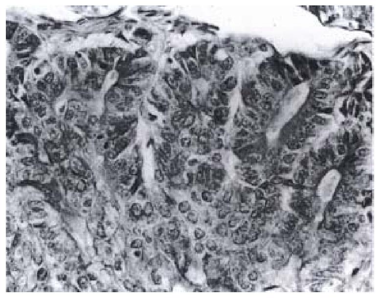
Twenty-seven (45.8%) of the 50 gastric carcinomas showed immunoreactivity for Fas antigen in gastric carcinoma cells. The Fas antigen immunoreactivity appeared brown or dark brown, which was mainly located in the cytoplasm (Figure 1), a few speci-mens simultaneously expressed Fas antigen on the cell membrane of tumor cells. Some of the mature lymphocytes infiltrating in the stroma of gastric car-cinoma had Fas antigen expression with a strong staining intensity.
Fas antigen expression was related to clinical patho-logical staging of gastric carcinoma. The rate of Fas antigen expression was not correlated with patient age, sex, tumor size, grades of differentiation and depth of invasion (P > 0.05 ). The immunoreactivity of Fas antigen was significantly associated with lymph node status and clinical stages of gastric car-cinoma. Sixteen (61.5%) of 26 gastric carcinomas without lymph node metastasis were immunoreactive versus 11 (33.3%) of 33 cases with lymph node metastasis (P < 0.05). Twenty-one (58.3%) of 36 gastric carcinomas in clinical stages I and II were immunoreactive versus 6 (26.1%) of 23 gastric car-cinomas in clinical stages III and IV (P < 0.05).
The survival rate of patients with Fas antigen ex-pression was compared with that of those without Fas antigen expression. Patients with Fas antigen expression in gastric carcinomas showed a significantly longer survival period as compared with those without Fas antigen expression (P < 0.05).
Fas antigen is a type I transmembrane protein, its molecular weight is 45000, and it belongs to the tumor necrosis factor/nerve growth factor receptor family[1]. Fas antigen as a receptor exists in the body and can induce a poptosis in target cells. In recent studies, Fas antigen expression has been identified in various human organs, e.g., heart, liver, lung, kidney, and ovary[2,3]. But, little is known about Fas antigen expression and its relationship with the biological behavior and prognosis of human gastric carcinoma.
In this study, we found that Fas antigen also expressed in gastric carcinoma tissues. Since Fas antigen is a transmembrane protein, it should appear both on the surface and in the cytoplasm of gastric carcinoma cells. But, in our study, most of the specimens expressed Fas antigen only in the cytoplasm of tumor cells, a few specimens expressed Fas antigen both on the surface and in the cytoplasm of tumor cells. There are several possible explana-tions for this. First, under pathological conditions, normal Fas antigen expression may be down-regulated, but expressi on of soluble Fas antigen is up regulated[4]. Second, Fas antigen may be affected by mutation on its DNA, or certain abnormalities may occur in the maturation process of this protein. Third, the structure of Fas antigen, which originally expressed on the surface, may be destroyed through a certain mechanism, e.g., its binding site on the membrane undergoes proteolysis, and only cytoplasmic Fas antigen expression remains. However, the mechanism remains to be elucidated in future in vitro studies.
Our findings concerning the relationship between Fas antigen expression and the pathological characteristics of gastric carcinoma showed that Fas antigen expression could relate to lymph node status and clinical stages. The rate of Fas antigen expression was significantly higher in gastric carcinomas without lymph node metastasis than in those with lymph node metastasis, and in clinical stages I and II than in clinical stages III and IV gastric carcinomas. This indicated that aberrant Fas antigen expression may be involved in lymph node metastasis of gastric carcinoma. In addition, the survival period of patients with Fas antigen expression was longer than those without Fas antigen expression. The results demonstrated that Fas antigen expression may be of some value in predicting prognosis in patients with gastric carcinoma.
Key project of the 9th 5-Year Plan for Medicine and Health of Army, No.96Z047.
Edited by Jing-Yun Ma
| 1. | Nagata S, Golstein P. The Fas death factor. Science. 1995;267:1449-1456. [RCA] [PubMed] [DOI] [Full Text] [Cited by in Crossref: 3053] [Cited by in RCA: 2991] [Article Influence: 99.7] [Reference Citation Analysis (0)] |
| 2. | Higaki K, Yano H, Kojiro M. Fas antigen expression and its relationship with apoptosis in human hepatocellular carcinoma and noncancerous tissues. Am J Pathol. 1996;149:429-437. [PubMed] |
| 3. | Leithäuser F, Dhein J, Mechtersheimer G, Koretz K, Brüderlein S, Henne C, Schmidt A, Debatin KM, Krammer PH, Möller P. Constitutive and induced expression of APO-1, a new member of the nerve growth factor/tumor necrosis factor receptor superfamily, in normal and neoplastic cells. Lab Invest. 1993;69:415-429. [PubMed] |
| 4. | Cheng J, Zhou T, Liu C, Shapiro JP, Brauer MJ, Kiefer MC, Barr PJ, Mountz JD. Protection from Fas-mediated apoptosis by a soluble form of the Fas molecule. Science. 1994;263:1759-1762. [RCA] [PubMed] [DOI] [Full Text] [Cited by in Crossref: 806] [Cited by in RCA: 826] [Article Influence: 26.6] [Reference Citation Analysis (0)] |


