Published online Dec 15, 1998. doi: 10.3748/wjg.v4.i6.519
Revised: August 20, 1998
Accepted: November 5, 1998
Published online: December 15, 1998
AIM: To establish an experimental model of stress ulcer produced by explosive noise, and to probe into its mechanism and protection.
METHODS: The country standard Wistar white rats were randomly divided into control group (n = 8), which were neither stimulated nor protected, and stimulating group (divided into subgroups A, B and C, including 8 rats each which were decapitated to draw blood for test immediately, 12 h and 24 h after stimulation) and prevention group (divided into subgroups A, B and C, having 8 rats each, subgroup A was given cimetidine, B anisodamine and C both drugs). Firing noises of submachine guns were used as inflicting factor. The rats were fasted for 24 h and stimulated by firing noise for 12 h. The change of ulcer index, gastric mucosal and related serum hormones were observed.
RESULTS: Stress ulcer was significant in the stimulating group, and its ulcer index (8.6 ± 0.6) was remarkably higher than that in both the control group and prevention group (0.3 ± 0.1, P < 0.01). Its serum gastrin (Gas ng/L, 294 ± 163 vs 63 ± 40, P < 0.01) and endothelin (ET ng/L, 181 ± 57 vs 135 ± 42, P < 0.1) were apparently higher than those in the control group, and its serum nitric oxide (NO) level was conspicuously lower than that in the control group (ng/L, 0.2 ± 0.1 vs 0.8 ± 0.5 P < 0.5), while the serum gastrin level (ng/L, 556 ± 225) in prevention group was distinctly higher than that in both the control (P < 0.01) and stimulating group (P < 0.05). There were no significant differences in the changes of ET and NO between the control and the stimulating groups.
CONCLUSION: Stress ulcer model of rats can be successfully established by the stimulation of explosive noise. Gas, ET and NO are related to the formation of stress ulcer, and play an important role in its mechanism. Hepatic function affected by noise is observed in this experiment.
- Citation: Liu GS, Huang YX, Li SW, Pan BR, Wang X, Sun DY, Wang QL. Experimental study on mechanism and protection of stress ulcer produced by explosive noise. World J Gastroenterol 1998; 4(6): 519-523
- URL: https://www.wjgnet.com/1007-9327/full/v4/i6/519.htm
- DOI: https://dx.doi.org/10.3748/wjg.v4.i6.519
Explosive noise may produce an enormous adverse effect on human body and mind. It has been known that peptic ulcer occurred much more frequently in war time than in peace time[1]. However, there have been no reports about the relationship between explosive sound and stress ulcer in literature. The aim of this study is to establish an animal model of stress ulcer produced by explosive noise and probe into its mechanism, prevention and treatment.
The healthy country standard Wistar rats were provided by the Animal Center of the Fourth Military Medical University. Submachine guns, a precision pulse counter, and a frequency spectrum analyser were provided by the Chinese PLA 371 Hospital. A PJ-2003/50 G-type γ radioimmunity counter (made in Xi’an), 754 spectrophotometer (made in Hefei), a high-speed and low-temperature centrifuge (made in Tumen), a light microscope and a JEM-2000EX-type transmission electron microscope (made in Japan) were used in this experiment.
Preparation of ulcer model. Having been fasted for 24 h, the experimental rats were confined in an isolated room to be tested. Firing noise of submachine guns acted as an inducing factor which had been recorded on a tape and was played to the rats through a loudspeaker at a distance of 20 cm-30 cm. Examined with the precision pulse counter and the frequency spectrum analyser, the intensity of the firing noise was measured as 110 dB(A) and its frequency as 0.25 kHz-4.00 kHz. After the rats were stimulated successively for 12 h by gun shot sound, their stomachs were opened and the formation of their stress ulcer was observed.
The rats were randomly divided into three groups: ① Control group, consisting of 8 rats, which were neither stimulated nor protected; ② stimulating group, subdivided into groups A, B and C, including 8 rats each. These subgroups were decapitated to draw blood for test immediately, 12 h, and 24 h after stimulation respectively; and ③ prevention group, also subdivided into groups A, B and C, having 8 rats each. Group A was injected 40 mg ip with cimetidine 30 min before the stimulation; group B given 2 mg ip of anisodamine 30 min before and 6 h after the stimulating; group C administered with above mentioned combined drugs. All the rats used in this study were anesthetized in their abdominal cavities with 1.2 mg of 5 g/L amobabital sodium after treatment and their abdomens were opened for observation on the changes of their gastric mucosa. After decapitation, two blood samples were taken: one for centrifugation of serum, the other added with EDTA and retardant peptidase for separation of plasma and kept at -40 °C in a refrigerator ready for the detection of hormone and hepatic and renal functions.
Measurements of mucosal damage. According to Guth’s method[2], the length of gastric mucosal damage area < 1 mm was scored 1 point; 1 mm-2 mm 2 points; 2 mm-3 mm 3 points; 3 mm-4 mm 4 points, > 4 mm scored segmentally. The sum total of the scores of the whole stomach constituted the ulcer index. Tissue and cell structures of all groups were observed and photoed under light microscope and electroscope respectively.
Detection of plasma Gas, ET, NO and hepatic-renal functions. Gas was detected before and after stimulation using the kit produced by the Northern Immunity Agents Research Institute in accordance with the instructions. ET was detected using the kit by the East Asian Immunity Technique Research Institute according to the operational manual. NO was tested by means of the kit provided by the Military Medical Academy according to the manual. Hepatic and renal functions were tested by the laboratory from the serum samples we sent.
Statistics. Both t test and analysis of variance were used for this study.
In control group, after the gastric wall was opened, the mucosa was seen intact, tidy and smooth without any ulcer or erosion except for a couple of bleeding spots under the mucosa of 2 rats.
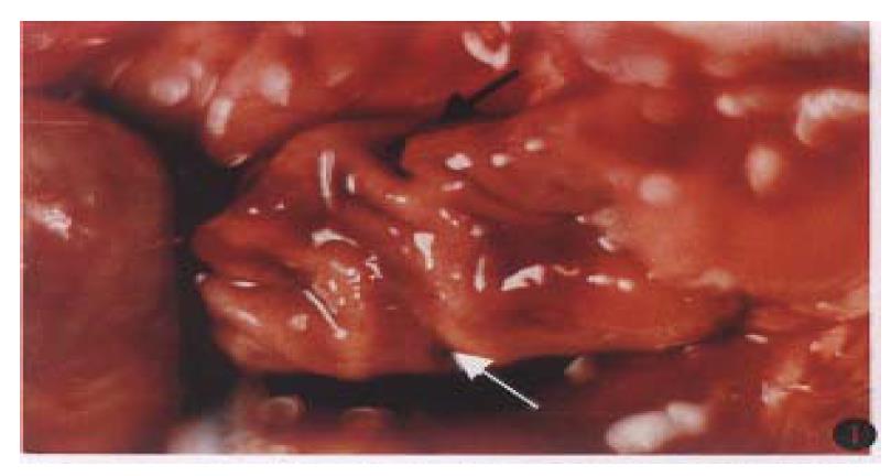
In stimulating group, on the abdominal cavity, different degrees of congestion and edema were found in seruous layers and on the gastric walls, congestion, edema, erosion and ulcer formation came into sight (Figure 1), especially in group B (ulcer index, 8.4 ± 0.6), the ulcer index being remarkably different (P < 0.01) between group B and the prevention group ( ulcer index, 0.3 ± 0.1) and control group (ulcer index, 0).
In prevention group, although there were still some congestion, edema, erosion and ulcer formation, these were conspicuously lower than in the stimulating group (P < 0.01). The preventive effects proved significantly better in group C’ (0.3 ± 0.1) than those in groups A’ and B’(P < 0.05).
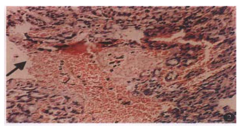
In Control group, mucosal layers were smooth and tidy, glands were arranged in order, and no tendency of inflammatory cell infiltration and submucosal hemorrage appeared. In stimulating group, in sub gropus A, B and C, there were interruptions of mucosa, enlarged glandular gaps and damaged parts of gland. Meanwhile, a large amount of RBCs were accumulated among the glands. Within submucous eosinophil, infiltration and capillary thrombosis were detected (Figure 2). In prevention group, all of the 3 subgroups were found intact in mucos, well-structured in glands slightly broadened in gaps with some RBCs sparsely existing beneath mucosa.
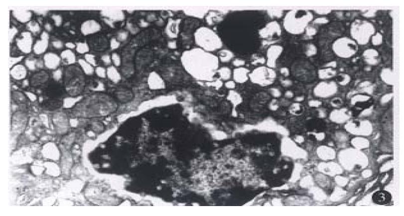
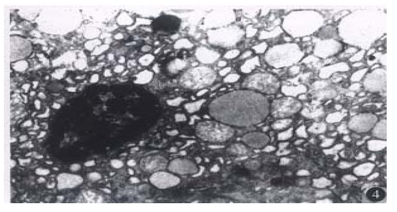
The control group showed intact cell structure, regular secretory granules, and no widened gaps among nuclei. In addition to these, mitochondrion and endoplasmic reticulum were also conspicuous. The structure of microvillus was perfect. On the contrary, the stimulation group exhihited irregular cell structure, with nuclei withered, gaps among nuclei broadened, endoplasmic reticulum expanded, mitochondrion hypertrophic, inflammatory cell infiltration in interstitial, and secretory granuals increased (Figure 3). The prevention group in appeared differently. Although there were slight withered nuclei, broadened nuclear gaps, and more or less expanded endoplasmic reticulum, all these were noticeably insignificant as compared with those in the stimulating group (Figure 4).
Gas was apparently higher in the stimulating and prevention groups than in control group (P < 0.05, Table 1), being most pronounced in stimulating group B.
ET was obviously higher in the stimulating group than in control group, while there were no significant changes between the prevention and control groups (P < 0.05), nor between stimulating and prevention groups (P < 0.05).
NO was lower in the stimulating group than in control group (P < 0.05), especially in group C (Table 1). No substantial difference was seen between the prevention and control groups (P < 0.05). ALT and BUN showed no remarkable changes among all groups (P < 0.05), while AST was found higher in the stimulating and prevention groups than in controls (P < 0.05, Table 2).
There are five types of stress ulcers[3], i.e., single bound stress, cold bound stress, socking stress, shock stress and spinal cord injury stress. It has been reported that noise may damage the human body and mind by causing disturbance in the stomach and intestines[4] and that ulcer occurred conspicuously more frequently in war time than in peace time[1]. Based on these findings and considering that rats are characterized by well-developed adrenalin function, sensitive response to stress and susceptiveness to stress ulcer[5], we designed and prepared the stress ulcer model of rats in order to explore its mechanism and prevention and treatment. Our results showed that a typical ulcer model of rats can be successfully established by stimulation with a strong explosive sound continuously for 12 h. Using Guth’s methods[2], the ulcer index was found to be significantly different between stimulating group and both the control group and the prevention group (P < 0.01). Such changes as interruptions of mucosa and damage of glands were detected under light microscope (Figure 1). This, therefore, has opened a new path to establish an animal model of stress ulcer.
In an attempt to probe into the underlying causes, we laid special stress on analysing the role of ET, NO and Gas in this disease. To our knowledge, ET serves as a strongest vasoconstrictive substance, while NO functions as a vasodilative one. They are contradictory regulating factors in the blood[6]. When balance between the two is lost, the blood flow in gastric mucosa may change and an acute mucosal damage occurs. ET has potent ulcerogenic and vasoconstrictive actions in the stomach where it induces gastric mucosal damage and increases gastric vascular tone[7]. In our study, after sound stimulation, serum ET level became conspicuously higher in stimulation group than in control group (P < 0.05), indicating ET played an important part in stress ulcer. This was probably because the gun-shot sound overexcited the sympathetic nerves to cause submucosal capillaries to constrict, thus stimulating endodermical cells to produce excessive ET, which, in turn aggravated vasoconstriction and decreased blood flow under mucosa, and, finally, resulted in the erosion and formation of ulcer. NO is produced when larginine guanidine combined with oxygen is converted intolcitriline under the catalysis of nitric oxide synthetase (NOS), which is the key factor for the production of NO. Kanno et al[8] reported that an induced NO (iNOS) can be brought about by a variety of cell factors and lipopolysaccharide (LPS) in vascular endothelia and smooth muscles and then produce more and more NO. However, in case of stress, because of vasoconstriction of gastric mucosa and decrease of blood flow, the production INOS in vascular endothelia can be inhibited, leading to lowered NO level. In addition, erosion and bleeding in mucosa after stress can also cause the synthesized NO to be inactivated by combining with hemoglobin[9], thereby lowering NO level. The decrease of NO level, together with the weakening of mucosal protection and vasodilation, further aggravates erosion of mucosa and formation of ulcer. Our results showed that NO level was noticeably lower in the stimulating group, especially in group C than in control group, which conforms to their findings. After stimulation by explosive noise, with sympathetic nerves overxcited, vagus nerves inhibited[10], cortisol secretion is, of course, increased, resulting in dysfunction of peptidergic nerves in non-adrenergic and non-cholinergic nerves within sympathetic and parasympathetic nerve systems[11], thereby, it causes G-cells to secrete more and more Gas. This may stimulate wall cells to produce excessive gastric acid, pH value in stomach lowered, and H+ back oozing, leading to inflamation, congestion, edema, erosion in gastric mucosa and even the formation of ulcer. Our results showed that Gas was apparently higher in the stimulating group than in control group (P < 0.05). It was discovered under dynamic observation that Gas secretion increased gradually after treatment, reached its peak at the 12th hour, and then returned to normal after 24 h. All these indicated that Gas not only took part in but played an important role in the process of formation of stress ulcer.
Explosive noise also exerts some harmful effects upon functions of the liver and kidneys. Liu et al[10] noted that following stimulation by noise, the tension of sympathetic nerves became stronger, and tissue metabolism exuberated, accompanied by vasospasma and tissue ischemia, with the result that the renewal of liver cells hastened and ALT of blood increased. Our results were in agreement with theirs, but insignificant in statistics. This may be due to limited number of samples. AST was remarkably higher in the stimulating group, especially higher group A than in control group (P < 0.05). However groups B and C showed a gradual decline. The reason why AST became higher may be that during the stress the sympthetic nerves of rats were so excited that their muscles all over the body kept constricting, and their hearts beat faster for a short time, which caused damages in their skeleton muscle and cardiac muscle cells, and led to the increase of AST release. After removal of stimulation, these actions weakened, cell functions recovered and AST in blood decreased gradually. The strong noise for a short time, therefore, did little damage to the function of the kidneys.
Our study also proved that among the three prevention groups, group C’ for which drugs were given in combination brought best results. Next to this were groups B’ and C’ treated with anisodamine and cimetidine respectively. Measured by analysis of varience, their ulcer index was significantly different from group A’ (P < 0.05), corresponding to the previous documents. Following medication, plasma Gas leve rose more remarkably in the prevention group than in the stimulating and control groups. This might be related to the fact that after using H2 receptor-antagonist and vasodilator agents, the secretion of gastric acid by wall cells was inhibited, and by feedback the secretion of Gas by G-cells at gastric antrium increased. Serum NO and ET did not change conspicuously before and after medication (P > 0.05), presumably because H2 receptor-blocking agents and vasodilator agents reduced the secretion of gastric acid, and abated the stimulation to constriction of submucous blood vessel. On the other hand, it brought about dilatic factors in capillaries, thus the releasing of ET by endodermical cells was weakened. As a result, the producing effect of iNOS was also suppressed so that the formation of NO remained unaffected. Another reason why ET and NO level stayed unchanged was supposedly that the alliviated mucosal erosion and ulcer reduced the mucosal bleeding and combination of NO with hemoglobin.
From these results, we came to the conclusion that although explosive noise can stimulate the production of stress ulcer, it can be effectively prevented and cured by such medicine as H2 receptor antagonist and vasodilator agents. Therefore, we believe that use of preventive medicines before war will be of significance for diminution and protection of stress ulcer in wartime.
| 1. | Zhang XY, Zhang NZ. Modern field of internal medicine (in Chinese). Beijing: People's Military Medicine Publishing House. 1997;249-251. |
| 2. | Guth PH, Aures D, Paulsen G. Topical aspirin plus HCl gastric lesions in the rat. Cytoprotective effect of prostaglandin, cimetidine, and probanthine. Gastroenterology. 1979;76:88-93. [PubMed] |
| 3. | Wan JL. Mechanism of formation of stress ulcer (in Chinese). Chin J Pathophysiol. 1993;9:664-666. |
| 4. | Huang XQ, Wang YZ. Damage of noise to body and mind of patient (in Chinese). Med Information. 1995;8:279-280. |
| 5. | Shi XY. Medical experimental zoology (in Chinese). Xi'an: Shaanxi Sci. & Tech Pub House. 1989;56-62. |
| 6. | Yao Z. Two newfound blood-regulating factors-endothelin and nitric oxide (in Chinese). Nippon Igaku No Shoukai. 1994;15:527-530. |
| 7. | Battal NM, Hata Y, Ito O, Matsuda H, Yoshida Y, Kawazoe T, Nagao M. Reduction of burn-induced gastric mucosal injury by an endothelin receptor antagonist in rats. Burns. 1997;23:295-299. [RCA] [PubMed] [DOI] [Full Text] [Cited by in Crossref: 3] [Cited by in RCA: 3] [Article Influence: 0.1] [Reference Citation Analysis (0)] |
| 8. | Kanno K, Hirata Y, Imai T, Marumo F. Induction of nitric oxide synthase gene by interleukin in vascular smooth muscle cells. Hypertension. 1993;22:34-39. [RCA] [PubMed] [DOI] [Full Text] [Cited by in Crossref: 96] [Cited by in RCA: 95] [Article Influence: 3.0] [Reference Citation Analysis (0)] |
| 9. | Lowenstein CJ, Snyder SH. Nitric Oxide: a novel biologic messenger. Cell. 1992;70:705-711. [RCA] [DOI] [Full Text] [Cited by in Crossref: 546] [Cited by in RCA: 527] [Article Influence: 16.0] [Reference Citation Analysis (0)] |
| 10. | Liu ZF, Zhong X, Wu XS, Jiang W, Zhou ZM, Song X. Effects of strong noise on blood viscolity, blood sugar and several enzymes in rats (in Chinese). Chin J Labor Hyg Occupation Dis. 1992;10:213-216. |
| 11. | Wank SA, Pisegna JR, de Weerth A. Brain and gastrointestinal cholecystokinin receptor family: structure and functional expression. Proc Natl Acad Sci USA. 1992;89:8691-8695. [RCA] [PubMed] [DOI] [Full Text] [Cited by in Crossref: 297] [Cited by in RCA: 286] [Article Influence: 8.7] [Reference Citation Analysis (0)] |


