Published online Sep 15, 1997. doi: 10.3748/wjg.v3.i3.200
Revised: May 13, 1996
Accepted: June 11, 1997
Published online: September 15, 1997
- Citation: Wang YG, Binmoeller K, Li ZL, Soehendra N. Endoscopic haemoclip ligation of pedunculated polyp before polypectomy. World J Gastroenterol 1997; 3(3): 200-200
- URL: https://www.wjgnet.com/1007-9327/full/v3/i3/200.htm
- DOI: https://dx.doi.org/10.3748/wjg.v3.i3.200
Metallic haemoclips have been endoscopically placed in the gastrointestinal tract for treating bleeding lesions and closing perforations[1,2]. A further potential application is ligating pedunculated polyps prior to polypectomy as a prophylactic measure to prevent bleeding. We used metallic haemoclips to treat two patients with pedunculated colonic polyps and bloody diarrhoea successfully.
A 58-year-old white man with a colonic polyp was referred to our unit for polypectomy. He had a history of chronic renal failure secondary to chronic glomerulonephritis for 14 years and required regular dialysis with 1 × 5000 IU heparin. Colonic examination was performed with a colonoscope (CF-IT 200, Olympus Corp., Tokyo), and three pedunculated polyps were found. Two non-bleeding polyps, both 1.5 cm, were in the sigmoid flexure; the other was 0.8 cm and had a longer stalk, measured about 0.8 cm in length and 0.4 cm in diameter, and was in the descending colon. There was active oozing (Forrest Ib) over the top of the polyp with the longer stalk. A standard polypectomy snare was used to snare the polyps, and metallic clips with a delivery catheter (MD-850, HX-3L, Olympus Corp.) were used for clip ligation. Routine polypectomy was performed for the two non-bleeding polyps. In one, a small vessel fistula (Forrest IIa) was seen and a clip was applied to ligate it. To prevent post-polypectomy bleeding, the stalk was ligated completely with one clip before the procedure was performed in the remaining polyp. There was no bleeding after the procedure. For the bleeding polyp, the stalk was ligated to stop the bleeding: first, the clip and delivery catheter were inserted through the endoscope and then the clip was opened, and the handle of the delivery catheter was rotated to change the direction of the clip prongs to grasp the stalk.
Thereafter, the delivery catheter was pushed and the clip was closed. Two clips were required to ligate the stalk completely. There was no massive bleeding after the polypectomy. No complications were observed by clipping and there was no recurrent bleeding during the 4-week follow-up.
A 48-year-old Chinese woman with recurrent bloody diarrhoea for four years and fresh bloody diarrhoea again for four days was admitted to our hospital for colonoscopic examination. She had no history of fever, weight loss, or haemorrhoids. The total colon was examined with a colonoscope (CF-30I, Olympus Corp.), and a 2.0-cm polyp with a long stalk, about 1.0 cm in length and 0.3 cm in diameter, was found in the sigmoid colon. There was a fresh blood clot on the top of the polyp. We used the same technique as in case 1 and ligated the stalk completely with one clip.
No massive bleeding occurred after the polypectomy, but a blood vessel fistula without active bleeding was found. No complications or recurrence of bleeding were observed during the 8-week follow-up.
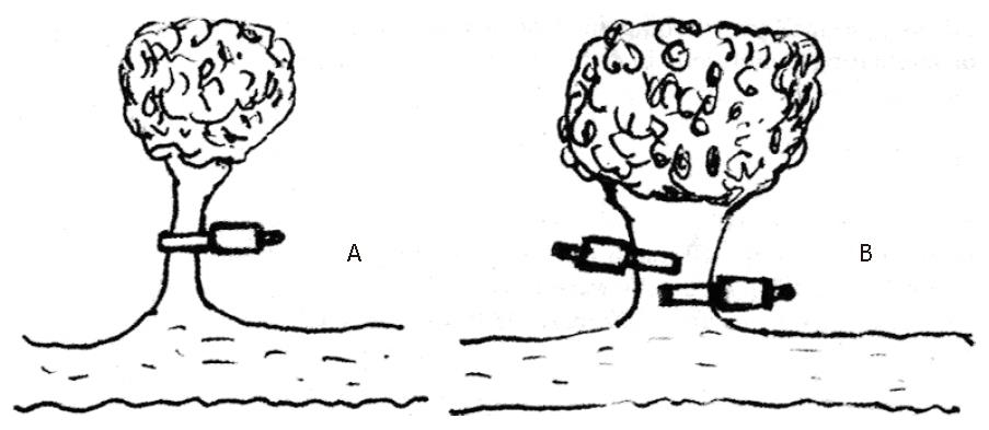
Ligation using suture or metallic clips is a basic surgical technique for preventing postoperative bleeding. Generally, there are nourishing blood vessels in the stalk of the pedunculated polyp, and their diameter depends on the size of the polyp and the diameter of the stalk. It is essential to ligate the vessels completely or to prevent postoperative bleeding in pedunculated polyps with or without active bleeding. The principle of clip ligation for pedunculated polyps is shown in Figure 1. The number of clips required for complete ligation of the stalk depends on the diameter of the stalk.
The improved metallic haemoclip and clip delivery system have made clips easier to apply. Clip ligation of the bleeding vessel achieves immediate haemostasis comparable to surgical ligation. A previous study has shown that endoscopic haemoclip placement is a highly effective and safe method for treating non-variceal gastrointestinal bleeding[1]. We have used the clips and succeeded in closing a 0.5-cm perforation after snare excision of a 3.7 cm × 3.4 cm gastric leiomyoma[2]. As case 1 was on dialysis with heparin and had long-stalked polyps with and without bleeding, he was at high risk for polypectomy-induced bleeding and secondary massive bleeding. We used metallic clips to ligate the stalk, arresting the bleeding immediately and avoiding massive post-polypectomy bleeding. The stalk of the pedunculated polyp should be clipped completely.
Recently, the detachable snare was introduced as a new haemostatic device for preventing postoperative bleeding in pedunculated polypectomy, and obtained good results[3]. A comparative study of haemoclips and detachable snares for ligating pedunculated polyps before polypectomy is needed in the near future.
In conclusion, as a prophylactic measure, haemoclips can be used to ligate the stalk of a polyp as a new indication either with or without bleeding before polypectomy.
Original title:
S- Editor: Filipodia L- Editor: Jennifer E- Editor: Hu S
| 1. | Binmoeller KF, Thonke F, Soehendra N. Endoscopic hemoclip treatment for gastrointestinal bleeding. Endoscopy. 1993;25:167-170. [RCA] [PubMed] [DOI] [Full Text] [Cited by in Crossref: 179] [Cited by in RCA: 163] [Article Influence: 5.1] [Reference Citation Analysis (0)] |
| 2. | Binmoeller KF, Grimm H, Soehendra N. Endoscopic closure of a perforation using metallic clips after snare excision of a gastric leiomyoma. Gastrointest Endosc. 1993;39:172-174. [RCA] [PubMed] [DOI] [Full Text] [Cited by in Crossref: 137] [Cited by in RCA: 126] [Article Influence: 3.9] [Reference Citation Analysis (0)] |
| 3. | Lee MS, Park CW, Lee JS, Cho SW, Shim CS. Endoscopic polypectomy by use of the detachable snare for prevention of post polypectomy bleeding (Abstract). Gastrointest Endosc. 1995;41:307. |


