Published online Sep 14, 2018. doi: 10.3748/wjg.v24.i34.3871
Peer-review started: June 21, 2018
First decision: July 18, 2018
Revised: July 24, 2018
Accepted: August 1, 2018
Article in press: August 1, 2018
Published online: September 14, 2018
Processing time: 85 Days and 13.8 Hours
To study the influence of different doses of tacrolimus (FK506) on gut microbiota after liver transplantation (LT) in rats.
Specific pathogen-free Brown Norway (BN) rats and Lewis rats were separated into five groups: (1) Tolerance group (BN-BN LT, n = 8); (2) rejection group (Lewis-BN LT, n = 8); (3) high dosage FK506 (FK506-H) group (Lewis-BN LT, n = 8); (4) middle dosage FK506 (FK506-M) group (Lewis-BN LT, n = 8); and (5) low dosage FK506 (FK506-L) group (Lewis-BN LT, n = 8). FK506 was administered to recipients at a dose of 1.0 mg/kg, 0.5 mg/kg, and 0.1 mg/kg body weight for 29 d after LT to the FK506-H, FK506-M, and FK506-L groups, respectively. On the 30th day after LT, all rats were sampled and euthanized. Blood samples were harvested for liver function and plasma endotoxin testing. Hepatic graft and ileocecal tissues were collected for histopathology observation. Ileocecal contents were used for DNA extraction, Real-time quantitative polymerase chain reaction (RT-PCR) and digital processing of denaturing gradient gel electrophoresis (DGGE) profiles and analysis.
Compared to the FK506-H and FK506-L groups, FK506-M was optimal for maintaining immunosuppression and inducing normal graft function; the FK506-M maintained gut barrier integrity and low plasma endotoxin levels; furthermore, DGGE results showed that FK506-M induced stable gut microbiota. Diversity analysis indicated that FK506-M increased species richness and rare species abundance, and cluster analysis confirmed the stable gut microbiota induced by FK506-M. Phylogenetic tree analysis identified crucial bacteria associated with FK506-M; seven of the nine bacteria that were decreased corresponded to Bacteroidetes, while increased bacteria were of the Bifidobacterium species. FK506-M increased Faecalibacterium prausnitzii and Bifidobacterium spp. and decreased Bacteroides-Prevotella and Enterobacteriaceae, as assessed by RT-PCR, which confirmed the crucial bacterial alterations identified through DGGE.
Compared to the low or high dosage of FK506, an optimal dosage of FK506 induced immunosuppression, normal graft function and stable gut microbiota following LT in rats. The stable gut microbiota presented increased probiotics and decreased potential pathogenic endotoxin-producing bacteria. These findings provide a novel strategy based on gut microbiota for immunosuppressive dosage assessment for recipients following LT.
Core tip: This is the first study to illustrate the effects of different dosages of Tacrolimus (FK506) on gut microbiota following liver transplantation (LT) and indicates that an optimal dosage of FK506 induces effective immunosupression, good graft function and stable gut microbiota after LT in rats. Based on the relationship between gut microbiota and the immunosuppressive dosage in this study, we can not only illustrate precise changes of gut microbiota given by different dosages of FK-506 following LT, but we also provide a novel monitoring strategy based on changes in gut microbiota for immunosuppressive dosage assessment in patients following LT.
- Citation: Jiang JW, Ren ZG, Lu HF, Zhang H, Li A, Cui GY, Jia JJ, Xie HY, Chen XH, He Y, Jiang L, Li LJ. Optimal immunosuppressor induces stable gut microbiota after liver transplantation. World J Gastroenterol 2018; 24(34): 3871-3883
- URL: https://www.wjgnet.com/1007-9327/full/v24/i34/3871.htm
- DOI: https://dx.doi.org/10.3748/wjg.v24.i34.3871
Liver transplantation (LT) is considered the only definitive therapy for end-stage liver diseases, including liver cirrhosis, liver failure and hepatocellular carcinoma[1,2]. Currently, the 1-year survival rate in patients following LT has reached 90% and the 5-year survival has reached 75%[3]. However, rejection after LT remains the major cause of hepatic graft dysfunction and is a life-threatening complication[4]. Treatment with inadequate immunosuppression might lead to a higher risk of rejection and even graft dysfunction, whereas excessive immunosuppression has been closely associated with a greater incidence of infection, sepsis, drug toxicity, cancer, chronic graft dysfunction, renal dysfunction, and even increased mortality[5-7], with bacterial infection being the most prevalent type of infection[8]. Thus, the optimal dosage of immunosuppressive medication is crucial for the prevention of rejection, lessening of immunosuppression toxicity and treatment of patients following LT[9].
The gut microbiota has been considered the most important micro-ecosystem that possesses a symbiotic relationship within the body[10-12]. The gut microbiota plays a crucial role in the development of fatty liver disease[13], liver cirrhosis[14], hepatocellular carcinoma[15], liver ischemic injury[16] and liver graft rejection[17]. Our previous study indicated that the healing of hepatic injury could ameliorate gut barrier function and promote gut microbial restoration following LT in rats[16]. We also found that graft rejection after LT could induce gut microbial alterations, thereby inducing subsequent gut barrier dysfunction, which presented a decrease of fecal secretory immunoglobulin A (sIgA) and an increase in blood endotoxin and tumor necrosis factor-α. RT-PCR results showed that the genus Faecalibacterium prausnitzii and Lactobacillus were decreased, whereas Clostridium bolteae was increased, which might in turn aggravate hepatic rejection[18].
Nevertheless, the influence of immunosuppressive medication on gut microbiota following LT remains unclear, and the association between the dosage of immunosuppressants and gut microbial alterations requires urgent elucidation. In this study, we studied the influence of different dosages of FK506 on hepatic graft function and gut microbiota following LT in rats. Furthermore, we identified the gut microbial profile and crucial bacterial community constituents using Denaturing gradient gel electrophoresis (DGGE), and further verified the alterations of dominant gut bacterial populations with RT-PCR.
Specific pathogen-free (SPF) male inbred Brown Norway (BN) and Lewis rats (weight 220-250 g, age 12-15 wk) were purchased from Beijing Vital River Laboratories (Beijing, China). They were housed in a clean-level animal house located at the First Affiliated Hospital, School of Medicine, Zhejiang University. All rats were housed at 22-24 °C in 12 h light/dark cycles and fed sterilized standard rat chow and water.
The study animals were divided into five groups as follows: (1) Tolerance group (BN-BN LT, n = 8); (2) rejection group (Lewis-BN LT, n = 8); (3) high dosage FK506 (FK506-H) group (Lewis-BN LT, n = 8); (4) middle dosage FK506 (FK506-M) group (Lewis-BN LT, n = 8); (5) low dosage FK506 (FK506-L) group (Lewis-BN LT, n = 8). In the Tolerance group, both donors and recipients were BN rats; in the other four groups, both donors were Lewis and recipients were BN rats. FK506 was administered to recipients at a dose of 1.0, 0.5, and 0.1 mg/kg weight for 1 mo after LT to the FK506-H, FK506-M, and FK506-L groups, respectively. To mimic the clinical application, FK506 was administrated via abdominal subcutaneous injection once every 12 h for 7 d after LT, and then via intragastric administration, once per day for the following 8-29 d. The sustained-release FK506 was administered intragastrically to maintain a consistent dosage effect for 24 h.
The study was conducted in accordance with the ‘‘Guide for the Care and Use of Laboratory Animals’’ published by the National Institutes of Health (NIH publication 86-23, revised 1985). The experimental protocol was approved by the Animal Care and Use Committee of the First Affiliated Hospital, School of Medicine, Zhejiang University.
The LT surgery was performed according to our previous methods[16,19], with slight modifications. The anesthesia was performed by intraperitoneal injection of Ketamine Hydrochloride (100 mg/kg) and Atropine (1 mg/kg) (Shanghai No. 1 Biochemical and Pharmaceutical, China), and then ether was inhaled to maintain anesthesia[6]. All recipients were revived shortly after the procedure and no further treatment was administered.
All rats were sampled on the 30th day after LT. The abdominal aorta was punctured, and blood samples were harvested for liver function and plasma endotoxin testing. Hepatic graft and ileum tissues near the ileocecus were collected for morphological observation. Ileocecal contents were harvested and stored at -80 °C for gut microbial analysis. All rats were then euthanized via an overdose of anesthetic.
Plasma alanine aminotransferase (ALT) and aspartate aminotransferase (AST) levels were detected using an automatic biochemical analyzer (Hitachi 7600, Tokyo, Japan).Plasma endotoxin level was determined using a colorimetric Limulus Test (Shanghai Yihua Medical Technology Co., Ltd, China) in accordance with the manufacturer’s instructions.
The hepatic graft and an ileum tissue sample taken 3 cm from the ileocecus were fixed in 40 g/L neutral formaldehyde and embedded in paraffin, cut into 4-μm slices, stained with hematoxylin and eosin (HE), and then analyzed under light microscopy. At the same time, approximately 1 mm3 of the same samples were fixed in a 2.5% glutaraldehyde solution and prepared in accordance with the standard technical procedures for transmission electron microscopy (TEM), as previously described[20]. Hepatic and ileal ultrastructures were analyzed at the Imaging Facility of Core Facilities, Zhejiang University School of Medicine. Histopathology and TEM evaluation were investigated by a pathologist blinded to the treatment group.
DNA extraction of a frozen aliquot of each ileocecal fecal sample was performed as in our previous studies[16]. DNA concentration was measured by NanoDrop (Thermo Scientific), and DNA integrity was verified using agarose gel electrophoresis.
The primers used for quantitative RT-PCR (RT-qPCR) are listed in Supplementary Table 1. All oligonucleotide primers were synthesized by TAKARA (Dalian, China). RT-qPCR was performed as reported previously[21]. The copy number of 16s rDNA operons per microliter of crude DNA template was determined by comparing serially diluted plasmid DNA standards running on the same plate.
The V3 variable region of 16S rDNA was amplified using a hot-start touchdown protocol with specific primers for the conserved regions of 16S rDNA. DGGE was performed using the D-Code universal mutation detection system apparatus (Bio-Rad, Hercules, CA, United States) with 16 cm × 18 cm × 1.5 mm gels according to the manufacturer’s protocol. On each DGGE gel, standard references in the middle and at each end were used for digital gel normalization and comparison of gels.
DGGE profiles were digitally processed using BioNumerics software version 6.01 (Applied Maths, St-Martens-Latem, Belgium) in a multistep procedure following the manufacturer’s instructions. The parameters for band-class allocation followed those of our previous study[16]. Quantitative information of a given band was calculated using the Gel-Pro analyzer 4 software (Media Cybernetics, United States). Diversity was calculated using Shannon’s diversity, Simpson index, species richness, Chao-1, Fisher alpha, and Menhinick index with Past software (http://folk.uio.no/ohammer/past/)[21]. Cluster analysis of the DGGE was conducted with an unweighted pair-group method with arithmetic means (UPGMA) based on the Dice similarity coefficient (band-based). Multidimensional scaling (MDS) and principal components analysis (PCA) were utilized following the instructions of the BioNumerics software.
Specific DGGE bands of interest were excised and sequenced. Positive clones were verified and sequenced using Sanger’s method on an ABI 3730 automated sequencing system (Invitrogen, Shanghai, China). Homology searches of the GenBank DNA database were performed using the BLAST tool. Reference sequences of phylogenetic neighbor species (up to 97% similarity) were included to construct a phylogenetic tree with MEGA 5.0 program based on the neighbor-joining method[15].
Continuous variables were summarized as mean ± standard deviation (SD). One-way variance analysis followed by Dunn’s multiple comparison tests was utilized to evaluate statistical differences among different groups. Student t-test was used to examine parametric data between the two groups. Statistical analyses were performed using SPSS version 19.0 for Windows (SPSS Inc., Chicago, IL, United States). A P-value of less than 0.05 was considered statistically significant.
An overdose or insufficient dosage of FK506 may lead to graft dysfunction, thus we first observed survival of all recipients. All rats following LT survived except one LT recipient in the FK506-H group that died of severe abdomen infection, which suggested an over-suppression of the immune status, thereby inducing infection and increasing mortality.
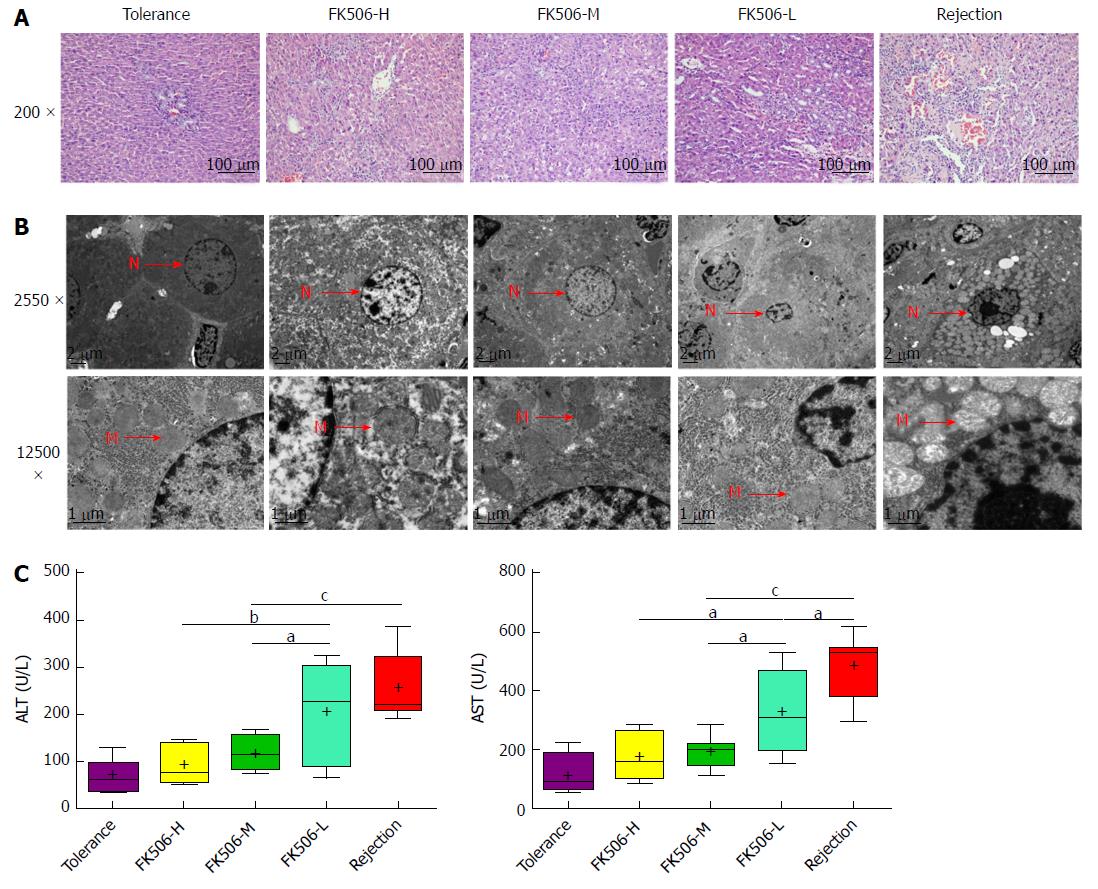
To illustrate the influence of different dosages of FK506 on hepatic grafts, we observed graft morphology. Under light microscopy (Figure 1A), hepatic grafts in the tolerance group presented a regular structure with well-arranged hepatocyte cords. In the rejection group, graft histology showed no obvious hepatocyte cords, accumulation of numerous red blood cells, and marked hepatocyte necrosis. However, FK506-M treatment preserved an approximately normal hepatic structure without hepatic acute rejection. Notably, hepatic graft resulted in hepatocyte cord disruption, widened sinusoids, and middle rejection injury in the FK506-L dosage group, suggesting an insufficient suppression of the immune status, thereby inducing rejection. Thus, compared to the FK506-H and FK506-L groups, FK506-M was the optimal dosage for maintaining immunosuppression in rats following LT.
We next observed hepatic graft ultrastructure with TEM (Figure 1B). In the tolerance group, a normal hepatocyte structure with intact mitochondria and endoplasmic reticulum was observed. Hepatic graft presented significant karyopyknosis, marked mitochondrial vacuolar degeneration, and organelle breakdown in the rejection group. Nevertheless, FK506-M treatment noticeably improved hepatocyte structure, mitochondria morphology, and other organelle structures. Notably, hepatocyte apoptosis and mild organelle damage were noted in the FK506-L group. Plasma ALT and AST levels reflected graft function. Compared with the rejection group, plasma ALT and AST were significantly decreased in the FK506-M treatment group (both P < 0.001). In addition, plasma ALT and AST were reduced in the FK506-M group versus the FK506-L group (both P < 0.05). There was no statistically significant difference between the tolerance and FK506-M-treated groups (Figure 1C).
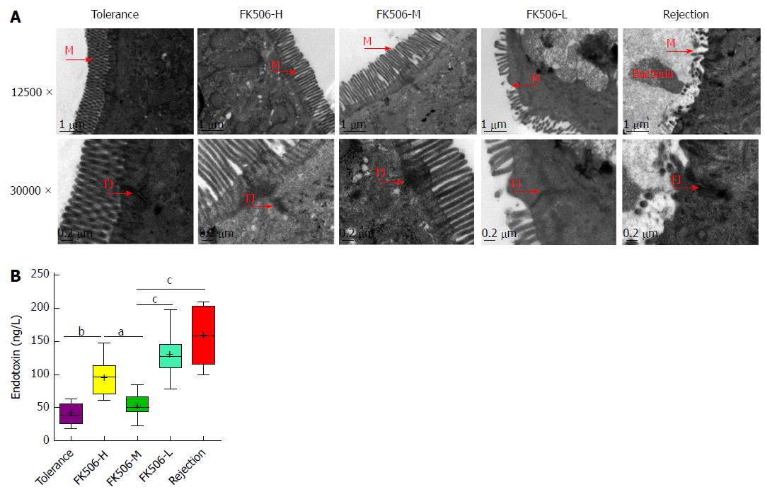
The liver-gut circulation and the gut-liver axis are closely associated with gut and liver function following LT. Thus, we next observed the ileal mucosa ultrastructure using TEM (Figure 2A). In the tolerance group, intestinal epithelial cells presented a normal morphology with many homogenously distributed microvilli and integrated tight junctions. In contrast, microvilli loss, tight junction damage, and bacterial invasion in epithelial cells were observed in the rejection group. However, FK506-M treatment significantly improved intestinal epithelial structure and kept microvilli and tight junctions intact. Notably, epithelial cell damage, microvilli disruption, and wider lateral spaces between neighboring cells were observed in the FK506-L treatment group.
Plasma endotoxin reflects gut barrier function. Compared with the tolerance and FK506-M groups, plasma endotoxin was significantly increased in the FK506-H group (P < 0.01 and 0.05, respectively), suggesting that over-suppression of immune status might lead to higher endotoxin levels. Meanwhile, plasma endotoxins were remarkably elevated in the rejection and FK506-L groups compared to the tolerance group (both P < 0.001). However, FK506-M administration significantly decreased endotoxin levels compared to the rejection and FK506-L groups (both P < 0.001) (Figure 2B).
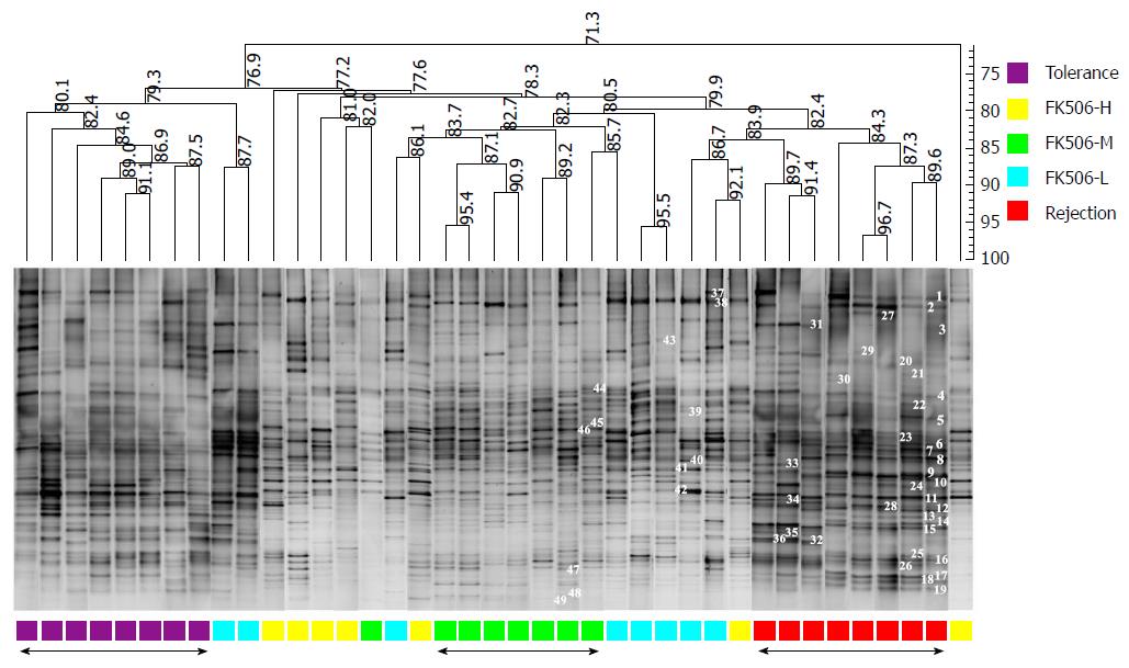
Endotoxins are the product of common gram-negative bacteria and can initiate various pathophysiological cascades[22]. Plasma endotoxin changes are mainly derived from changes in the gut microbiota. Based on the DGGE method, we analyzed the distribution of the ileocecal bacterial community (Figure 3). To characterize the DGGE profiles, we utilized the Dice coefficient and UPGMA as a cluster method to indicate band similarity. These profiles formed two primary clusters. The left cluster mainly consisted of samples from the tolerance group, in which total similarity was 80.1%, with the similarity among lanes ranging from 80.1% to 91.1%. The other cluster contained samples mainly from the rejection group and the different doses of the FK506 groups. Importantly, the similarity among lanes ranged from 82.4% to 96.7% in the rejection group and from 82.7% to 95.4% in the FK506-M treatment group, suggesting a significant uniqueness and stability of the gut microbiota.
In contrast, the DGGE lanes from the FK506-H and FK506-L treatment groups presented an inordinate distribution and an evident difference. The similarity of these lanes was relatively low and could not be clustered together. These results based on the DGGE profile indicated that an optimal dosage of FK506 induced a unique and stable gut microbiota in rats following LT.
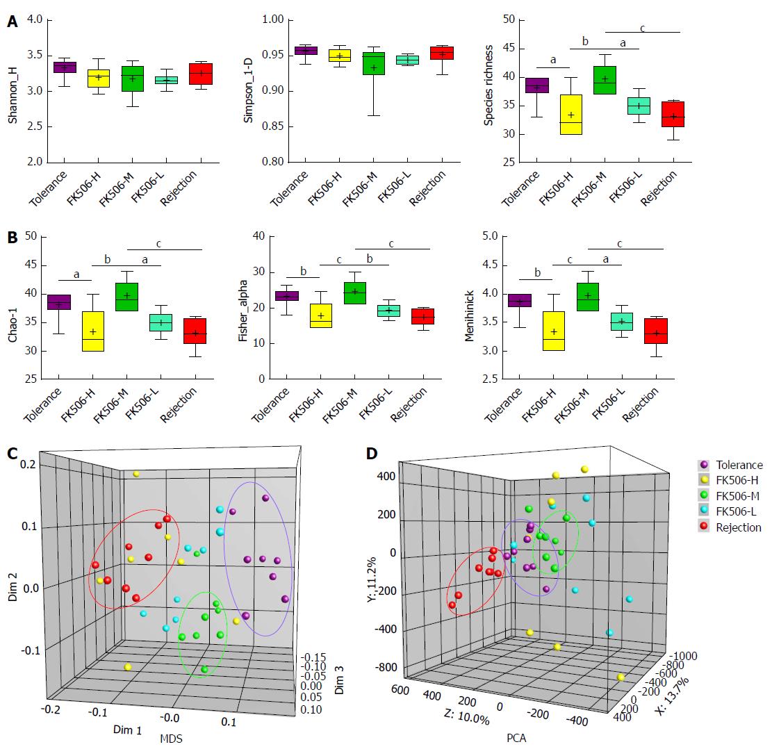
Gut microbial diversity and species richness are important indexes used to evaluate gut microbial balance and stability. Thus, we calculated the gray amount of each band in each lane of the DGGE profiles with Gel-Pro analyzer to compare microbial diversity. There were no statistical differences in the Shannon and Simpson indexes among the five groups (Figure 4A). Compared to the tolerance group, species richness was significantly decreased in the FK506-H, FK506-L, and rejection groups (all P < 0.05). In contrast, FK506-M treatment markedly increased species richness compared to the FK506-H, FK506-L, and rejection groups (P < 0.01, 0.05 and 0.001, respectively) (Figure 4A).
We also analyzed the abundance of rare species, estimated using the Chao-1, Fisher alpha, and Menhinick indexes. Compared with the tolerance group, Chao-1, Fisher alpha and Menhinick indexes were remarkably reduced in the FK506-H, FK506-L, and rejection groups (all P < 0.05). However, FK506-M treatment significantly increased the abundance of rare species in contrast to the FK506-H, FK506-L, and rejection groups (all P < 0.05) (Figure 4B).
Furthermore, based on DGGE profiles, we conducted MDS and PCA analysis. The Euclidean distance between two points indicates the similarity between the two. Gut microbial structures from samples of the tolerance, FK506-M, and rejection groups were respectively clustered together, and showed separation from each other using MDS analysis (Dim 1, Dim 2, and Dim 3) (Figure 4C), suggesting a relatively unique and stable gut microbiota. PCA is an alternative approach to visualizing relationships among lanes using lane data (band classes). Our PCA analysis also gave similar results based on PCA axes X/Y/Z (13.7%, 11.2%, and 10.0%, respectively) (Figure 4D).
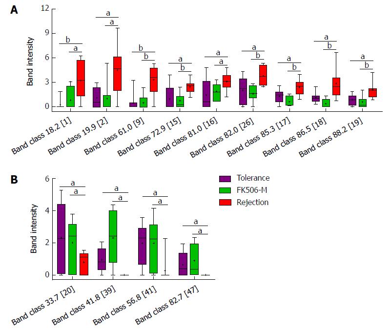
Of the 49 PCR-DGGE bands analyzed in the study, 48 band classes were selected. To identify crucial bacterial populations induced by FK506-M treatment, we compared band intensity between the FK506-M and the rejection groups. Of the 41 band classes selected, 28 showed slight variation in band intensity between the two groups. In contrast to the rejection group, the intensities of nine band classes [18.2 (band 1), 19.9 (band 2), 61.0 (band 9), 72.9 (band 15), 81.0 (band 16), 82.0 (band 26), 85.3 (band 17), 86.5 (band 18), and 88.2 (band 19)] were significantly decreased (Figure 5A), while four band classes [33.7 (band 20), 41.8 (band 39), 56.8 (band 41), and 82.7 (band 47)] were increased in the FK506-M treated group (Figure 5B).
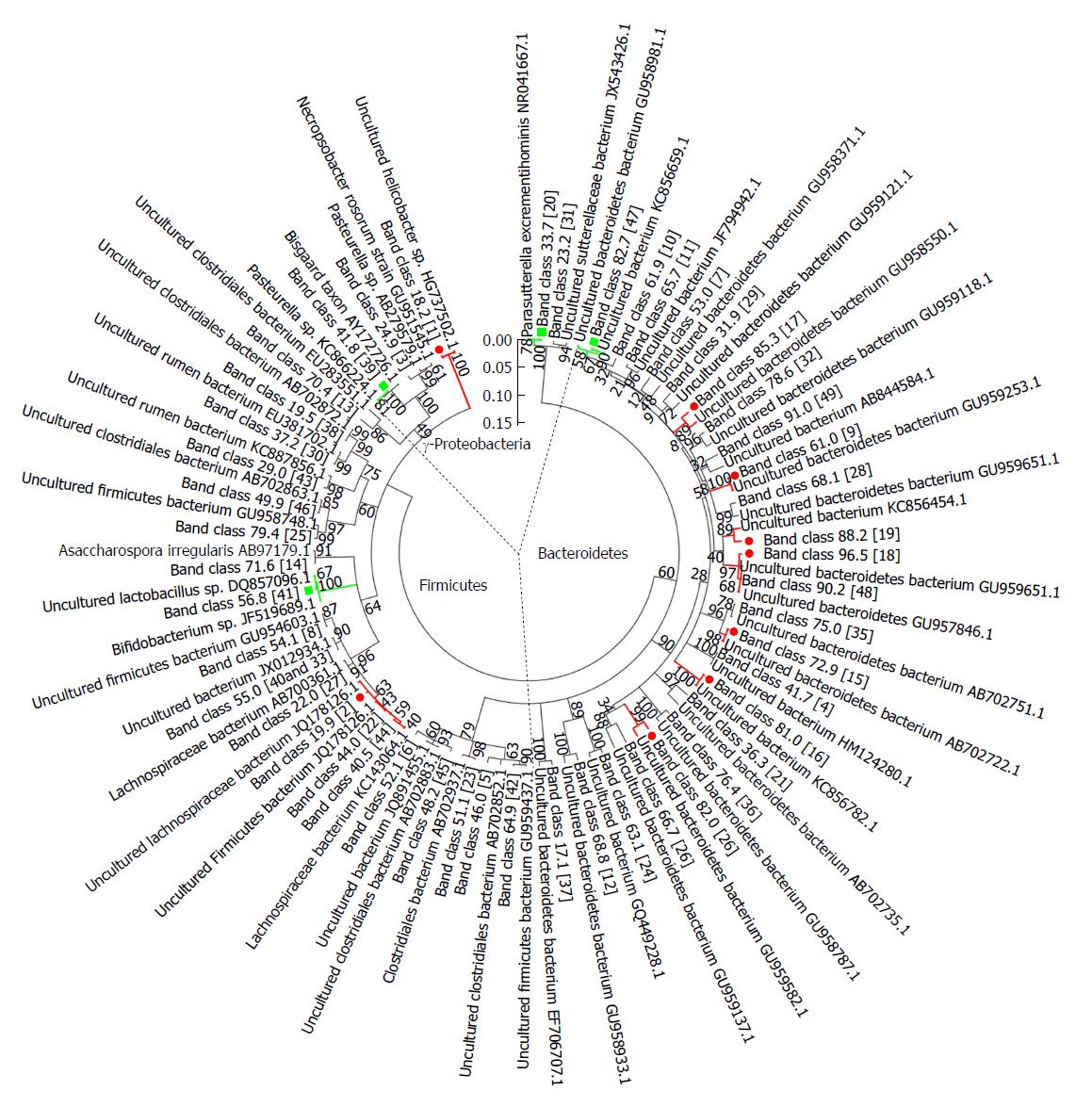
To determine the bacterial phylogenetic relationships and identify key bacteria involved in the microbial changes induced by FK506-M, the phylogenetic tree of sequences derived from DGGE bands was analyzed using the MEGA 5 software (Figure 6). Almost all the matched bacteria of the 48 band classes were assigned to three phyla: Bacteroidetes (50.0%), Firmicutes (39.6%), and Gammaproteobacteria (10.4%). The closest matched bacteria shown on the phylogenetic tree corresponded to 13 crucial band classes associated with FK506-M treatment. Details of the 13 crucial band classes are presented in Supplementary Table 2. In these crucial bacteria associated with FK506-M treatment, 7 of the 9 decreased band classes were matched to Bacteroidetes bacterium, belonging to phylum Bacteroidetes, but the increased band classes were matched to the Bifidobacterium species.
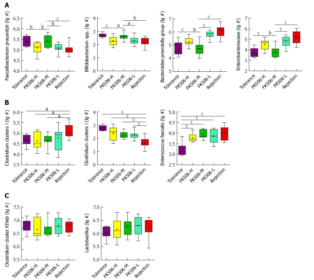
To verify crucial bacteria alterations induced by FK506-M treatment, we further analyzed dominant bacterial populations using RT-qPCR (Figure 7). Compared with the tolerance group, Faecalibacterium prausnitzii and Bifidobacterium spp. were significantly decreased, whereas the Bacteroides-Prevotella group and Enterobacteriaceae were increased in the FK506-L treated and rejection groups (all P < 0.05). However, FK506-M treatment significantly increased Faecalibacterium prausnitzii and Bifidobacterium spp. and decreased the Bacteroides-Prevotella group and Enterobacteriaceae when compared to the FK506-L and rejection groups (all P < 0.05) (Figure 7A). These results essentially verified the changes observed in the levels of 13 crucial bacteria associated with FK506-M treatment based on the DGGE profiles.
Moreover, in the rejection group, the Clostridium clusters I was elevated, whereas the Clostridium clusters XI was reduced, in contrast to the other groups (Figure 7B). Enterococcus faecalis was specifically decreased in the tolerance group (all P < 0.001). There were no statistical differences in the Clostridium cluster XIVab and Lactobacillus between the different groups (Figure 7C).
Liver transplantation is a life-saving technique for patients with end-stage liver disease. Appropriate dosage of immunosuppressive medications including FK506 could effectively prevent and treat allograft rejection in patients following LT. FK506 is a calcineurin inhibitor, which has become the most used immunosuppressant following LT[23]. Compared to cyclosporine, FK506 is more effective in reducing rejection, leading to better graft function and overall survival in LT patients[24]. Insufficient dosage of FK506 may lead to rejection and even graft dysfunction, while over dosage is closely associated with infection, increased complications, and even mortality. Unfortunately, FK506-related toxicity, mainly due to overload usage is frequent in LT patients[25].
In patients following LT, an optimal immunosuppressive therapy should balance the risk of infection, cancer, and drug toxicity caused by excessive immunosuppression, and the risk of rejection caused by an inadequately suppressed immune system[5]. To explore the optimal immunosuppressive dosage is very important for the long-term survival of the recipient and the liver graft. Jia et al[9] proved that compared to high FK506 blood concentration (10-15 ng/L) with adverse effects such as infection, early renal impairment, hepatocellular carcinoma recurrence, even death, reasonably low FK506 blood concentrations (5-10 ng/mL) by less oral dosage especially in the early phase, namely “Minimizing tacrolimus”, could prevent graft rejection, avoid related toxicity, protect the graft function, and promote long-term survival of the recipient. Moreover, Geng[26] proved that lower FK506 trough levels (< 5 ng/mL) in the late period of living donor LT are safe and improve the long-term outcomes.
Our previous studies indicated that bacterial community alterations in the gut were associated with liver cirrhosis[14], hepatic graft rejection[18], hepatic injury[19], and pancreatic carcinoma[27]. However, the influence of immunosuppressive medication on the gut microbiota and the association between the dosage of immunosuppressive drugs and alterations in gut microbiota have not been reported to date. In our study, all recipients survived following LT, except one recipient in the FK506-H dosage group that died of severe abdomen infection, suggesting an over-suppression of the immune status. Hepatic graft presented intermediate rejection injury in the FK506-L group, suggesting insufficient immunosuppression. In contrast, the FK506-M dosage maintained approximately normal hepatic structure and function. Survival results and graft morphology indicated that FK506-M treatment was optimal for maintaining immunosuppression. Meanwhile, the FK506-M dosage kept the gut barrier intact and maintained low levels of plasma endotoxin. Thus, the FK506-M dosage was considered optimal for maintaining immunosuppression in rats following LT.
Furthermore, we observed the influence of FK506 with different dosages on the gut microbiota. We further analyzed gut microbiota using a DGGE approach and found that the FK506-M dosage induced a unique and stable gut microbiota. MDS and PCA analysis validated the stable gut microbiota identified by DGGE. Phylogenetic tree analysis identified crucial bacteria associated with the FK506-M treatment. In addition, treatment with FK506-M significantly increased the growth of Faecalibacterium prausnitzii and Bifidobacterium spp. and decreased that of the Bacteroides-Prevotella group and Enterobacteriaceae as determined via RT-qPCR, which essentially verified the alterations in the population of crucial bacteria observed from the DGGE profiles.
Endotoxin, also known as lipopolysaccharide (LPS), is the main product of common gram-negative bacteria and is a crucial factor for the association of gut microbiota with liver inflammation[22]. In a pathogen-associated molecular pattern of microbiota-liver axis, LPS may possess the capacity to activate inflammation and initiate various pathophysiological cascades. High levels of LPS activate the NF-κB pathway, produce proinflammatory cytokines [tumor necrosis factor (TNF)-α, interleukin (IL)-6, and IL-1], and lead to liver inflammation and aggravation of hepatic oxidative damage[28]. In our study, plasma endotoxins were remarkably elevated in the rejection and FK506-L dosage groups, but the FK506-M dosage significantly decreased endotoxin levels. To explore the causes of the observed endotoxin changes, we analyzed gut microbial alterations and identified the crucial bacteria involved. Importantly, compared to the rejection and FK506-L dosage groups, RT-qPCR results verified that FK506-M treatment significantly increased Faecalibacterium prausnitzii and Bifidobacterium spp. and decreased the growth of the Bacteroides-Prevotella group and Enterobacteriaceae, which are the main common gram-negative bacteria producing LPS. Thus, we speculated that the optimal dosage of FK506 might lead to an increase in probiotics and a decrease in the potential pathogenic endotoxin-producing bacteria.
In patients following LT, blood concentrations of immunosuppressant and its stability are important. To mimic clinical application, FK506 was administered via abdominal subcutaneous injection for 7 d and then via intragastric administration during the subsequent 8-28 d after LT. In patients, routine monitoring of blood tacrolimus concentration (TC) was performed using the PRO-TracTMIITacrolimus Elisa Kit (Diasorin, United States), however, there is no kit for rat blood TC testing. According to our experimental experience, it may be due to immunological reasons, as the blood TC in rats can not be tested by the above kit used for humans. Fortunately, according to clinical practice of LT, different dosages of FK506 are positively correlated to different serum concentrations across a specific dosage range, meanwhile, intragastric dosages of FK506 were more feasible and easier to control the blood TC. In this experiment, the over-low dosage of FK506 led to a higher risk of rejection, whereas over-high dosage was closely associated with a greater incidence of infection, sepsis, and even increased mortality. Therefore, treatment with different dosages of FK506 instead of relying on the examination of blood TC was feasible and credible in rats after LT.
Gut microbiota alterations are closely associated with liver function in health and disease due to liver-gut circulation and the gut microbiota-liver axis[29]. Gut microbiota is involved in a variety of human liver diseases, such as alcoholic liver disease[30,31], non-alcoholic fatty liver disease[32], non-alcoholic steatohepatitis[33], hepatitis B infection[21], liver cirrhosis[14,34], and hepatocellular carcinoma[15,28,35]. Liver diseases always reflect changes due to alterations in intestinal permeability and gut microbial composition[29], while an imbalance in the gut microbiota in turn, has been reported to be an important contributor to the progression of liver disease or liver injury[36]. Notably, the improvement of hepatic injury might ameliorate gut barrier function and promote gut microbial restorations following LT, which might further benefit hepatic graft by positive feedback of the “gut-liver axis”[16,18]. Thus, the gut microbiota becomes an additional therapeutic target to improve hepatic injury after LT. In the present study, the optimal dosage of FK506 led to good hepatic graft function, kept the gut barrier intact, and induced a unique and stable gut microbiota, which further verify the effects of the “gut microbiota-liver” axis.
In conclusion, an optimal dosage of FK506 induces effective immunosuppression, normal graft function and stable gut microbiota after LT in rats. A stable gut microbiota resulted in increased probiotics, including Faecalibacterium prausnitzii and Bifidobacterium spp. and decreased potential pathogenic endotoxin-producing bacteria, such as the Bacteroides-Prevotella group and Enterobacteriaceae. These findings provide a novel strategy involving the use of the gut microbiota for the assessment of the dosage of immunosuppressive medications and its effects in recipients following LT.
Liver transplantation (LT) is the only definitive therapy for end-stage liver diseases. The optimal dosage of immunosuppressive medication is crucial to prevent rejection, lessen the side effects of immunosuppressors (IS) and treatment of patients following LT. The gut microbiota plays a crucial role in the development of obesity, diabetes, liver diseases and so on. Nevertheless, the influence of IS on gut microbiota following LT remains unclear, the association between the dosage of IS and gut microbial alterations requires urgent elucidation.
Acute rejection is still a leading cause of hepatic graft dysfunction following LT. Each recipient of LT must take IS to prevent acute rejection, but the effect of IS on gut microbiota remains unclear. Tacrolimus (FK506) is the main IS following LT, so we studied the influence of different dosages of FK506 on gut microbiota after LT in rat.
In this study, we studied the influence of different dosages of FK506 on hepatic graft function and gut microbiota following LT in rats. Furthermore, we will identify the precise changes of the gut microbiota given by different doses of FK-506 and provide a new monitoring strategy for IS dosage assessment in recipients after LT.
We performed LT experiments in rats by taking different dosage of FK506 for 29 d, and on the 30th day after LT, all rats were sampled and then euthanized. We studied the hepatic graft function using serum alanine aminotransferase and aspartate aminotransferase examination and morphology change using histopathology and transmission electron microscopy evaluation. We also identified the gut microbial profile and crucial bacterial community constituents using digital processing of denaturing gradient gel electrophoresis (DGGE), and further verified the alterations of dominant gut bacterial populations with RT-PCR.
Compared to the FK506-H and FK506-L groups, FK506-M was optimal for maintaining immunosuppression and induced normal graft function; the FK506-M maintained the integrity of gut barrier and low plasma endotoxin levels. Furthermore, FK506-M induced stable gut microbiota, increased species richness and rare species abundance by DGGE and Cluster analysis. The FK506-M increased Faecalibacterium prausnitzii and Bifidobacterium spp. and decreased Bacteroides-Prevotella and Enterobacteriaceae by Phylogenetic tree analysis and RT-PCR verification.
An optimal dosage of FK506 induces effective immunosuppression, normal graft function and stable gut microbiota after LT in rat. A stable gut microbiota resulted in increased probiotics and decreased potential pathogenic endotoxin-producing bacteria. These findings provide a novel strategy involving the use of the gut microbiota for the assessment of the dosage of immunosuppressive medications and their effects on recipients following LT.
This study is the first to illustrate the effects of FK506 with different dosages on gut microbiota following LT and indicates that an optimal dosage of FK506 induces effective immunosuppression, good graft function and stable gut microbiota after LT in rats. Based on the relationship between gut microbiota and immunosuppressive dosage in this study, we can not only illustrate precise changes of gut microbiota given by FK-506 with different dosages following LT, but we also provide a novel monitoring strategy based on changes in gut microbiota for IS assessment in recipients following LT. With the improvement of metagenomics and metabolomics techniques, the integrative study can further reveal how the gut microbiota participates in the side effects of IS on recipients and gut microbiota may be a target to reduce side effects of IS. In addition, in order to further study the mechanism of the involvement of gut microbiota on the side effects of IS on recipients, it is essential to do clinical research.
Manuscript source: Unsolicited manuscript
Specialty type: Gastroenterology and hepatology
Country of origin: China
Peer-review report classification
Grade A (Excellent): 0
Grade B (Very good): B
Grade C (Good): C
Grade D (Fair): 0
Grade E (Poor): 0
P- Reviewer: Cholongitas E, Venu RP S- Editor: Wang XJ L- Editor: Filipodia E- Editor: Bian YN
| 1. | Hübscher SG. What is the long-term outcome of the liver allograft? J Hepatol. 2011;55:702-717. [RCA] [PubMed] [DOI] [Full Text] [Cited by in Crossref: 101] [Cited by in RCA: 96] [Article Influence: 6.9] [Reference Citation Analysis (0)] |
| 2. | Sayegh MH, Carpenter CB. Transplantation 50 years later--progress, challenges, and promises. N Engl J Med. 2004;351:2761-2766. [RCA] [PubMed] [DOI] [Full Text] [Cited by in Crossref: 301] [Cited by in RCA: 309] [Article Influence: 14.7] [Reference Citation Analysis (0)] |
| 3. | Wiesner RH, Menon KV. Late hepatic allograft dysfunction. Liver Transpl. 2001;7:S60-S73. [RCA] [PubMed] [DOI] [Full Text] [Cited by in Crossref: 29] [Cited by in RCA: 29] [Article Influence: 1.2] [Reference Citation Analysis (0)] |
| 4. | Hu J, Wang Z, Tan CJ, Liao BY, Zhang X, Xu M, Dai Z, Qiu SJ, Huang XW, Sun J. Plasma microRNA, a potential biomarker for acute rejection after liver transplantation. Transplantation. 2013;95:991-999. [RCA] [PubMed] [DOI] [Full Text] [Cited by in Crossref: 49] [Cited by in RCA: 59] [Article Influence: 4.9] [Reference Citation Analysis (0)] |
| 5. | Ravaioli M, Neri F, Lazzarotto T, Bertuzzo VR, Di Gioia P, Stacchini G, Morelli MC, Ercolani G, Cescon M, Chiereghin A. Immunosuppression Modifications Based on an Immune Response Assay: Results of a Randomized, Controlled Trial. Transplantation. 2015;99:1625-1632. [RCA] [PubMed] [DOI] [Full Text] [Cited by in Crossref: 57] [Cited by in RCA: 63] [Article Influence: 6.3] [Reference Citation Analysis (0)] |
| 6. | Jiang JW, Ren ZG, Cui GY, Zhang Z, Xie HY, Zhou L. Chronic bile duct hyperplasia is a chronic graft dysfunction following liver transplantation. World J Gastroenterol. 2012;18:1038-1047. [RCA] [PubMed] [DOI] [Full Text] [Full Text (PDF)] [Cited by in CrossRef: 5] [Cited by in RCA: 5] [Article Influence: 0.4] [Reference Citation Analysis (0)] |
| 7. | Zhang W, Fung J. Limitations of current liver transplant immunosuppressive regimens: renal considerations. Hepatobiliary Pancreat Dis Int. 2017;16:27-32. [RCA] [PubMed] [DOI] [Full Text] [Cited by in Crossref: 16] [Cited by in RCA: 21] [Article Influence: 2.6] [Reference Citation Analysis (0)] |
| 8. | Humar A, Michaels M; AST ID Working Group on Infectious Disease Monitoring. American Society of Transplantation recommendations for screening, monitoring and reporting of infectious complications in immunosuppression trials in recipients of organ transplantation. Am J Transplant. 2006;6:262-274. [RCA] [PubMed] [DOI] [Full Text] [Cited by in Crossref: 389] [Cited by in RCA: 378] [Article Influence: 19.9] [Reference Citation Analysis (0)] |
| 9. | Jia JJ, Lin BY, He JJ, Geng L, Kadel D, Wang L, Yu DD, Shen T, Yang Z, Ye YF. “Minimizing tacrolimus” strategy and long-term survival after liver transplantation. World J Gastroenterol. 2014;20:11363-11369. [RCA] [PubMed] [DOI] [Full Text] [Full Text (PDF)] [Cited by in CrossRef: 29] [Cited by in RCA: 29] [Article Influence: 2.6] [Reference Citation Analysis (0)] |
| 10. | Eckburg PB, Bik EM, Bernstein CN, Purdom E, Dethlefsen L, Sargent M, Gill SR, Nelson KE, Relman DA. Diversity of the human intestinal microbial flora. Science. 2005;308:1635-1638. [RCA] [PubMed] [DOI] [Full Text] [Cited by in Crossref: 5700] [Cited by in RCA: 5591] [Article Influence: 279.6] [Reference Citation Analysis (2)] |
| 11. | Nenci A, Becker C, Wullaert A, Gareus R, van Loo G, Danese S, Huth M, Nikolaev A, Neufert C, Madison B. Epithelial NEMO links innate immunity to chronic intestinal inflammation. Nature. 2007;446:557-561. [RCA] [PubMed] [DOI] [Full Text] [Cited by in Crossref: 792] [Cited by in RCA: 945] [Article Influence: 52.5] [Reference Citation Analysis (0)] |
| 12. | Cani PD, Delzenne NM. The role of the gut microbiota in energy metabolism and metabolic disease. Curr Pharm Des. 2009;15:1546-1558. [RCA] [PubMed] [DOI] [Full Text] [Cited by in Crossref: 625] [Cited by in RCA: 651] [Article Influence: 40.7] [Reference Citation Analysis (0)] |
| 13. | Shen F, Zheng RD, Sun XQ, Ding WJ, Wang XY, Fan JG. Gut microbiota dysbiosis in patients with non-alcoholic fatty liver disease. Hepatobiliary Pancreat Dis Int. 2017;16:375-381. [RCA] [PubMed] [DOI] [Full Text] [Cited by in Crossref: 435] [Cited by in RCA: 411] [Article Influence: 51.4] [Reference Citation Analysis (1)] |
| 14. | Chen Y, Yang F, Lu H, Wang B, Chen Y, Lei D, Wang Y, Zhu B, Li L. Characterization of fecal microbial communities in patients with liver cirrhosis. Hepatology. 2011;54:562-572. [RCA] [PubMed] [DOI] [Full Text] [Cited by in Crossref: 674] [Cited by in RCA: 797] [Article Influence: 56.9] [Reference Citation Analysis (3)] |
| 15. | Dapito DH, Mencin A, Gwak GY, Pradere JP, Jang MK, Mederacke I, Caviglia JM, Khiabanian H, Adeyemi A, Bataller R. Promotion of hepatocellular carcinoma by the intestinal microbiota and TLR4. Cancer Cell. 2012;21:504-516. [RCA] [PubMed] [DOI] [Full Text] [Cited by in Crossref: 854] [Cited by in RCA: 1028] [Article Influence: 79.1] [Reference Citation Analysis (0)] |
| 16. | Ren Z, Cui G, Lu H, Chen X, Jiang J, Liu H, He Y, Ding S, Hu Z, Wang W. Liver ischemic preconditioning (IPC) improves intestinal microbiota following liver transplantation in rats through 16s rDNA-based analysis of microbial structure shift. PLoS One. 2013;8:e75950. [RCA] [PubMed] [DOI] [Full Text] [Full Text (PDF)] [Cited by in Crossref: 40] [Cited by in RCA: 40] [Article Influence: 3.3] [Reference Citation Analysis (0)] |
| 17. | Xie Y, Luo Z, Li Z, Deng M, Liu H, Zhu B, Ruan B, Li L. Structural shifts of fecal microbial communities in rats with acute rejection after liver transplantation. Microb Ecol. 2012;64:546-554. [RCA] [PubMed] [DOI] [Full Text] [Cited by in Crossref: 33] [Cited by in RCA: 36] [Article Influence: 2.8] [Reference Citation Analysis (0)] |
| 18. | Ren Z, Jiang J, Lu H, Chen X, He Y, Zhang H, Xie H, Wang W, Zheng S, Zhou L. Intestinal microbial variation may predict early acute rejection after liver transplantation in rats. Transplantation. 2014;98:844-852. [RCA] [PubMed] [DOI] [Full Text] [Full Text (PDF)] [Cited by in Crossref: 78] [Cited by in RCA: 78] [Article Influence: 7.1] [Reference Citation Analysis (0)] |
| 19. | Ren ZG, Liu H, Jiang JW, Jiang L, Chen H, Xie HY, Zhou L, Zheng SS. Protective effect of probiotics on intestinal barrier function in malnourished rats after liver transplantation. Hepatobiliary Pancreat Dis Int. 2011;10:489-496. [RCA] [PubMed] [DOI] [Full Text] [Cited by in Crossref: 29] [Cited by in RCA: 31] [Article Influence: 2.2] [Reference Citation Analysis (0)] |
| 20. | Ren Z, Chen X, Cui G, Yin S, Chen L, Jiang J, Hu Z, Xie H, Zheng S, Zhou L. Nanosecond pulsed electric field inhibits cancer growth followed by alteration in expressions of NF-κB and Wnt/β-catenin signaling molecules. PLoS One. 2013;8:e74322. [RCA] [PubMed] [DOI] [Full Text] [Full Text (PDF)] [Cited by in Crossref: 38] [Cited by in RCA: 36] [Article Influence: 3.0] [Reference Citation Analysis (0)] |
| 21. | Lu H, Wu Z, Xu W, Yang J, Chen Y, Li L. Intestinal microbiota was assessed in cirrhotic patients with hepatitis B virus infection. Intestinal microbiota of HBV cirrhotic patients. Microb Ecol. 2011;61:693-703. [RCA] [PubMed] [DOI] [Full Text] [Cited by in Crossref: 187] [Cited by in RCA: 179] [Article Influence: 12.8] [Reference Citation Analysis (0)] |
| 22. | Lamping N, Dettmer R, Schröder NW, Pfeil D, Hallatschek W, Burger R, Schumann RR. LPS-binding protein protects mice from septic shock caused by LPS or gram-negative bacteria. J Clin Invest. 1998;101:2065-2071. [RCA] [PubMed] [DOI] [Full Text] [Cited by in Crossref: 243] [Cited by in RCA: 252] [Article Influence: 9.3] [Reference Citation Analysis (0)] |
| 23. | US Multicenter FK506 Liver Study Group. A comparison of tacrolimus (FK 506) and cyclosporine for immunosuppression in liver transplantation. N Engl J Med. 1994;331:1110-1115. [RCA] [PubMed] [DOI] [Full Text] [Cited by in Crossref: 784] [Cited by in RCA: 756] [Article Influence: 24.4] [Reference Citation Analysis (0)] |
| 24. | Haddad EM, McAlister VC, Renouf E, Malthaner R, Kjaer MS, Gluud LL. Cyclosporin versus tacrolimus for liver transplanted patients. Cochrane Database Syst Rev. 2006;CD005161. [RCA] [PubMed] [DOI] [Full Text] [Cited by in Crossref: 8] [Cited by in RCA: 9] [Article Influence: 0.5] [Reference Citation Analysis (0)] |
| 25. | Wiesner RH, Fung JJ. Present state of immunosuppressive therapy in liver transplant recipients. Liver Transpl. 2011;17 Suppl 3:S1-S9. [RCA] [PubMed] [DOI] [Full Text] [Cited by in Crossref: 81] [Cited by in RCA: 84] [Article Influence: 6.0] [Reference Citation Analysis (0)] |
| 26. | Geng L, Wang LD, Huang JJ, Shen T, Wang ZY, Lin BY, Ye YF, Zheng SS. Lower tacrolimus trough levels in the late period after living donor liver transplantation contribute to improvements in long-term clinical outcomes. Hepatobiliary Pancreat Dis Int. 2018;17:204-209. [RCA] [PubMed] [DOI] [Full Text] [Cited by in Crossref: 8] [Cited by in RCA: 8] [Article Influence: 1.1] [Reference Citation Analysis (0)] |
| 27. | Ren Z, Jiang J, Xie H, Li A, Lu H, Xu S, Zhou L, Zhang H, Cui G, Chen X. Gut microbial profile analysis by MiSeq sequencing of pancreatic carcinoma patients in China. Oncotarget. 2017;8:95176-95191. [RCA] [PubMed] [DOI] [Full Text] [Full Text (PDF)] [Cited by in Crossref: 173] [Cited by in RCA: 163] [Article Influence: 20.4] [Reference Citation Analysis (0)] |
| 28. | Darnaud M, Faivre J, Moniaux N. Targeting gut flora to prevent progression of hepatocellular carcinoma. J Hepatol. 2013;58:385-387. [RCA] [PubMed] [DOI] [Full Text] [Cited by in Crossref: 45] [Cited by in RCA: 63] [Article Influence: 5.3] [Reference Citation Analysis (0)] |
| 29. | Chassaing B, Etienne-Mesmin L, Gewirtz AT. Microbiota-liver axis in hepatic disease. Hepatology. 2014;59:328-339. [RCA] [PubMed] [DOI] [Full Text] [Cited by in Crossref: 219] [Cited by in RCA: 256] [Article Influence: 23.3] [Reference Citation Analysis (0)] |
| 30. | Yan AW, Fouts DE, Brandl J, Stärkel P, Torralba M, Schott E, Tsukamoto H, Nelson KE, Brenner DA, Schnabl B. Enteric dysbiosis associated with a mouse model of alcoholic liver disease. Hepatology. 2011;53:96-105. [RCA] [PubMed] [DOI] [Full Text] [Full Text (PDF)] [Cited by in Crossref: 668] [Cited by in RCA: 638] [Article Influence: 45.6] [Reference Citation Analysis (0)] |
| 31. | Hartmann P, Chen P, Wang HJ, Wang L, McCole DF, Brandl K, Stärkel P, Belzer C, Hellerbrand C, Tsukamoto H. Deficiency of intestinal mucin-2 ameliorates experimental alcoholic liver disease in mice. Hepatology. 2013;58:108-119. [RCA] [PubMed] [DOI] [Full Text] [Cited by in Crossref: 162] [Cited by in RCA: 201] [Article Influence: 16.8] [Reference Citation Analysis (0)] |
| 32. | Le Roy T, Llopis M, Lepage P, Bruneau A, Rabot S, Bevilacqua C, Martin P, Philippe C, Walker F, Bado A. Intestinal microbiota determines development of non-alcoholic fatty liver disease in mice. Gut. 2013;62:1787-1794. [RCA] [PubMed] [DOI] [Full Text] [Cited by in Crossref: 601] [Cited by in RCA: 715] [Article Influence: 59.6] [Reference Citation Analysis (0)] |
| 33. | Zhu L, Baker SS, Gill C, Liu W, Alkhouri R, Baker RD, Gill SR. Characterization of gut microbiomes in nonalcoholic steatohepatitis (NASH) patients: a connection between endogenous alcohol and NASH. Hepatology. 2013;57:601-609. [RCA] [PubMed] [DOI] [Full Text] [Cited by in Crossref: 1015] [Cited by in RCA: 1281] [Article Influence: 106.8] [Reference Citation Analysis (1)] |
| 34. | Qin N, Yang F, Li A, Prifti E, Chen Y, Shao L, Guo J, Le Chatelier E, Yao J, Wu L. Alterations of the human gut microbiome in liver cirrhosis. Nature. 2014;513:59-64. [RCA] [PubMed] [DOI] [Full Text] [Cited by in Crossref: 1230] [Cited by in RCA: 1537] [Article Influence: 139.7] [Reference Citation Analysis (40)] |
| 35. | Zhang HL, Yu LX, Yang W, Tang L, Lin Y, Wu H, Zhai B, Tan YX, Shan L, Liu Q. Profound impact of gut homeostasis on chemically-induced pro-tumorigenic inflammation and hepatocarcinogenesis in rats. J Hepatol. 2012;57:803-812. [RCA] [PubMed] [DOI] [Full Text] [Cited by in Crossref: 180] [Cited by in RCA: 214] [Article Influence: 16.5] [Reference Citation Analysis (0)] |
| 36. | Schnabl B, Brenner DA. Interactions between the intestinal microbiome and liver diseases. Gastroenterology. 2014;146:1513-1524. [RCA] [PubMed] [DOI] [Full Text] [Cited by in Crossref: 615] [Cited by in RCA: 743] [Article Influence: 67.5] [Reference Citation Analysis (0)] |


