Published online Aug 7, 2016. doi: 10.3748/wjg.v22.i29.6683
Peer-review started: March 9, 2016
First decision: April 14, 2016
Revised: April 29, 2016
Accepted: June 2, 2016
Article in press: June 2, 2016
Published online: August 7, 2016
Processing time: 142 Days and 22.5 Hours
Esophageal squamous cell carcinoma (ESCC) is known for its rapid progression and poor outcomes. China has the highest incidence and mortality in the world. Diagnoses made at early stages and accurate staging are associated with better outcomes, all of which can play a significant role in the selection of treatment protocols. ESCC is staged according to the widely accepted TNM system. Common imaging modalities used in staging ESCC before treatment include endoscopy, computed tomography (CT), positron emission tomography (PET) and magnetic resonance imaging (MRI). Endoscopic ultrasound is useful for staging tumor depth and nodal status. Narrow band imaging is valuable for early stage disease assessment. CT and PET provide additional valuable information regarding node and metastasis staging. The ability of MRI to delineate ESCC is continuously being improved and adds information regarding locoregional status to routine examinations.
Core tip: Esophageal cancer is an aggressive malignant disease, as well as a growing health concern, with regard to mortality and prognosis. Esophageal squamous cell carcinoma is the prevailing histological type of esophageal cancer, with a high incidence in China. Staging of esophageal squamous cell carcinoma is based on the TNM system. Current imaging modalities include endoscopy, computed tomography, positron emission tomography and magnetic resonance imaging.
- Citation: Luo LN, He LJ, Gao XY, Huang XX, Shan HB, Luo GY, Li Y, Lin SY, Wang GB, Zhang R, Xu GL, Li JJ. Evaluation of preoperative staging for esophageal squamous cell carcinoma. World J Gastroenterol 2016; 22(29): 6683-6689
- URL: https://www.wjgnet.com/1007-9327/full/v22/i29/6683.htm
- DOI: https://dx.doi.org/10.3748/wjg.v22.i29.6683
Esophageal cancer is an aggressive malignant disease, as well as a growing health concern, with regard to mortality and prognosis. Esophageal squamous cell carcinoma (ESCC) is the prevailing histological type of esophageal cancer worldwide but has a great degree of regional variability, with a high incidence in eastern countries. Ninety percent of cases of esophageal cancer are squamous cell carcinomas in the so-called “esophageal cancer belt”, which covers the area from Northern Iran through the Central Asian Republics to North-Central China[1]. Northern and central China have an incidence of ESCC as high as 100 cases per 100000 persons per year; ESCC is 3 to 4 times more common in men than in women, and the peak incidence occurs in the seventh decade of life[2].
Risks factors for ESCC include alcoholism, smoking, diet, human papillomavirus (HPV) infection, esophageal diseases (e.g., achalasia) and a personal history of head and neck cancer[3-5]. ESCC typically appears in the lower two-thirds of the esophagus, with only 10%-15% of cases occurring in the upper one-third of the esophagus[6]. Poor outcomes in patients with ESCC are related to diagnosis at an advanced stage and the tendency to spread at an early stage[7]. To improve the prognosis of ESCC, accurate staging is necessary for guiding treatment protocols.
In this paper, we review the common imaging modalities used in staging preoperative ESCC that are reported in the literature.
Generally, patients with squamous intraepithelial lesions and half of patients with superficial ESCC show no symptoms at all when diagnosed[8]. Symptoms are present only when tumors are large enough to interfere with esophageal function or to have spread through the esophageal wall. Usually, intermittent dysphagia is the first symptom. As the frequency and severity of dysphagia increase, weight loss follows. Patients with advanced disease often suffer from odynophagia, chest pain radiating to the back and anemia and may even develop a tracheoesophageal fistula.
The diagnostic work-up for ESCC should involve various aspects, including symptoms, history, physical examination findings, imaging results and pathological findings. Once a diagnosis is made, accurate staging must be performed before treatment to guarantee that the most appropriate treatment schemes are applied, the results of which can be sufficiently assessed[2,9].
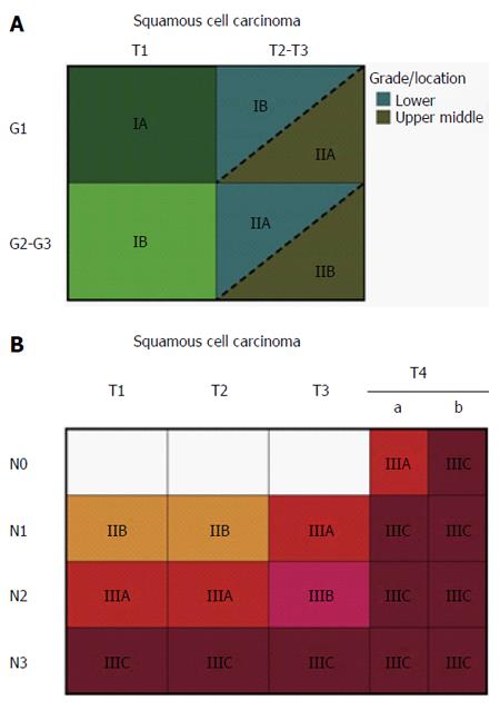
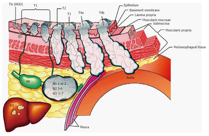
At present, the widely accepted staging criteria are those of the TNM system developed by the American Joint Committee on Cancer (AJCC). The TNM staging system takes into consideration the depth of tumor invasion (T-staging), the nodal status (N-staging) and metastatic spread (M-staging). This system has been updated over time, and the current version is shown in Figure 1[10]. It is worth noting that in the latest version, T4 lesions are classified as resectable (T4a) or unresectable (T4b), and the division of N-staging is based on the lymph nodes involved. Tis lesions (i.e., intraepithelial neoplasias) have been redefined as high-grade dysplasia, and M-staging is classified as no distant metastases (M0) or distant metastases (M1; Figure 2[10]).
The common imaging techniques for staging ESCC that are currently in use are endoscopy, especially endoscopic ultrasound (EUS) and its innovations, computed tomography (CT), positron emission tomography (PET) and magnetic resonance imaging (MRI).
On white light endoscopy, squamous dysplasias may be flat, erythematous, nodular, plaque-like or erosive appearing. The majority of lesions are abnormal under endoscopic examination[11].
In the TNM system, the T-stage is determined by the depth of tumor invasion. EUS is a highly regarded technique for diagnosing and staging ESCC before treatment because it is able to distinguish the layers of the esophageal wall with endoscopic sonographic transducers (7.5-12 MHz). The accuracy of T-staging varies based on the stage. A meta-analysis of 49 studies showed that EUS had better accuracy in advanced disease than in early stage disease, with a pooled sensitivity of 92% in T4 and 82% in T1[12]. Further studies showed that EUS had an excellent performance in differentiating sub-stages of early disease. Thosani et al[13] conducted a meta-analysis of 19 studies and determined that EUS had a pooled sensitivity and specificity of 85% and 87%, respectively, for T1a, and 86% and 86%, respectively, for T1b. Recently, submucosal saline injection combined with EUS was reported to have better performance in staging T1b than EUS alone (86.7% vs 60%)[14]. It was reported that the echogenicity differences in tumor lesions and the submucosa were enhanced after the submucosa was filled with saline, which made it easier to identify lesions and the surrounding normal tissue. However, few studies have focused on sub-staging, especially for an advanced stage, to differentiate a resectable stage (T4a) from an unresectable stage (T4b).
With regard to N-staging, EUS is reported to have an accuracy of up to 84% in node-positive patients, but its accuracy falls to 69% when node-negative patients are taken into consideration[15]. In clinical practice, EUS has a limitation in detecting node status because some adjacent lymph nodes may be beyond its area of detection. In a meta-analysis of 49 studies, Puli et al[12] compared EUS alone and EUS-guided fine needle aspiration (EUS-FNA) and found that EUS-FNA (pooled sensitivity and specificity of 96.7% and 95.5%, respectively) was superior to EUS alone (pooled sensitivity and specificity of 84.7% and 84.6%, respectively).
Narrow band imaging (NBI) uses spectral characteristics of endoscopic light to enhance the visibility of vasculature in the mucosa[16-18]. An optical filter with a narrow band spectrum is applied to correspond to the peak absorption spectrum of hemoglobin to enhance the visualization of mucosal and submucosal microvascular patterns. A panel of nine experts from Asian-Pacific countries agreed that NBI could replace chromoendoscopy, which distinguishes normal esophageal mucosa by applying Lugol’s iodine stain during routine examination, because it is easy to use and imparts considerable information[19]. On NBI, early lesions appear brown and well demarcated, and it is reported that microvascular patterns correspond to the depth of tumor invasion, which is classified into five types and several subtypes[20]. Thus, NBI is being increasingly used to stage superficial ESCC.
Several clinical studies have been conducted to assess the effectiveness of NBI for detecting superficial lesions, but the results have led to varying conclusions. In a randomized controlled multicenter trial, NBI was able to detect superficial lesions more frequently than white light imaging in the esophagus (97% vs 55%, P < 0.01)[21]. In contrast, Sharma et al[22] indicated that the performances of NBI and white light imaging were the same. To obtain more detail, magnification endoscopy in conjunction with NBI has been employed. Goda et al[23] investigated 72 patients with 101 superficial ESCCs and found that magnification endoscopy with NBI has the potential to reduce the overestimation risk of high-resolution endoscopy. However, NBI does have an advantage in detecting superficial lesions, and it is believed that with protocol adjustment, NBI is promising for the staging of preoperative ESCC.
Esophageal wall thickness is a basic finding when assessing the esophagus by CT, and a lack of symmetry with respect to wall thickness is another classic but not specific CT finding in ESCC. Li et al[24] reported that CT had a sensitivity of 77.4% and a specificity of 74.8% for distinguishing T1/2 from T3 in ESCC with whole esophageal circumferential tumor involvement. Recently, a prospective, nonrandomized study showed that CT had higher accuracy than EUS in the precise assessment of superficial ESCC limited to the epithelium or lamina propria mucosa (94.6% vs 80.6%, P < 0.05)[25].
However, CT has a limited ability to accurately distinguish T1, T2 and T3 diseases. The combination of CT and EUS has an overall staging accuracy of approximately 85%[26]. The general protocol after the diagnosis of ESCC is to perform an initial CT because EUS shows no advantage in detecting distant metastases. If no distant metastases are present, then EUS is performed.
Therefore, the most useful aspect of CT in detecting tumor depth is in evaluating advanced stages (T4), when adjacent structures are invaded. Several studies have reported the effectiveness of multidetector computed tomography (MDCT), a great modality for performing multiplanar reformation imaging to confirm a true lesion from three or more plane images in the case of mediastinal invasion. A retrospective study involving 21 patients with ESCC reported that the accuracies for MDCT to differentiate T3 and T4 disease were 77.3% and 81.8%, respectively, and that the sensitivity and specificity were generally high (over 75% for all)[27]. In addition, Ba-Ssalamah et al[28] believe that with the specific assessment criteria of MDCT, two different readers can achieve a sensitivity as high as 95% and a high positive predictive value of 96%.
CT assessment of nodal status depends on the size of the lymph nodes. Most studies consider the common size criterion of 1 cm to be enlarged. It has been reported that CT has a sensitivity of 30%-60% and a specificity of 60%-80% for identifying enlarged lymph nodes[29]. Tio et al[30] reported that the accuracy of CT in staging nodal status was 46% to 58%. However, an obvious limitation of CT in N-staging involves metastatic nodes without obvious enlargement in size, as well as the fact that enlarged nodes may contain no metastasis. Thus, regular application of EUS-FNA and CT for patients with ESCC may show advantages in N-staging compared with CT alone.
18F-fluorodeoxyglucose PET (FDG-PET), a non-invasive modality for management, has been increasingly used to stage ESCC, but it is not useful for detecting tumor depth (T-staging) and its accuracy in assessing nodal status varies widely. Little et al[31] investigated 58 patients with superficial esophageal cancer and found that FDG-PET was not indicated for staging superficial lesions because it could not differentiate Tis (5/11, 45%) from T1 (26/47, 55%, P = 0.03). FDG-PET is believed to be the most important modality for detecting unsuspected metastatic disease[32] (M-staging) because of its ability to distinguish physiologically normal tissues from malignant tumors due to enhanced glucose transport in tumors. Early use of PET in the staging of patients with ESCC may facilitate treatment strategies and identify unsuspected metastases in up to 20% of patients. Metastatic spread from ESCC can occur in unusual and unexpected locations, and whole body PET allows the detection of these metastases[33,34]. In a meta-analysis, PET had a moderate sensitivity of 67% and a high specificity of 97% in diagnosing distant metastatic disease[35].
Several clinical studies indicate that FDG-PET has a high sensitivity in identifying lesions in the entire body. Meltzer et al[36] conducted an independent, blinded retrospective evaluation of FDG-PET findings obtained in 47 patients referred for initial preoperative staging of ESCC. They found that PET had a great specificity in detecting nodal status (approximately 90%) and a sensitivity of 70% (n = 10) in detecting distant metastases when it was applied in the high-sensitivity mode. Furthermore, in comparison with CT, PET correctly identified one more site of metastasis that was missed by CT. A multicenter study involving 349 patients with ESCC reported that PET had false-positive results in only 5 (1.43%) patients in describing the extent of the disease[37].
In addition to PET scans, MRI with the functional feature of diffusion-weighted imaging (DWI) is another advancing imaging technology, which has current and future potential to overcome the limitations of conventional staging methods in patients with ESCC. MRI is often considered technically challenging because of artifacts due to organ motion and blood flow[38]. However, the usefulness of MRI in ESCC may expand as technical improvements allow proper visualization of the esophagus. van Rossum et al[39] believe that an anticipated MRI protocol consisting of T2-weighted and DWI sequences will be effective in future clinical practice by guiding treatment options for patients with ESCC.
In early studies of MRI, the results of T-staging were disappointing, especially for early stage[40]. However, as technology has improved, combining T2-weighted and DWI scans has yielded detection rates for MRI in T-staging of ESCC of 33% for T1, 58% for T2, 96% for T3 and 100% for T4[41]. Recent studies reported that high-resolution MRI for T-staging was able to achieve an accuracy as high as that for standard EUS (81% vs 81%-92%)[42,43]. With regard to lymph node staging, a prospective study with 24 patients reported that T2-weighted MRI in combination with DWI had a sensitivity of 77.8% and a specificity of 55.6%[41]. Moreover, a more recent prospective study that performed PET and whole-body MRI in 49 patients found that whole-body MRI and PET had similar accuracies in T–staging (98% vs 94%) and N-staging (67% vs 59%), and even for metastatic disease[44].
As the incidence of ESCC increases worldwide, substantial work is required to improve diagnostic and staging accuracy. Imaging plays a pivotal role in the management of ESCC. At present, there is no single staging modality that is superior to another in preoperative TNM staging for ESCC. Local staging is usually best accomplished using a combination of EUS and CT, while distant metastatic disease is better assessed by PET. In the near future, MRI has the potential to improve staging, thereby compensating for the limitations of current imaging modalities. However, staging techniques still need to be refined to better assess prognosis, as well as to achieve the best possible outcomes by individualizing treatment. Technological advances in EUS and its innovations are being investigated and will ideally improve the outcomes of patients with ESCC.
Manuscript source: Invited manuscript
Specialty type: Gastroenterology and hepatology
Country of origin: China
Peer-review report classification
Grade A (Excellent): 0
Grade B (Very good): 0
Grade C (Good): C
Grade D (Fair): 0
Grade E (Poor): 0
P- Reviewer: Miyata H S- Editor: Qi Y L- Editor: Wang TQ E- Editor: Ma S
1. | Torre LA, Bray F, Siegel RL, Ferlay J, Lortet-Tieulent J, Jemal A. Global cancer statistics, 2012. CA Cancer J Clin. 2015;65:87-108. [PubMed] [DOI] [Cited in This Article: ] [Cited by in Crossref: 18694] [Cited by in F6Publishing: 21126] [Article Influence: 2112.6] [Reference Citation Analysis (2)] |
2. | Pennathur A, Luketich JD. Resection for esophageal cancer: strategies for optimal management. Ann Thorac Surg. 2008;85:S751-S756. [PubMed] [DOI] [Cited in This Article: ] [Cited by in Crossref: 85] [Cited by in F6Publishing: 98] [Article Influence: 5.8] [Reference Citation Analysis (0)] |
3. | Vioque J, Barber X, Bolumar F, Porta M, Santibáñez M, de la Hera MG, Moreno-Osset E. Esophageal cancer risk by type of alcohol drinking and smoking: a case-control study in Spain. BMC Cancer. 2008;8:221. [PubMed] [DOI] [Cited in This Article: ] [Cited by in Crossref: 48] [Cited by in F6Publishing: 50] [Article Influence: 2.9] [Reference Citation Analysis (0)] |
4. | Iyer RB, Silverman PM, Tamm EP, Dunnington JS, DuBrow RA. Diagnosis, staging, and follow-up of esophageal cancer. AJR Am J Roentgenol. 2003;181:785-793. [PubMed] [DOI] [Cited in This Article: ] [Cited by in Crossref: 45] [Cited by in F6Publishing: 47] [Article Influence: 2.1] [Reference Citation Analysis (0)] |
5. | Hewer T, Rose E, Ghadirian P, Castegnaro M, Malaveille C, Bartsch H, Day N. Ingested mutagens from opium and tobacco pyrolysis products and cancer of the oesophagus. Lancet. 1978;2:494-496. [PubMed] [Cited in This Article: ] |
6. | Neumann WL, Genta RM. Tumors of the Digestive System.Tumours of the esophagus. Cancer Grading Manual. Berlin, Heidelberg: Springer Berlin Heidelberg 2013; 51-63. [Cited in This Article: ] |
7. | Pennathur A, Farkas A, Krasinskas AM, Ferson PF, Gooding WE, Gibson MK, Schuchert MJ, Landreneau RJ, Luketich JD. Esophagectomy for T1 esophageal cancer: outcomes in 100 patients and implications for endoscopic therapy. Ann Thorac Surg. 2009;87:1048-154; discussion 1048-154;. [PubMed] [DOI] [Cited in This Article: ] [Cited by in Crossref: 194] [Cited by in F6Publishing: 201] [Article Influence: 12.6] [Reference Citation Analysis (0)] |
8. | Chen LQ, Hu CY, Ghadirian P, Duranceau A. Early detection of esophageal squamous cell carcinoma and its effects on therapy: an overview. Dis Esophagus. 1999;12:161-167. [PubMed] [Cited in This Article: ] |
9. | Krasna MJ, Reed CE, Nedzwiecki D, Hollis DR, Luketich JD, DeCamp MM, Mayer RJ, Sugarbaker DJ. CALGB 9380: a prospective trial of the feasibility of thoracoscopy/laparoscopy in staging esophageal cancer. Ann Thorac Surg. 2001;71:1073-1079. [PubMed] [Cited in This Article: ] |
10. | Rice TW, Blackstone EH, Rusch VW. A cancer staging primer: esophagus and esophagogastric junction. J Thorac Cardiovasc Surg. 2010;139:527-529. [PubMed] [DOI] [Cited in This Article: ] [Cited by in Crossref: 32] [Cited by in F6Publishing: 25] [Article Influence: 1.7] [Reference Citation Analysis (0)] |
11. | Roth MJ, Liu SF, Dawsey SM, Zhou B, Copeland C, Wang GQ, Solomon D, Baker SG, Giffen CA, Taylor PR. Cytologic detection of esophageal squamous cell carcinoma and precursor lesions using balloon and sponge samplers in asymptomatic adults in Linxian, China. Cancer. 1997;80:2047-2059. [PubMed] [Cited in This Article: ] |
12. | Puli SR, Reddy JB, Bechtold ML, Antillon D, Ibdah JA, Antillon MR. Staging accuracy of esophageal cancer by endoscopic ultrasound: a meta-analysis and systematic review. World J Gastroenterol. 2008;14:1479-1490. [PubMed] [DOI] [Cited in This Article: ] [Cited by in CrossRef: 243] [Cited by in F6Publishing: 230] [Article Influence: 13.5] [Reference Citation Analysis (2)] |
13. | Thosani N, Singh H, Kapadia A, Ochi N, Lee JH, Ajani J, Swisher SG, Hofstetter WL, Guha S, Bhutani MS. Diagnostic accuracy of EUS in differentiating mucosal versus submucosal invasion of superficial esophageal cancers: a systematic review and meta-analysis. Gastrointest Endosc. 2012;75:242-253. [PubMed] [DOI] [Cited in This Article: ] [Cited by in Crossref: 148] [Cited by in F6Publishing: 157] [Article Influence: 12.1] [Reference Citation Analysis (0)] |
14. | Li JJ, Shan HB, Gu MF, He L, He LJ, Chen LM, Luo GY, Xu GL. Endoscopic ultrasound combined with submucosal saline injection for differentiation of T1a and T1b esophageal squamous cell carcinoma: a novel technique. Endoscopy. 2013;45:667-670. [PubMed] [DOI] [Cited in This Article: ] [Cited by in Crossref: 9] [Cited by in F6Publishing: 13] [Article Influence: 1.1] [Reference Citation Analysis (0)] |
15. | Rösch T. Endosonographic staging of esophageal cancer: a review of literature results. Gastrointest Endosc Clin N Am. 1995;5:537-547. [PubMed] [Cited in This Article: ] |
16. | Gheorghe C. Narrow-band imaging endoscopy for diagnosis of malignant and premalignant gastrointestinal lesions. J Gastrointestin Liver Dis. 2006;15:77-82. [PubMed] [Cited in This Article: ] |
17. | Gono K, Obi T, Yamaguchi M, Ohyama N, Machida H, Sano Y, Yoshida S, Hamamoto Y, Endo T. Appearance of enhanced tissue features in narrow-band endoscopic imaging. J Biomed Opt. 2004;9:568-577. [PubMed] [DOI] [Cited in This Article: ] [Cited by in Crossref: 634] [Cited by in F6Publishing: 588] [Article Influence: 28.0] [Reference Citation Analysis (0)] |
18. | Kuznetsov K, Lambert R, Rey JF. Narrow-band imaging: potential and limitations. Endoscopy. 2006;38:76-81. [PubMed] [DOI] [Cited in This Article: ] [Cited by in Crossref: 156] [Cited by in F6Publishing: 148] [Article Influence: 7.8] [Reference Citation Analysis (0)] |
19. | Uedo N, Fujishiro M, Goda K, Hirasawa D, Kawahara Y, Lee JH, Miyahara R, Morita Y, Singh R, Takeuchi M. Role of narrow band imaging for diagnosis of early-stage esophagogastric cancer: current consensus of experienced endoscopists in Asia-Pacific region. Dig Endosc. 2011;23 Suppl 1:58-71. [PubMed] [DOI] [Cited in This Article: ] [Cited by in Crossref: 72] [Cited by in F6Publishing: 72] [Article Influence: 5.1] [Reference Citation Analysis (0)] |
20. | Kumagai Y, Inoue H, Nagai K, Kawano T, Iwai T. Magnifying endoscopy, stereoscopic microscopy, and the microvascular architecture of superficial esophageal carcinoma. Endoscopy. 2002;34:369-375. [PubMed] [DOI] [Cited in This Article: ] [Cited by in Crossref: 165] [Cited by in F6Publishing: 159] [Article Influence: 6.9] [Reference Citation Analysis (0)] |
21. | Muto M, Minashi K, Yano T, Saito Y, Oda I, Nonaka S, Omori T, Sugiura H, Goda K, Kaise M. Early detection of superficial squamous cell carcinoma in the head and neck region and esophagus by narrow band imaging: a multicenter randomized controlled trial. J Clin Oncol. 2010;28:1566-1572. [PubMed] [DOI] [Cited in This Article: ] [Cited by in Crossref: 427] [Cited by in F6Publishing: 499] [Article Influence: 33.3] [Reference Citation Analysis (0)] |
22. | Sharma P, Hawes RH, Bansal A, Gupta N, Curvers W, Rastogi A, Singh M, Hall M, Mathur SC, Wani SB. Standard endoscopy with random biopsies versus narrow band imaging targeted biopsies in Barrett’s oesophagus: a prospective, international, randomised controlled trial. Gut. 2013;62:15-21. [PubMed] [DOI] [Cited in This Article: ] [Cited by in Crossref: 229] [Cited by in F6Publishing: 240] [Article Influence: 20.0] [Reference Citation Analysis (0)] |
23. | Goda K, Tajiri H, Ikegami M, Yoshida Y, Yoshimura N, Kato M, Sumiyama K, Imazu H, Matsuda K, Kaise M. Magnifying endoscopy with narrow band imaging for predicting the invasion depth of superficial esophageal squamous cell carcinoma. Dis Esophagus. 2009;22:453-460. [PubMed] [DOI] [Cited in This Article: ] [Cited by in Crossref: 69] [Cited by in F6Publishing: 61] [Article Influence: 3.8] [Reference Citation Analysis (0)] |
24. | Li H, Chen TW, Zhang XM, Li ZL, Chen XL, Tang HJ, Huang XH, Chen N, Yang Q, Hu J. Computed tomography scan as a tool to predict tumor T category in resectable esophageal squamous cell carcinoma. Ann Thorac Surg. 2013;95:1749-1755. [PubMed] [DOI] [Cited in This Article: ] [Cited by in Crossref: 13] [Cited by in F6Publishing: 10] [Article Influence: 0.8] [Reference Citation Analysis (0)] |
25. | Hatta W, Uno K, Koike T, Iijima K, Asano N, Imatani A, Shimosegawa T. A prospective comparative study of optical coherence tomography and EUS for tumor staging of superficial esophageal squamous cell carcinoma. Gastrointest Endosc. 2012;76:548-555. [PubMed] [DOI] [Cited in This Article: ] [Cited by in Crossref: 30] [Cited by in F6Publishing: 32] [Article Influence: 2.5] [Reference Citation Analysis (0)] |
26. | Botet JF, Lightdale CJ, Zauber AG, Gerdes H, Urmacher C, Brennan MF. Preoperative staging of esophageal cancer: comparison of endoscopic US and dynamic CT. Radiology. 1991;181:419-425. [PubMed] [DOI] [Cited in This Article: ] [Cited by in Crossref: 273] [Cited by in F6Publishing: 270] [Article Influence: 7.9] [Reference Citation Analysis (0)] |
27. | Pongpornsup S, Posri S, Totanarungroj K. Diagnostic accuracy of multidetector computed tomography (MDCT) in evaluation for mediastinal invasion of esophageal cancer. J Med Assoc Thai. 2012;95:704-711. [PubMed] [Cited in This Article: ] |
28. | Ba-Ssalamah A, Matzek W, Baroud S, Bastati N, Zacherl J, Schoppmann SF, Hejna M, Wrba F, Weber M, Herold CJ. Accuracy of hydro-multidetector row CT in the local T staging of oesophageal cancer compared to postoperative histopathological results. Eur Radiol. 2011;21:2326-2335. [PubMed] [DOI] [Cited in This Article: ] [Cited by in Crossref: 24] [Cited by in F6Publishing: 28] [Article Influence: 2.0] [Reference Citation Analysis (0)] |
29. | Kato H, Kuwano H, Nakajima M, Miyazaki T, Yoshikawa M, Ojima H, Tsukada K, Oriuchi N, Inoue T, Endo K. Comparison between positron emission tomography and computed tomography in the use of the assessment of esophageal carcinoma. Cancer. 2002;94:921-928. [PubMed] [Cited in This Article: ] |
30. | Tio TL, Cohen P, Coene PP, Udding J, den Hartog Jager FC, Tytgat GN. Endosonography and computed tomography of esophageal carcinoma. Preoperative classification compared to the new (1987) TNM system. Gastroenterology. 1989;96:1478-1486. [PubMed] [Cited in This Article: ] |
31. | Little SG, Rice TW, Bybel B, Mason DP, Murthy SC, Falk GW, Rybicki LA, Blackstone EH. Is FDG-PET indicated for superficial esophageal cancer? Eur J Cardiothorac Surg. 2007;31:791-796. [PubMed] [DOI] [Cited in This Article: ] [Cited by in Crossref: 43] [Cited by in F6Publishing: 44] [Article Influence: 2.4] [Reference Citation Analysis (1)] |
32. | Meyers BF, Downey RJ, Decker PA, Keenan RJ, Siegel BA, Cerfolio RJ, Landreneau RJ, Reed CE, Balfe DM, Dehdashti F. The utility of positron emission tomography in staging of potentially operable carcinoma of the thoracic esophagus: results of the American College of Surgeons Oncology Group Z0060 trial. J Thorac Cardiovasc Surg. 2007;133:738-745. [PubMed] [DOI] [Cited in This Article: ] [Cited by in Crossref: 105] [Cited by in F6Publishing: 81] [Article Influence: 4.5] [Reference Citation Analysis (0)] |
33. | Erasmus JJ, Munden RF. The role of integrated computed tomography positron-emission tomography in esophageal cancer: staging and assessment of therapeutic response. Semin Radiat Oncol. 2007;17:29-37. [PubMed] [DOI] [Cited in This Article: ] [Cited by in Crossref: 35] [Cited by in F6Publishing: 39] [Article Influence: 2.2] [Reference Citation Analysis (0)] |
34. | Bruzzi JF, Truong MT, Macapinlac H, Munden RF, Erasmus JJ. Integrated CT-PET imaging of esophageal cancer: unexpected and unusual distribution of distant organ metastases. Curr Probl Diagn Radiol. 2007;36:21-29. [PubMed] [DOI] [Cited in This Article: ] [Cited by in Crossref: 29] [Cited by in F6Publishing: 31] [Article Influence: 1.7] [Reference Citation Analysis (0)] |
35. | van Westreenen HL, Westerterp M, Bossuyt PM, Pruim J, Sloof GW, van Lanschot JJ, Groen H, Plukker JT. Systematic review of the staging performance of 18F-fluorodeoxyglucose positron emission tomography in esophageal cancer. J Clin Oncol. 2004;22:3805-3812. [PubMed] [DOI] [Cited in This Article: ] [Cited by in Crossref: 273] [Cited by in F6Publishing: 242] [Article Influence: 11.5] [Reference Citation Analysis (1)] |
36. | Meltzer CC, Luketich JD, Friedman D, Charron M, Strollo D, Meehan M, Urso GK, Dachille MA, Townsend DW. Whole-body FDG positron emission tomographic imaging for staging esophageal cancer comparison with computed tomography. Clin Nucl Med. 2000;25:882-887. [PubMed] [Cited in This Article: ] |
37. | Vomackova K, Neoral C, Aujeský R, Vrba R, Stasek M, Myslivecek M, Formánek R. The benefit of PET/CT in the diagnosis and treatment of esophageal cancer. Rozhl Chir. 2015;94:8-16. [PubMed] [Cited in This Article: ] |
38. | Giovagnoni A, Valeri G, Ferrara C. MRI of esophageal cancer. Abdom Imaging. 2002;27:361-366. [PubMed] [DOI] [Cited in This Article: ] [Cited by in F6Publishing: 1] [Reference Citation Analysis (0)] |
39. | van Rossum PS, van Lier AL, Lips IM, Meijer GJ, Reerink O, van Vulpen M, Lam MG, van Hillegersberg R, Ruurda JP. Imaging of oesophageal cancer with FDG-PET/CT and MRI. Clin Radiol. 2015;70:81-95. [PubMed] [DOI] [Cited in This Article: ] [Cited by in Crossref: 44] [Cited by in F6Publishing: 49] [Article Influence: 4.9] [Reference Citation Analysis (0)] |
40. | Nakashima A, Nakashima K, Seto H, Kakishita M, Sakamoto T, Yamada A, Fujimaki M. Thoracic esophageal carcinoma: evaluation in the sagittal section with magnetic resonance imaging. Abdom Imaging. 1997;22:20-23. [PubMed] [Cited in This Article: ] |
41. | Sakurada A, Takahara T, Kwee TC, Yamashita T, Nasu S, Horie T, Van Cauteren M, Imai Y. Diagnostic performance of diffusion-weighted magnetic resonance imaging in esophageal cancer. Eur Radiol. 2009;19:1461-1469. [PubMed] [DOI] [Cited in This Article: ] [Cited by in Crossref: 78] [Cited by in F6Publishing: 58] [Article Influence: 3.6] [Reference Citation Analysis (0)] |
42. | van Vliet EP, Eijkemans MJ, Kuipers EJ, Poley JW, Steyerberg EW, Siersema PD. Publication bias does not play a role in the reporting of the results of endoscopic ultrasound staging of upper gastrointestinal cancers. Endoscopy. 2007;39:325-332. [PubMed] [DOI] [Cited in This Article: ] [Cited by in Crossref: 14] [Cited by in F6Publishing: 15] [Article Influence: 0.8] [Reference Citation Analysis (0)] |
43. | Riddell AM, Allum WH, Thompson JN, Wotherspoon AC, Richardson C, Brown G. The appearances of oesophageal carcinoma demonstrated on high-resolution, T2-weighted MRI, with histopathological correlation. Eur Radiol. 2007;17:391-399. [PubMed] [DOI] [Cited in This Article: ] [Cited by in Crossref: 67] [Cited by in F6Publishing: 71] [Article Influence: 3.7] [Reference Citation Analysis (0)] |
44. | Malik V, Harmon M, Johnston C, Fagan AJ, Claxton Z, Ravi N, O’Toole D, Muldoon C, Keogan M, Reynolds JV. Whole Body MRI in the Staging of Esophageal Cancer--A Prospective Comparison with Whole Body 18F-FDG PET-CT. Dig Surg. 2015;32:397-408. [PubMed] [DOI] [Cited in This Article: ] [Cited by in Crossref: 17] [Cited by in F6Publishing: 18] [Article Influence: 1.8] [Reference Citation Analysis (0)] |