Published online May 7, 2016. doi: 10.3748/wjg.v22.i17.4421
Peer-review started: December 22, 2015
First decision: January 13, 2016
Revised: January 21, 2016
Accepted: February 22, 2016
Article in press: February 22, 2016
Published online: May 7, 2016
Processing time: 132 Days and 23.8 Hours
Laparoscopic cholecystectomy has become a standard treatment of symptomatic gallstone disease. Although spilled gallstones are considered harmless, unretrieved gallstones can result in intra-abdominal abscess. We report a case of abscess formation due to spilled gallstones after laparoscopic cholecystectomy mimicking a retroperitoneal sarcoma on radiologic imaging. A 59-year-old male with a surgical history of a laparoscopic cholecystectomy complicated by gallstones spillage presented with a 1 mo history of constant right-sided abdominal pain and tenderness. Computed tomography and magnetic resonance imaging demonstrated a retroperitoneal sarcoma at the sub-hepatic space. On open exploration a 5 cm × 5 cm retroperitoneal mass was excised. The mass contained purulent material and gallstones. Final pathology revealed abscess formation and foreign body granuloma. Vigilance concerning the possibility of lost gallstones during laparoscopic cholecystectomy is important. If possible, every spilled gallstone during surgery should be retrieved to prevent this rare complication.
Core tip: Gallstone abscess resulting from spilled gallstones is a rare complication following laparoscopic cholecystectomy. We report a rare presentation of gallstone abscess due to spilled gallstones following laparoscopic cholecystectomy, mimicking a retroperitoneal sarcoma in a 59-year-old male. Recognizing the patient information about a history of laparoscopic cholecystectomy and sharing the patient information with radiologists can make an accurate diagnosis and avoid misinterpretation when the diagnosis is equivocal in radiologic imaging. Clear documentation of gallbladder perforation and gallstone can help with diagnosis of the complication and for correct management. Five noteworthy features are discussed in this paper.
- Citation: Kim BS, Joo SH, Kim HC. Spilled gallstones mimicking a retroperitoneal sarcoma following laparoscopic cholecystectomy. World J Gastroenterol 2016; 22(17): 4421-4426
- URL: https://www.wjgnet.com/1007-9327/full/v22/i17/4421.htm
- DOI: https://dx.doi.org/10.3748/wjg.v22.i17.4421
Laparoscopic cholecystectomy is a standard treatment for symptomatic gallstone disease. Gallbladder perforation and gallstones spillage during laparoscopic cholecystectomy occurs in up to 40% of cases[1-3].The incidence of unretrieved spilled gallstones has been estimated at 16% to 50%[3-5]. Gallstone abscess following spilled gallstones is an extremely rare delayed complication of laparoscopic cholecystectomy[3,6]. Common locations of the abscess are in the abdominal wall followed by intra-abdominal cavity usually in the sub-hepatic or retroperitoneum inferior to sub-hepatic space[3]. We report a rare case of abscess formation due to spilled gallstones mimicking a retroperitoneal sarcoma following laparoscopic cholecystectomy.
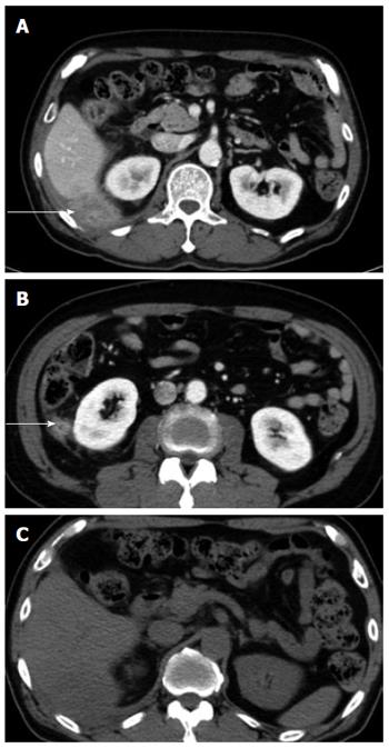
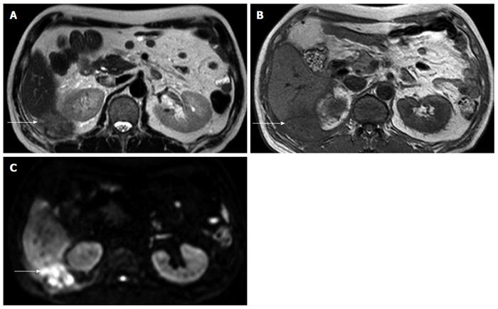
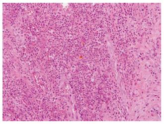
Our institutional review board approved the reporting of this case. A 59-year-old male was admitted to our clinic with a 1-mo history of worsening right-sided constant abdominal pain. Outside abdominal sonography showed a 4.5 cm mass-like lesion in the sub-hepatic space. He had no significant past medical history apart from a history of laparoscopic cholecystectomy about 5 mo previously. Laboratory workup revealed a white blood cell count of 7500/mm3. All other laboratory findings including liver function tests, amylase and lipase were within normal limits. Tumor makers were normal. The hepatitis serologic markers were all negative. Computed tomography (CT) scan of the abdomen revealed a 4.9 cm × 4.6 cm right retroperitoneal mass invading the muscle wall and a 2 cm mass abutting the right kidney (Figure 1A-C). Magnetic resonance imaging (MRI) detected an ill-defined mass in the sub-hepatic space with subtle high signal intensity in T2 weighted image, low signal intensity in T1 weighted image and high signal intensity in diffusion weighted image (Figure 2A-C). With the suspicion that this mass was a retroperitoneal sarcoma, an exploratory laparotomy was performed. It revealed a severe adhesion around the mass containing purulent material and gallstones in the sub-hepatic space. Examination of frozen sections revealed acute and chronic inflammation with abscess and foreign body granuloma. Small abscess cavity found in the sub-phrenic space was destroyed using suction. Purulent materials were removed and several remaining stones were retrieved and copious irrigation was performed. Final pathology showed acute and chronic inflammation with abscess and foreign body granuloma (Figure 3). Postoperative recovery was uneventful. Six months later, the patient remains in good condition without any complaints.
Gallbladder perforation and gallstone spillage during laparoscopic cholecystectomy occurs in up to 40% of cases[1-3]. The risk of gallbladder perforation leading to bile and gallstone spillage is more common during laparoscopic cholecystectomy than during open cholecystectomy. Gallbladder perforation most often occurs during the traction of the gallbladder or dissection from the gallbladder bed or extraction of the gallbladder through the port site, and it is more common in the presence of adhesions, inflammation around the gallbladder and in the early years of a surgical career[6,7]. The most important risk factor of gallbladder perforation is acute inflammation of the gallbladder with an edematous, friable or gangrenous wall[7,8]. However, gallstones spill into the abdominal cavity in only 7.3% of these cases[9,10].
Zehenter et al[10] reported all possible complications due to spilled gallstones during laparoscopic cholecystectomy. The most common complication is intra-abdominal abscesses that are located most often in the sub-hepatic space or its retroperitoneal region, as occurred presently. Fistula formations, hernia sacs, ovary and fallopian tubes containing lost gallstones are among some of the rare complications reported for gallstone abscess. These complications are rarely fatal. Thus, converting to an open procedure is not indicated, because only 8.5% of patients will lead to a complication[10].
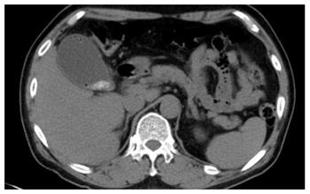
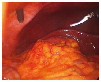
An inflammatory response can be induced, which features walling off by omentum and local fibrosis. Inflammation and infection are more common with pigment stones[11]. This is postulated to be due to the release of bacteria from within the stones as the body breaks down the stone matrix[12]. In our case, preoperative CT in the primary procedure showed emphysematous cholecystitis with multiple stones (Figure 4). Operatively, gallbladder empyema with a friable wall was found (Figure 5). The edematous and friable wall hindered grasping of the gallbladder during the traction and the dissection of the gallbladder from the liver, which lead to gallbladder perforation and gallstones and infected bile spillage. Despite every effort to remove remaining stones including extensive peritoneal lavage and aspiration with a large suction tube (Figure 6), gallstone abscess due to spilled stones occurred 5 mo after laparoscopic cholecystectomy. This highlights that care must be taken not to spread gallstones into more inaccessible sites, making retrieval even more difficult. Spilled gallstones can be retrieved easily in open cholecystectomy by irrigating the abdominal cavity and aspirating with a large suction tube or by collecting the stones with a sponge, which is difficult in laparoscopic cholecystectomy[13-15].
CT and MRI features of gallstones often include the presence of gallstones within the abscess, which is essential for a diagnosis[16]. Pigment stones with high calcium content are easily diagnosed with CT, whereas pure cholesterol stones and those with low calcium content may go undetected, as in our case[17]. Presently, CT and MRI were equivocal with the findings of retroperitoneal sarcoma. Spilled stones were not evident in radiologic imaging. Presence of the highly reflective echoes with posterior shadowing in the abscess cavity can be pathognomonic for gallstone abscess in patients with a history of laparoscopic cholecystectomy.
Abscess formation secondary to spilled stones mimicking a retroperitoneal sarcoma is rare. Despite their extreme rarity, the characteristic appearance of intra-abdominal abscesses from gallstones should be recognized because their radiographic appearance can mimic more severe disease, such as peritoneal metastasis[16,18]. Abscess formation from spilled stones after laparoscopic cholecystectomy has been reported to have an average duration of 4 mo to 10 years[19,20]. Presently, despite the history of laparoscopic cholecystectomy 5 mo before, gallstone abscess mimicking a retroperitoneal sarcoma in radiologic finding made the diagnosis difficult. Spilled gallstones around the liver may be confused with peritoneal metastasis or lymph nodes, having a similar appearance of round soft-tissue perihepatic nodules with a high attenuation peripheral rim on CT. Unenhanced CT may be useful in elucidating the calcified nature of these nodules. However, although the presence of calcification favours the diagnosis of spilled gallstones, some mucin producing tumours, such as tumours from the ovary and colon, can contain calcium[21]. Therefore, spilled gallstones should be considered in a patient with a history of LC presenting with an abdominal abscess.
The current case highlights five noteworthy features. Gallstone abscess resulting from spilled gallstones is a rare complication following laparoscopic cholecystectomy, but spilled gallstones should always be considered in a patient with a history of laparoscopic cholecystectomy presenting with an abdominal abscess. Secondly, a surgeon needs to share the patient information with radiologists to make an accurate diagnosis and avoid misinterpretation when the diagnosis is equivocal in radiologic imagings. Thirdly, careful manipulation of gallbladder with empyema, wall thickening and many gallstones during laparoscopic cholecystectomy should be made. Fourth, every effort to retrieve all spilled stones during laparoscopic cholecystectomy should be made to avoid late complications and the peritoneum should be irrigated with copious saline. Finally, it is important for the surgeon to clearly document in the operative record whether the gallbladder was perforated and whether stones spilled during laparoscopic cholecystectomy to help with diagnosis of the complication and for correct management.
In conclusion, retroperitoneal abscess formation secondary to spilled gallstones following laparoscopic cholecystectomy is a rare condition. If possible, every spilled gallstone during laparoscopic cholecystectomy should be retrieved to prevent such a rare complication. As in our case, preoperative radiological investigations may be equivocal. Clear documentation of gallbladder perforation and gallstone spillage is important, as is being vigilant to the possibility of stones during surgery.
A 59-year-old male with a history of laparoscopic cholecystectomy 5 mo prior presented with a 1-mo history of worsening right-sided constant abdominal pain.
The patient presented with a 1-mo history of worsening right-sided constant abdominal pain.
Retroperitoneal sarcoma, metastatic cancer.
All labs were within normal limits.
Computed tomography showed a 4.9 cm × 4.6 cm right retroperitoneal mass invading the muscle wall and a 2 cm mass abutting the right kidney.
Acute and chronic inflammation with abscess and foreign body granuloma.
Abscess cavity was destroyed using suction. Purulent materials were removed and several remaining stones were retrieved and copious irrigation was performed.
Intra-abdominal abscesses secondary to spilled gallstones are located most often in the sub-hepatic space or its retroperitoneal region. Fistula formations, hernia sacs, ovary and fallopian tubes containing lost gallstones are among some of the rare complications reported for gallstone abscess.
The characteristic appearance of intra-abdominal abscesses from gallstones should be recognized because their radiographic appearance can mimic more severe disease, such as peritoneal metastasis and retroperitoneal sarcoma.
Retroperitoneal abscess formation due to spilled gallstones following laparoscopic cholecystectomy is a rare condition. If possible, every spilled gallstone during laparoscopic cholecystectomy should be retrieved to prevent such a rare complication. Clear documentation of gallbladder perforation and gallstone spillage is important, as is being vigilant to the possibility of stones during surgery.
This case report is very useful to doctors in clinical practice. It is helpful to remind doctors to avoid the same mistake in clinical work.
P- Reviewer: Hu H, Sinha R S- Editor: Gong ZM L- Editor: A E- Editor: Liu XM
| 1. | Soper NJ, Dunnegan DL. Does intraoperative gallbladder perforation influence the early outcome of laparoscopic cholecystectomy? Surg Laparosc Endosc. 1991;1:156-161. [PubMed] |
| 2. | Woodfield JC, Rodgers M, Windsor JA. Peritoneal gallstones following laparoscopic cholecystectomy: incidence, complications, and management. Surg Endosc. 2004;18:1200-1207. [RCA] [PubMed] [DOI] [Full Text] [Cited by in Crossref: 98] [Cited by in RCA: 114] [Article Influence: 5.4] [Reference Citation Analysis (0)] |
| 3. | Brockmann JG, Kocher T, Senninger NJ, Schürmann GM. Complications due to gallstones lost during laparoscopic cholecystectomy. Surg Endosc. 2002;16:1226-1232. [RCA] [PubMed] [DOI] [Full Text] [Cited by in Crossref: 95] [Cited by in RCA: 98] [Article Influence: 4.3] [Reference Citation Analysis (0)] |
| 4. | Sarli L, Pietra N, Costi R, Grattarola M. Gallbladder perforation during laparoscopic cholecystectomy. World J Surg. 1999;23:1186-1190. [RCA] [PubMed] [DOI] [Full Text] [Cited by in Crossref: 55] [Cited by in RCA: 63] [Article Influence: 2.4] [Reference Citation Analysis (0)] |
| 5. | Diez J, Arozamena C, Gutierrez L, Bracco J, Mon A, Sanchez Almeyra R, Secchi M. Lost stones during laparoscopic cholecystectomy. HPB Surg. 1998;11:105-108; discuss 108-109. [PubMed] |
| 6. | Rice DC, Memon MA, Jamison RL, Agnessi T, Ilstrup D, Bannon MB, Farnell MB, Grant CS, Sarr MG, Thompson GB. Long-term consequences of intraoperative spillage of bile and gallstones during laparoscopic cholecystectomy. J Gastrointest Surg. 1997;1:85-90; discussion 90-91. [RCA] [PubMed] [DOI] [Full Text] [Cited by in Crossref: 76] [Cited by in RCA: 77] [Article Influence: 2.8] [Reference Citation Analysis (0)] |
| 7. | Sathesh-Kumar T, Saklani AP, Vinayagam R, Blackett RL. Spilled gall stones during laparoscopic cholecystectomy: a review of the literature. Postgrad Med J. 2004;80:77-79. [RCA] [PubMed] [DOI] [Full Text] [Cited by in Crossref: 105] [Cited by in RCA: 99] [Article Influence: 4.7] [Reference Citation Analysis (0)] |
| 8. | De Simone P, Donadio R, Urbano D. The risk of gallbladder perforation at laparoscopic cholecystectomy. Surg Endosc. 1999;13:1099-1102. [RCA] [PubMed] [DOI] [Full Text] [Cited by in Crossref: 21] [Cited by in RCA: 23] [Article Influence: 0.9] [Reference Citation Analysis (0)] |
| 9. | Schäfer M, Suter C, Klaiber C, Wehrli H, Frei E, Krähenbühl L. Spilled gallstones after laparoscopic cholecystectomy. A relevant problem? A retrospective analysis of 10,174 laparoscopic cholecystectomies. Surg Endosc. 1998;12:305-309. [RCA] [PubMed] [DOI] [Full Text] [Cited by in Crossref: 88] [Cited by in RCA: 87] [Article Influence: 3.2] [Reference Citation Analysis (0)] |
| 10. | Zehetner J, Shamiyeh A, Wayand W. Lost gallstones in laparoscopic cholecystectomy: all possible complications. Am J Surg. 2007;193:73-78. [RCA] [PubMed] [DOI] [Full Text] [Cited by in Crossref: 112] [Cited by in RCA: 117] [Article Influence: 6.5] [Reference Citation Analysis (0)] |
| 11. | Gürleyik E, Gürleyik G, Yücel O, Unalmiŝer S. Does chemical composition have an influence on the fate of intraperitoneal gallstone in rat? Surg Laparosc Endosc. 1998;8:113-116. [RCA] [PubMed] [DOI] [Full Text] [Cited by in Crossref: 18] [Cited by in RCA: 20] [Article Influence: 0.7] [Reference Citation Analysis (0)] |
| 12. | Stewart L, Smith AL, Pellegrini CA, Motson RW, Way LW. Pigment gallstones form as a composite of bacterial microcolonies and pigment solids. Ann Surg. 1987;206:242-250. [PubMed] |
| 13. | Horton M, Florence MG. Unusual abscess patterns following dropped gallstones during laparoscopic cholecystectomy. Am J Surg. 1998;175:375-379. [RCA] [PubMed] [DOI] [Full Text] [Cited by in Crossref: 79] [Cited by in RCA: 87] [Article Influence: 3.2] [Reference Citation Analysis (0)] |
| 14. | Frola C, Cannici F, Cantoni S, Tagliafico E, Luminati T. Peritoneal abscess formation as a late complication of gallstones spilled during laparoscopic cholecystectomy. Br J Radiol. 1999;72:201-203. [RCA] [PubMed] [DOI] [Full Text] [Cited by in Crossref: 22] [Cited by in RCA: 24] [Article Influence: 0.9] [Reference Citation Analysis (0)] |
| 15. | Offiah C, Robinson P, Keeling-Roberts CS. The one that got away. Br J Radiol. 2002;75:393-394. [RCA] [PubMed] [DOI] [Full Text] [Cited by in Crossref: 5] [Cited by in RCA: 5] [Article Influence: 0.2] [Reference Citation Analysis (0)] |
| 16. | Bennett AA, Gilkeson RC, Haaga JR, Makkar VK, Onders RP. Complications of “dropped” gallstones after laparoscopic cholecystectomy: technical considerations and imaging findings. Abdom Imaging. 2000;25:190-193. [RCA] [PubMed] [DOI] [Full Text] [Cited by in Crossref: 23] [Cited by in RCA: 24] [Article Influence: 1.0] [Reference Citation Analysis (0)] |
| 17. | Baron RL, Rohrmann CA, Lee SP, Shuman WP, Teefey SA. CT evaluation of gallstones in vitro: correlation with chemical analysis. AJR Am J Roentgenol. 1988;151:1123-1128. [RCA] [PubMed] [DOI] [Full Text] [Cited by in Crossref: 69] [Cited by in RCA: 58] [Article Influence: 1.6] [Reference Citation Analysis (0)] |
| 18. | Dasari BV, Loan W, Carey DP. Spilled gallstones mimicking peritoneal metastases. JSLS. 2009;13:73-76. [PubMed] |
| 19. | Morrin MM, Kruskal JB, Hochman MG, Saldinger PF, Kane RA. Radiologic features of complications arising from dropped gallstones in laparoscopic cholecystectomy patients. AJR Am J Roentgenol. 2000;174:1441-1445. [RCA] [PubMed] [DOI] [Full Text] [Cited by in Crossref: 62] [Cited by in RCA: 65] [Article Influence: 2.6] [Reference Citation Analysis (0)] |
| 20. | Van Brunt PH, Lanzafame RJ. Subhepatic inflammatory mass after laparoscopic cholecystectomy. A delayed complication of spilled gallstones. Arch Surg. 1994;129:882-883. [PubMed] |
| 21. | Nayak L, Menias CO, Gayer G. Dropped gallstones: spectrum of imaging findings, complications and diagnostic pitfalls. Br J Radiol. 2013;86:20120588. [RCA] [PubMed] [DOI] [Full Text] [Cited by in Crossref: 43] [Cited by in RCA: 61] [Article Influence: 5.1] [Reference Citation Analysis (0)] |


