Published online Mar 21, 2016. doi: 10.3748/wjg.v22.i11.3220
Peer-review started: May 6, 2015
First decision: August 26, 2015
Revised: September 3, 2015
Accepted: November 9, 2015
Article in press: November 9, 2015
Published online: March 21, 2016
Processing time: 319 Days and 13.6 Hours
AIM: To identify whether the forceps estimation is more useful than visual estimation in the measurement of colon polyp size.
METHODS: We recorded colonoscopy video clips that included scenes visualizing the polyp and scenes using open biopsy forceps in association with the polyp, which were used for an exam. A total of 40 endoscopists from the Busan Ulsan Gyeongnam Intestinal Study Group Society (BIGS) participated in this study. Participants watched 40 pairs of video clips of the scenes for visual estimation and forceps estimation, and wrote down the estimated polyp size on the exam paper. When analyzing the results of the exam, we assessed inter-observer differences, diagnostic accuracy, and error range in the measurement of the polyp size.
RESULTS: The overall intra-class correlation coefficients (ICC) of inter-observer agreement for forceps estimation and visual estimation were 0.804 (95%CI: 0.731-0.873, P < 0.001) and 0.743 (95%CI: 0.656-0.828, P < 0.001), respectively. The ICCs of each group for forceps estimation were higher than those for visual estimation (Beginner group, 0.761 vs 0.693; Expert group, 0.887 vs 0.840, respectively). The overall diagnostic accuracy for visual estimation was 0.639 and for forceps estimation was 0.754 (P < 0.001). In the beginner group and the expert group, the diagnostic accuracy for the forceps estimation was significantly higher than that of the visual estimation (Beginner group, 0.734 vs 0.613, P < 0.001; Expert group, 0.784 vs 0.680, P < 0.001, respectively). The overall error range for visual estimation and forceps estimation were 1.48 ± 1.18 and 1.20 ± 1.10, respectively (P < 0.001). The error ranges of each group for forceps estimation were significantly smaller than those for visual estimation (Beginner group, 1.38 ± 1.08 vs 1.68 ± 1.30, P < 0.001; Expert group, 1.12 ± 1.11 vs 1.42 ± 1.11, P < 0.001, respectively).
CONCLUSION: Application of the open biopsy forceps method when measuring colon polyp size could help reduce inter-observer differences and error rates.
Core tip: Using open biopsy forceps is known to be a useful technique to reduce error rates in colon polyp size measurements, but in practice most endoscopists just measure polyp size by visualization. There is little information about accuracy differences between these two methods. In this study, we showed that the inter-observer difference, diagnostic accuracy, and error range of forceps estimation were better than those of visual estimation in the measurement of the polyp size. We propose that forceps estimation should be considered to measure the colon polyp size before removing the polyp.
- Citation: Kim JH, Park SJ, Lee JH, Kim TO, Kim HJ, Kim HW, Lee SH, Baek DH, (BIGS) BUGISGS. Is forceps more useful than visualization for measurement of colon polyp size? World J Gastroenterol 2016; 22(11): 3220-3226
- URL: https://www.wjgnet.com/1007-9327/full/v22/i11/3220.htm
- DOI: https://dx.doi.org/10.3748/wjg.v22.i11.3220
Colorectal cancer is the third most common tumor in men and the second in women, accounting for 10% of all cancers worldwide. It is the fourth most common cancer-related cause of death in the world[1]. In Korea, according to data from the Korean National Cancer Center, the age-standardized incidence rate of colorectal cancer was 49.8 per 100000 men and 26.4 per 100000 women in 2010[2]. It is well known that most colorectal cancers arise from adenomatous polyps and patients with adenomatous polyps have greater risks of future development of advanced neoplasia[3-7]. This is the theoretical basis for the removal of all adenomatous polyps detected during colonoscopic examination[8,9]. Potential risks of malignant evolution are correlated with size, location, age, gender, growth pattern, and grade of dysplasia[10-14]. It has been well documented that larger polyps have more advanced histological features. About one-third of all polyps larger than 10 mm in size have advanced histology, but diminutive polyps (≤ 5 mm in size) rarely have advanced pathology[15,16]. Although the polypectomy technique for diminutive and small polyps is highly variable among endoscopists[17,18], polyps ≥ 6 mm have been removed by snare polypectomy as the technique of choice and diminutive polyps have been removed commonly by cold biopsy forceps[18-20]. Some recent studies found that the complete resection rate of cold forceps polypectomy for diminutive polyps was 90%-92%[21,22]. However, Lee et al[23] reported that snare polypectomy is superior to cold forceps polypectomy for the endoscopic removal of diminutive polyps with regard to completeness of polypectomy (93.2% vs 75.9%, P = 0.009), and Kim et al[22] reported that the complete resection rates for polyps sized 5 to 7 mm was significantly higher in the cold snare polypectomy group compared with the cold forceps polypectomy group (93.8% vs 70.3%, P = 0.013). In these regards, accurate measurement of polyp size is important, but it is not easy to accurately measure polyp size during colonoscopic examination. Chen et al[24] found significant inter-observer differences in the detection of adenomas larger than 1 cm. Even experienced endoscopists may make an error when estimating polyp size[25]. To estimate the polyp size, several methods have been suggested, including (1) visual estimation; (2) the use of open biopsy forceps; (3) the use of a linear measuring probe; or (4) the use of graduated injection needle and snare[26-29]. Among them, the use of open biopsy forceps is known to be a useful technique in colon polyp size measurements, considering its favorable time-to-effectiveness ratio, although it is not a completely reliable method to estimate polyp size[27,28]. However, in practice most endoscopists just measure polyp size by visualization, and there is not enough information available regarding the efficacy of these two methods.
The aim of our study was to determine the inter-observer differences and the error rates of the forceps estimation (using open biopsy forceps) and the visual estimation in the measurement of colon polyp size, and to identify whether the forceps estimation technique is more accurate and practical than visual estimation in the measurement of colon polyp size.
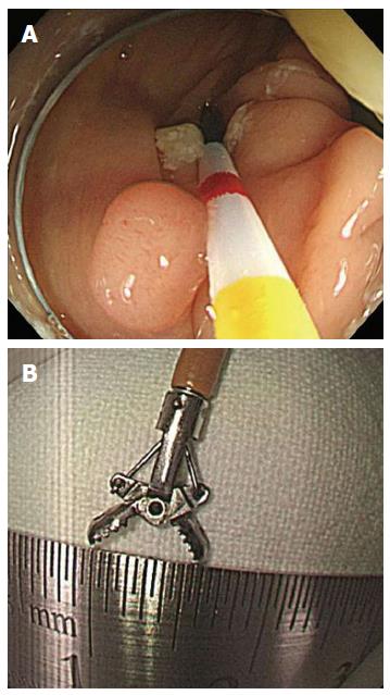
This study was approved by the Institutional Review Board of Kosin University Gospel Hospital. And all study participants, or their legal guardian, provided informed written consent prior to study enrollment. The colonic polyps detected by colonoscopy were recorded on video using Ancamcorder 2.5 (free recording software, Antools Inc., Korea). We recorded the scenes of polyp size measurement by visual estimation, using open biopsy forceps and using a graduated catheter. The polyp size measured by a graduated catheter (ERCP-catheter, 0130200, MTW, Germany) was considered to be the actual size of the polyp in vivo (Figure 1A). The length of the fully open biopsy forceps (Radial Jaw 4 Biopsy Forceps, Boston Scientific, United States) was 6 mm (Figure 1B). We recorded a total of 120 video clips, each of which consisted of three parts. The first part of all of the video clips included 40 scenes visualizing the polyp. These were captured to simulate a realistic diagnostic colonoscopy. The second part of the video clips included 40 scenes using open biopsy forceps in association with the polyp (forceps estimation). To avoid any optical illusions, the biopsy forceps were opened and then withdrawn in the open position toward the endoscope tip. Then, the endoscope tip was advanced to the polyp. The last part of the video clips showed the polyp being measured with the graduated catheter, after being optimally placed and aligned with the major axis of the polyp (Figure 1A). A study investigator measured the actual size of polyps and edited all of the video clips so that each section was between 7 and 15 s in length. The first and second parts of each video clip were used for the exam, and the third part of each video clip was used to determine the actual size of the polyp in vivo in order to calculate the diagnostic accuracy of the observers.
Forty endoscopists of the Busan Ulsan Gyeongnam Intestinal Study Group Society (BIGS) participated in this study. Sixteen were experienced endoscopists (experts) who had performed more than 1000 colonoscopies. The remaining 24 were endoscopic training fellows (beginners) who had performed fewer than 300 colonoscopies. On the day of the exam, participants watched 40 pairs of video clips. Each pair consisted of a first section (visual estimation section) showing only the polyp and the second section (forceps estimation section) showing the polyp with the open forceps. Upon viewing each video clip, subjects were instructed to write down the estimated polyp size on the exam paper. To avoid the possibility of the second clip influencing the initial visual estimation from the first clip, participants were informed before the exam that written estimates could not be corrected. When analyzing the results of the exam, we assessed inter-observer differences, diagnostic accuracy, and error ranges in the measurement of the polyp size.
Inter-observer differences (agreement) in the test were evaluated by calculating the intra-class correlation coefficient (ICC). Diagnostic accuracy was assessed using the actual size of the polyps as measured by the graduated catheter. If the subject made an estimate within 1 mm of the actual polyp size, the answer was considered to be correct. Error range was estimated by calculating the difference between the written estimate and the actual size of the polyp. An ICC below 0.59 was defined as poor agreement, an ICC of 0.60-0.79 was defined as moderate agreement, and an ICC greater than 0.80 was considered to be an excellent agreement[30]. The significance of difference between two groups (visual estimation vs forceps estimation) was analyzed by Wilcoxon-signed rank test. Statistical analyses were performed using SPSS version 20.0 (IBM, Armonk, New York, United States).
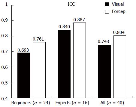
The characteristics of the colon polyps visualized in the exam are summarized in Table 1. The overall ICCs of the inter-observer agreement for the forceps estimation and the visual estimation were 0.804 (95%CI: 0.731-0.873, P < 0.001) and 0.743 (95%CI: 0.656-0.828, P < 0.001), respectively (Figure 2). The ICCs of each group for forceps estimation were higher than those for visual estimation. The ICCs of the beginner group for the forceps estimation and the visual estimation were 0.761 (95%CI: 0.676-0.842, P < 0.001) and 0.693 (95%CI: 0.596-0.792, P < 0.001), respectively. The ICCs of the expert group for the forceps estimation and the visual estimation were 0.887 (95%CI: 0.837-0.929, P < 0.001) and 0.840 (95%CI: 0.775-0.898, P < 0.001), respectively.
| n (%) | ||
| Size | < 5 mm | 19 (47.5) |
| 5 ≥ and < 10 mm | 12 (30.0) | |
| ≥ 10 mm | 9 (22.5) | |
| Type | Sessile | 23 (57.5) |
| Semi pedunculated | 8 (20.0) | |
| Pedunculated | 5 (12.5) | |
| LST | 4 (10.0) | |
| Location | Rectum | 3 (7.5) |
| Sigmoid colon | 8 (20.0) | |
| Descending colon | 7 (17.5) | |
| Transverse colon | 9 (22.5) | |
| Ascending colon | 11 (27.5) | |
| Cecum | 2 (5.0) | |
| Histology | TA with LGD | 27 (67.5) |
| TA with HGD | 3 (7.5) | |
| Chronic colitis | 6 (15.0) | |
| Serrated polyp | 3 (7.5) | |
| Inflammatory polyp | 1 (2.5) |
The overall diagnostic accuracy for the forceps estimation and the visual estimation were 0.754 and 0.639, respectively (P < 0.001) (Table 2). In the beginner group and the expert group, the diagnostic accuracy for the forceps estimation was significantly higher than that of the visual estimation (Beginner group, 0.734 vs 0.613, P < 0.001; Expert group, 0.784 vs 0.680, P < 0.001, respectively). The diagnostic accuracy for polyps ≤ 5 mm in size with the forceps estimation was significantly higher than that with visual estimation (P < 0.001). There were also significant differences in diagnostic accuracy for polyps ≥ 6 mm, ≤ 9 mm and ≥ 10 mm in size (P < 0.001 and P = 0.018, respectively) (Table 3).
| Visual estimation | Forceps estimation | P value | |
| Diagnostic accuracy | |||
| Beginners | 0.613 | 0.734 | < 0.001 |
| Experts | 0.680 | 0.784 | < 0.001 |
| Total | 0.639 | 0.754 | < 0.001 |
| Error range (mm) | |||
| Beginners | 1.68 ± 1.30 | 1.38 ± 1.08 | < 0.001 |
| Experts | 1.42 ± 1.11 | 1.12 ± 1.11 | < 0.001 |
| Total | 1.48 ± 1.18 | 1.20 ± 1.10 | < 0.001 |
| Visual estimation | Forceps estimation | P value | |
| Diagnostic accuracy | |||
| ≤ 5 mm | 0.834 | 0.929 | < 0.001 |
| 6-9 mm | 0.508 | 0.692 | < 0.001 |
| ≥ 10 mm | 0.403 | 0.469 | 0.018 |
| Error range (mm) | |||
| ≤ 5 mm | 0.90 ± 0.27 | 0.70 ± 0.17 | < 0.001 |
| 6-9 mm | 1.85 ± 0.82 | 1.30 ± 0.59 | < 0.001 |
| ≥ 10 mm | 2.71 ± 0.76 | 2.40 ± 0.67 | < 0.001 |
The overall error ranges for forceps estimation and visual estimation were 1.20 ± 1.10 and 1.48 ± 1.18, respectively (P < 0.001) (Table 2). The error ranges of each group for forceps estimation were significantly smaller than those for visual estimation (Beginner group, 1.38 ± 1.08 vs 1.68 ± 1.30, P < 0.001; Expert group, 1.12 ± 1.11 vs 1.42 ± 1.11, P < 0.001, respectively). The error range for polyps ≤ 5 mm in size with the forceps estimation was significantly smaller than that with visual estimation (P < 0.001). There were also significant differences in the error range for polyps ≥ 6 mm, ≤ 9 mm and ≥ 10 mm in size (P < 0.001 and P < 0.001, respectively) (Table 3).
We also evaluated the diagnostic accuracy according to the colon polyp type. With the sessile and lateral spreading tumor (LST) types, the diagnostic accuracy of forceps estimation was higher than that of visual estimation (P < 0.001 and P = 0.012, respectively). With semi-pedunculated and pedunculated types, there was no significant difference in the diagnostic accuracy between the forceps estimation and the visual estimation (P = 0.344 and P = 0.432, respectively) (Table 4).
| Visual estimation | Forceps estimation | P value | |
| Sessile type | 0.709 | 0.818 | < 0.001 |
| Semi pedunculated type | 0.627 | 0.703 | 0.344 |
| Pedunculated type | 0.608 | 0.638 | 0.432 |
| LST type | 0.556 | 0.757 | 0.012 |
In this study, a total of 40 endoscopists participated in the exam. When analyzing the results of the exam, we assessed inter-observer differences, diagnostic accuracy, and error ranges in the measurement of colon polyp size using open biopsy forceps vs visualization. Because a video clip provides a more realistic simulation of an in vivo colonoscopic experience compared to a photograph, we used video clips recorded from real polyp size measurements in the exam.
In this study, the overall ICC value for the forceps estimation was greater than 0.80, which indicates an excellent inter-observer agreement. However, the overall ICC value for the visual estimation was less than 0.80, which represents a moderate inter-observer agreement (Figure 2). ICC values for the forceps estimation and the visual estimation in the expert group were greater than 0.80. However, in the beginner group, ICC values for the forceps estimation and the visual estimation were less than 0.80. Our results suggest that measuring polyp size using open biopsy forceps is more consistent than by visual estimation alone, and the cumulative experience of the colonoscopic examinations affects the consistency measuring the polyp size.
In our study, the overall diagnostic accuracy of forceps estimation was better than that of visual estimation (0.754 vs 0.639, P < 0.001, respectively). The overall error range of forceps estimation was also smaller than that of visual estimation (1.20 ± 1.10 vs 1.48 ± 1.18, P < 0.001, respectively) (Table 2). These results indicate that the method using open biopsy forceps helps increase the accuracy and decrease the error range of polyp size measurement. Chang et al[26] reported that the overall diagnostic accuracy of visual estimation was 0.59 before education and 0.81 after education (using 30 educational video clips of visual estimation of polyp size). In comparison with their study, the value of the overall diagnostic accuracy of the forceps estimation in our study was lower than that of visual estimation after education in their study (0.754 vs 0.81). However, this difference may not be significant because the number of participants in our study was much greater than that in their study (forty vs twelve). Education in estimating the colon polyp size by visualization may be a good method, but according to the results of our study, it is favorable to use the open biopsy forceps to measure the size of the polyp.
Our study also showed that the diagnostic accuracy of the forceps estimation according to the polyp size was significantly greater than that of the visual estimation, and the error range of the forceps estimation according to the polyp size was significantly smaller than that of the visual estimation (Table 3). These results indicate that the method using open biopsy forceps can increase the accuracy of polyp size measurement and decrease the error range of polyp size measurement, regardless of polyp size.
In this study, there was no significant difference in the diagnostic accuracy between the visual estimation and the forceps estimation in semi-pedunculated and pedunculated polyp types (Table 4). However, in sessile and LST types, the diagnostic accuracy of the forceps estimation was greater than that of the visual estimation. Semi-pedunculated and pedunculated polyp types are relatively difficult to measure using open biopsy forceps, because these polyps tend to dangle in vivo when attempting to measure. The sessile and LST polyp types are mostly fixed in vivo; therefore, it is relatively easy to measure the size of these polyps using the open biopsy forceps.
This study has some limitations. First, the estimation of diagnostic accuracy was based on the size of colon polyps measured by graduated catheters, but this method is not completely accurate in estimating the actual size of colon polyps. In several studies on colon polyp size, investigators considered the ruler measurement of a removed polyp to be the true size[27,31,32]. When measuring colon polyps, it is important to consider that polypectomy using electrocauterization may lead to shrinkage of the polyp, and formalin fixation can also cause shrinkage of the polyp. The graduated catheter cannot always be aligned with the true diameter of the polyp, therefore it may also cause an inaccurate estimation. But, we tried to minimize this error by measuring the polyp several times at various alignments. Additionally, because we considered the answer to be correct if the error was less than 1 mm, the results of our study are believed to be quite reliable. Second, the distance between the colonoscope tip and the polyp in the video clips included 40 scenes visualizing the polyp without open biopsy forceps was variable, and it could cause the confusion of participants to estimate the size of polyp. However, we tried to record the scenes showing various distance from the colonoscope tip to the polyp in each video clip.
In conclusion, the application of the open biopsy forceps method could help reduce inter-observer differences and error rates when measuring colon polyp size. Although using open biopsy forceps can potentially result in an erroneous polyp size measurement, as shown in our results, the inter-observer agreement and the diagnostic accuracy of forceps estimation were significantly higher than those of visual estimation. In case of the polyp which is needed to be removed by endoscopic mucosal resection (EMR), the measurement of the polyp size by forceps estimation before removing the polyp by EMR is maybe tedious and more time consuming than visual estimation alone. However, because this additional step helps to increase the diagnostic accuracy and inter-observer agreement when measuring the polyp size, we propose that forceps estimation should be considered to measure the colon polyp size before removing the polyp.
Measuring the size of colon polyps is important during colonoscopy exams, because size is one of the factors which predict the potential risks of malignant evolution of colon polyps. Using open biopsy forceps is known to be a useful technique to reduce error rates in colon polyp size measurements, but in practice most endoscopists just measure polyp size by visualization. There is little information about accuracy differences between these two methods. In this study, the authors assessed the usefulness of the forceps estimation in the measurement of colon polyp size.
This study presents that the application of the open biopsy forceps method when measuring colon polyp size could help reduce inter-observer differences and error rates.
In this study, a total of 40 endoscopists participated in the exam. When analyzing the results of the exam, the authors assessed inter-observer differences, diagnostic accuracy, and error ranges in the measurement of colon polyp size using open biopsy forceps vs visualization.
The open biopsy forceps method can be applied to measure colon polyp size for reducing the inter-observer differences and error rates.
Forceps estimation is the measurement of colon polyp size using open biopsy forceps. Visual estimation is the measurement of colon polyp size by visualization.
The authors of this paper assessed the usefulness of forceps to measure the size of colon polyp, and showed that the application of the open biopsy forceps method could help reduce inter-observer differences and error rates when measuring colon polyp size. These results suggest that forceps estimation is more useful than visual estimation in the measurement of the colon polyp size.
P- Reviewer: Corte C S- Editor: Yu J L- Editor: A E- Editor: Zhang DN
| 1. | GLOBOCAN 2008. Accesed Jul 12, 2013. Available from: http://www.globocan.iarc.fr. |
| 2. | Jung KW, Won YJ, Kong HJ, Oh CM, Seo HG, Lee JS. Cancer statistics in Korea: incidence, mortality, survival and prevalence in 2010. Cancer Res Treat. 2013;45:1-14. [RCA] [PubMed] [DOI] [Full Text] [Full Text (PDF)] [Cited by in Crossref: 365] [Cited by in RCA: 363] [Article Influence: 30.3] [Reference Citation Analysis (0)] |
| 3. | Cottet V, Jooste V, Fournel I, Bouvier AM, Faivre J, Bonithon-Kopp C. Long-term risk of colorectal cancer after adenoma removal: a population-based cohort study. Gut. 2012;61:1180-1186. [RCA] [PubMed] [DOI] [Full Text] [Cited by in Crossref: 162] [Cited by in RCA: 184] [Article Influence: 14.2] [Reference Citation Analysis (0)] |
| 4. | Loeve F, van Ballegooijen M, Boer R, Kuipers EJ, Habbema JD. Colorectal cancer risk in adenoma patients: a nation-wide study. Int J Cancer. 2004;111:147-151. [RCA] [PubMed] [DOI] [Full Text] [Cited by in Crossref: 69] [Cited by in RCA: 70] [Article Influence: 3.3] [Reference Citation Analysis (0)] |
| 5. | Muto T, Bussey HJ, Morson BC. The evolution of cancer of the colon and rectum. Cancer. 1975;36:2251-2270. [PubMed] |
| 6. | Robertson DJ, Greenberg ER, Beach M, Sandler RS, Ahnen D, Haile RW, Burke CA, Snover DC, Bresalier RS, McKeown-Eyssen G. Colorectal cancer in patients under close colonoscopic surveillance. Gastroenterology. 2005;129:34-41. [PubMed] |
| 7. | Yamaji Y, Mitsushima T, Ikuma H, Watabe H, Okamoto M, Kawabe T, Wada R, Doi H, Omata M. Incidence and recurrence rates of colorectal adenomas estimated by annually repeated colonoscopies on asymptomatic Japanese. Gut. 2004;53:568-572. [PubMed] |
| 8. | Citarda F, Tomaselli G, Capocaccia R, Barcherini S, Crespi M. Efficacy in standard clinical practice of colonoscopic polypectomy in reducing colorectal cancer incidence. Gut. 2001;48:812-815. [PubMed] |
| 9. | Winawer SJ, Zauber AG, Ho MN, O’Brien MJ, Gottlieb LS, Sternberg SS, Waye JD, Schapiro M, Bond JH, Panish JF. Prevention of colorectal cancer by colonoscopic polypectomy. The National Polyp Study Workgroup. N Engl J Med. 1993;329:1977-1981. [RCA] [PubMed] [DOI] [Full Text] [Cited by in Crossref: 3107] [Cited by in RCA: 3126] [Article Influence: 97.7] [Reference Citation Analysis (1)] |
| 10. | Shinya H, Wolff WI. Morphology, anatomic distribution and cancer potential of colonic polyps. Ann Surg. 1979;190:679-683. [PubMed] |
| 11. | Fung CH, Goldman H. The incidence and significance of villous change in adenomatous polyps. Am J Clin Pathol. 1970;53:21-25. [PubMed] |
| 12. | Lotfi AM, Spencer RJ, Ilstrup DM, Melton LJ. Colorectal polyps and the risk of subsequent carcinoma. Mayo Clin Proc. 1986;61:337-343. [PubMed] |
| 13. | Panish JF. Management of patients with polypoid lesions of the colon--current concepts and controversies. Am J Gastroenterol. 1979;71:315-324. [PubMed] |
| 14. | Atkin WS, Morson BC, Cuzick J. Long-term risk of colorectal cancer after excision of rectosigmoid adenomas. N Engl J Med. 1992;326:658-662. [RCA] [PubMed] [DOI] [Full Text] [Cited by in Crossref: 761] [Cited by in RCA: 689] [Article Influence: 20.9] [Reference Citation Analysis (0)] |
| 15. | Lieberman D, Moravec M, Holub J, Michaels L, Eisen G. Polyp size and advanced histology in patients undergoing colonoscopy screening: implications for CT colonography. Gastroenterology. 2008;135:1100-1105. [RCA] [PubMed] [DOI] [Full Text] [Full Text (PDF)] [Cited by in Crossref: 355] [Cited by in RCA: 329] [Article Influence: 19.4] [Reference Citation Analysis (1)] |
| 16. | Pickhardt PJ. The natural history of colorectal polyps and masses: rediscovered truths from the barium enema era. AJR Am J Roentgenol. 2007;188:619-621. [RCA] [PubMed] [DOI] [Full Text] [Cited by in Crossref: 15] [Cited by in RCA: 19] [Article Influence: 1.1] [Reference Citation Analysis (0)] |
| 17. | Carter D, Beer-Gabel M, Zbar A, Avidan B, Bardan E. A survey of colonoscopic polypectomy practice amongst Israeli gastroenterologists. Ann Gastroenterol. 2013;26:135-140. [PubMed] |
| 18. | Singh N, Harrison M, Rex DK. A survey of colonoscopic polypectomy practices among clinical gastroenterologists. Gastrointest Endosc. 2004;60:414-418. [PubMed] |
| 19. | Hewett DG. Colonoscopic polypectomy: current techniques and controversies. Gastroenterol Clin North Am. 2013;42:443-458. [RCA] [PubMed] [DOI] [Full Text] [Cited by in Crossref: 28] [Cited by in RCA: 30] [Article Influence: 2.5] [Reference Citation Analysis (0)] |
| 20. | Uraoka T, Ramberan H, Matsuda T, Fujii T, Yahagi N. Cold polypectomy techniques for diminutive polyps in the colorectum. Dig Endosc. 2014;26 Suppl 2:98-103. [RCA] [PubMed] [DOI] [Full Text] [Cited by in Crossref: 43] [Cited by in RCA: 55] [Article Influence: 5.0] [Reference Citation Analysis (1)] |
| 21. | Jung YS, Park JH, Kim HJ, Cho YK, Sohn CI, Jeon WK, Kim BI, Sohn JH, Park DI. Complete biopsy resection of diminutive polyps. Endoscopy. 2013;45:1024-1029. [RCA] [PubMed] [DOI] [Full Text] [Cited by in Crossref: 44] [Cited by in RCA: 45] [Article Influence: 3.8] [Reference Citation Analysis (0)] |
| 22. | Kim JS, Lee BI, Choi H, Jun SY, Park ES, Park JM, Lee IS, Kim BW, Kim SW, Choi MG. Cold snare polypectomy versus cold forceps polypectomy for diminutive and small colorectal polyps: a randomized controlled trial. Gastrointest Endosc. 2015;81:741-747. [RCA] [PubMed] [DOI] [Full Text] [Cited by in Crossref: 136] [Cited by in RCA: 131] [Article Influence: 13.1] [Reference Citation Analysis (0)] |
| 23. | Lee CK, Shim JJ, Jang JY. Cold snare polypectomy vs. Cold forceps polypectomy using double-biopsy technique for removal of diminutive colorectal polyps: a prospective randomized study. Am J Gastroenterol. 2013;108:1593-1600. [RCA] [PubMed] [DOI] [Full Text] [Cited by in Crossref: 142] [Cited by in RCA: 168] [Article Influence: 14.0] [Reference Citation Analysis (0)] |
| 24. | Chen SC, Rex DK. Endoscopist can be more powerful than age and male gender in predicting adenoma detection at colonoscopy. Am J Gastroenterol. 2007;102:856-861. [RCA] [PubMed] [DOI] [Full Text] [Cited by in Crossref: 284] [Cited by in RCA: 306] [Article Influence: 17.0] [Reference Citation Analysis (0)] |
| 25. | Marshall JB. Polyps in the colon. Answers to key questions. Postgrad Med. 1992;92:53-54, 57-60, 65. [PubMed] |
| 26. | Chang CY, Chiu HM, Wang HP, Lee CT, Tai JJ, Tu CH, Tai CM, Chiang TH, Huang JK, Chang DC. An endoscopic training model to improve accuracy of colonic polyp size measurement. Int J Colorectal Dis. 2010;25:655-660. [RCA] [PubMed] [DOI] [Full Text] [Cited by in Crossref: 13] [Cited by in RCA: 15] [Article Influence: 1.0] [Reference Citation Analysis (0)] |
| 27. | Gopalswamy N, Shenoy VN, Choudhry U, Markert RJ, Peace N, Bhutani MS, Barde CJ. Is in vivo measurement of size of polyps during colonoscopy accurate? Gastrointest Endosc. 1997;46:497-502. [PubMed] |
| 28. | Hyun YS, Han DS, Bae JH, Park HS, Eun CS. Graduated injection needles and snares for polypectomy are useful for measuring colorectal polyp size. Dig Liver Dis. 2011;43:391-394. [PubMed] |
| 29. | Morales TG, Sampliner RE, Garewal HS, Fennerty MB, Aickin M. The difference in colon polyp size before and after removal. Gastrointest Endosc. 1996;43:25-28. [PubMed] |
| 30. | Altman DG. Practical statistics for medical research. London: Chapman & Hall 1991; . |
| 31. | Riner MA, Rankin RA, Guild RT, Kastens DJ. Accuracy of estimation of colon polyp size. Gastrointest Endosc. 1988;34:284. [PubMed] |
| 32. | Schoen RE, Gerber LD, Margulies C. The pathologic measurement of polyp size is preferable to the endoscopic estimate. Gastrointest Endosc. 1997;46:492-496. [PubMed] |


