Published online Sep 21, 2014. doi: 10.3748/wjg.v20.i35.12673
Revised: April 28, 2014
Accepted: June 12, 2014
Published online: September 21, 2014
Processing time: 170 Days and 5.9 Hours
Basaloid squamous carcinoma (BSC) is a rare variant of esophageal cancer. There are very few reports of “early” BSC. Here we report a case of early BSC with unusual findings by narrowband imaging magnified endoscopy (NBI-ME). A 70-year-old man with a middle thoracic esophageal tumor was referred to our hospital. White-light endoscopy revealed a reddish depressed lesion 5 mm in diameter having a subepithelial tumor-like prominence with a gentle rising slope. NBI-ME revealed irregular loop-shaped microvessels coexistent with thick irregularly branched non-looped vessels. Iodine staining revealed a pale brown lesion. We performed endoscopic submucosal dissection for diagnostic treatment. Histologic examination showed the proliferation of basal cell-like hyperchromatic tumor cells in the lamina propria and with slight invasion into the submucosa at a depth of 320 μm. The tumor cells formed solid nests and microcystic structures, containing an Alcian blue-positive mucoid matrix. The surface was covered with squamous epithelium without cellular atypia. Thin vessels were observed in the intra-epithelial papilla and thick vessels were observed around the solid nests beneath the epithelium. Based on these findings together, we diagnosed the lesion as BSC. In this case, the NBI-ME findings differed from those of typical squamous cell carcinoma in that both non-invasive cancer-like irregular loop-shaped microvessels coexisted with massively invasive cancer-like thick non-looped vessels. We speculate that the looped and non-looped vessels observed by NBI-ME histologically corresponded to thin vessels in the intra-epithelial papilla and thick vessels around the tumor nests, respectively. These NBI-ME findings might be a feature of early esophageal BSC.
Core tip: Basaloid squamous carcinoma (BSC) is a rare variant of esophageal cancer. In our case, narrowband imaging magnified endoscopy (NBI-ME) findings differed from those observed in typical squamous cell carcinoma in that non-invasive cancer-like irregular loop-shaped microvessels coexisted with massively invasive cancer-like thick non-looped vessels. We speculate that the looped and non-looped vessels observed by NBI-ME histologically corresponded to thin vessels in the intra-epithelial papilla and thick vessels around the tumor nests, respectively. These NBI-ME findings might be a feature of early BSC. To our knowledge, this is the first report in the English literature describing the NBI-ME findings of BSC.
- Citation: Kai Y, Kato M, Hayashi Y, Akasaka T, Shinzaki S, Nishida T, Tsujii M, Morii E, Takehara T. Esophageal early basaloid squamous carcinoma with unusual narrowband imaging magnified endoscopy findings. World J Gastroenterol 2014; 20(35): 12673-12677
- URL: https://www.wjgnet.com/1007-9327/full/v20/i35/12673.htm
- DOI: https://dx.doi.org/10.3748/wjg.v20.i35.12673
Basaloid squamous carcinoma (BSC) is a rare variant of esophageal cancer, with a frequency ranging from 0.4% to 3.6% of total esophageal malignant tumors[1]. The prognosis of BSC is poorer than that of typical squamous cell carcinoma (SCC) in advanced stages, but in early stages, the prognosis of BSC does not differ significantly from that of typical SCC[1]. Early diagnosis of BSC is therefore important to improve the prognosis.
Narrowband imaging (NBI) and NBI with magnified endoscopy (NBI-ME) are novel endoscopic imaging technologies useful for SCC diagnosis[2]. Few reports, however, provide a detailed description of the NBI-ME findings of BSC. Here, we report a rare case of early BSC with unusual NBI-ME findings. To our knowledge, this is the first report in English literature describing the NBI-ME findings of BSC.
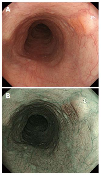
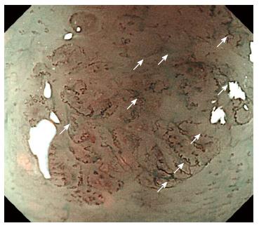
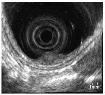
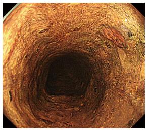
A 70-year-old man was referred to our hospital for evaluation of an esophageal tumor in the middle thoracic esophagus, 28 cm from the incisors. White-light conventional endoscopy revealed a reddish depressed lesion 5 mm in diameter having a subepithelial tumor-like prominence with a gentle rising slope (Figure 1A). The depressed area was observed as a brownish area by NBI (Figure 1B). NBI-ME revealed irregular loop-shaped microvessels coexisting with irregularly branched thick non-looped vessels (Figure 2). Endoscopic ultrasonography (20 MHz, Miniature Probe) revealed no irregularity or thinning of the third of five layers, which corresponded to the submucosal layer (Figure 3). Iodine staining revealed a pale brown lesion (Figure 4). The tumor was diagnosed as SCC from a biopsy specimen, but because of the subepithelial tumor-like prominence and atypical NBI-ME findings, we also considered poorly differentiated SCC or histologic special type of esophageal cancer, including BSC, adenosquamous carcinoma, small cell carcinoma, and adenoid cystic carcinoma in the differential diagnosis. Endoscopic submucosal dissection was performed as a diagnostic treatment.
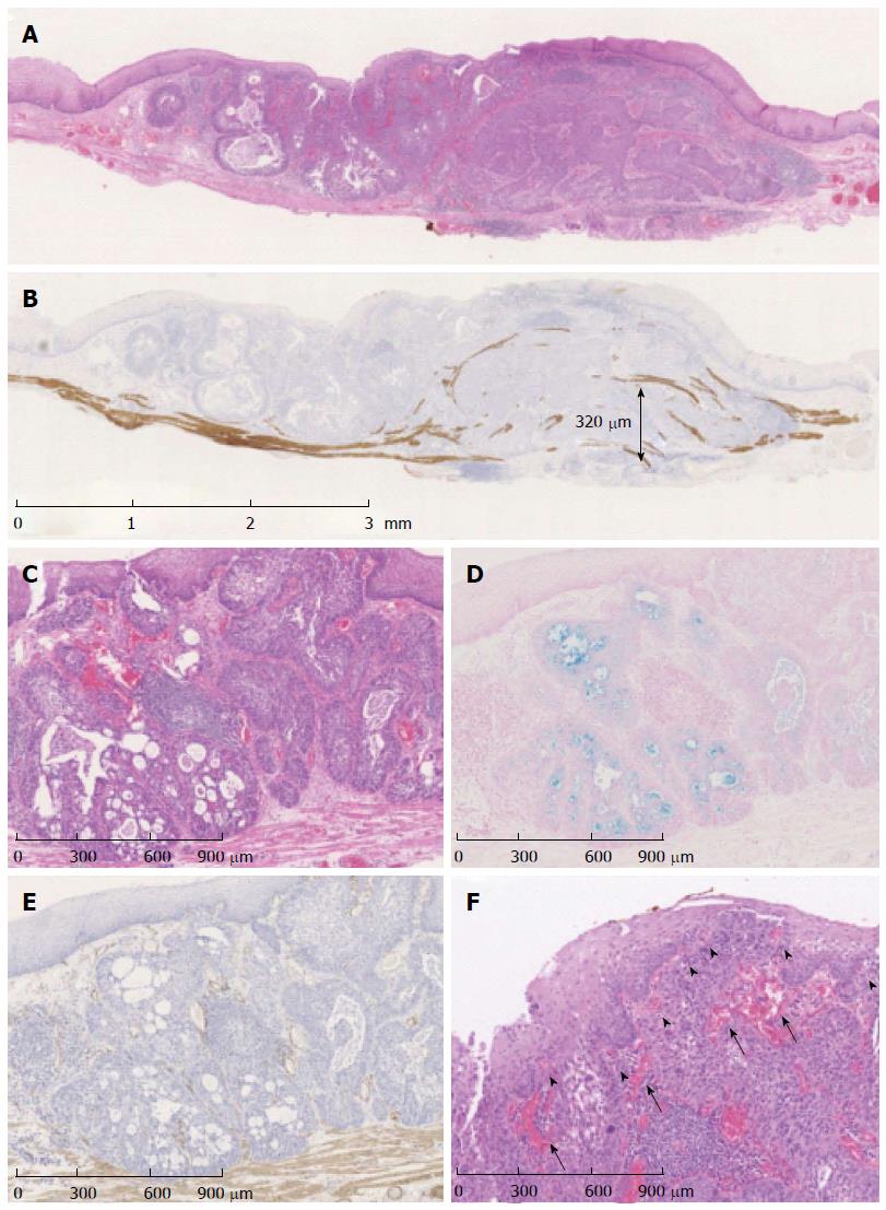
Histologic examination revealed the proliferation of basal cell-like hyperchromatic tumor cells in the lamina propria (Figure 5A). Immunostaining using desmin suggested that the tumor invaded the submucosa at a depth of 320 μm (Figure 5B). The tumor cells formed solid nests and microcystic structures (Figure 5C) containing an Alcian blue-positive mucoid matrix (Figure 5D). Immunostaining for α-smooth muscle actin (SMA) was negative (Figure 5E). The surface was covered with squamous epithelium without cellular atypia. Thin vessels were observed in intra-epithelial papilla and thick vessels were observed around the solid nests beneath the epithelium (Figure 5F). Based on these findings together, we diagnosed the lesion as BSC, T1bN0M0, stage IA, based on the Union for International Cancer Control classification.
Esophageal BSC is a rare but distinct variant of esophageal cancer, and was first reported by Wain et al[3] in 1986. BSC is histopathologically characterized by the proliferation of basaloid cancer cells, forming nests of various sizes and α-SMA negative microcystic structures containing an Alcian blue-positive mucoid matrix in the mucosal epithelium, as well as the deposition of eosinophilic substances in the stroma[4]. The prognosis for BSC is poorer than that for typical SCC in advanced stages, but in early stages the prognosis does not differ significantly from that of typical SCC[1]. Therefore, early detection and diagnosis is important for improving the prognosis of BSC. Common macroscopic characteristics of BSC are protruding lesions or submucosal tumor-like structures[5]. Other special types of esophageal cancer, however, including adenosquamous carcinoma, small cell carcinoma, and adenoid cystic carcinoma, have the same macroscopic features of BSC. Moreover, these tumors are often covered with a non-neoplastic surface, and it is difficult to diagnose accurately by biopsy. In fact, the diagnostic accuracy of BSC using biopsy specimens is only 4.7%-10%[6]. Therefore, conventional endoscopy and biopsy are insufficient for a precise differential diagnosis of BSC.
NBI has recently become widely used in clinical practice. NBI detects superficial SCC more frequently than white-light imaging, and has significantly higher sensitivity and accuracy compared with white-light[2]. In addition, NBI-ME is useful for differentiating cancerous from non-cancerous lesions and assessing tumor depth and invasion by analysis of the microvascular patterns[7,8]. Generally, the presence of irregular loop-shaped microvessels suggests noninvasive cancer in the mucosal layer, and thick non-looped vessels suggest invasive cancer in typical SCC[9]. In contrast to the usual type of SCC, loop-shaped vessels coexisted with thick non-looped vessels in the present case.
NBI-ME findings of BSC are rare and thus not well described. To our knowledge, there is no report in the English literature of the NBI-ME findings of BSC, and we found only a case report written in Japanese by Takeuchi et al[10] in which NBI-ME revealed irregularly branched and slightly dilated non-looped vessels. In the present case, NBI-ME revealed irregular loop-shaped microvessels that coexisted with irregularly branched thick non-looped vessels. These thick non-looped vessels were similar to those reported by Takeuchi et al[10] but no irregular loop-shaped microvessels were described by Takeuchi. Histologically, thick vessels were commonly observed around the solid tumor nests in the lamina propria in both cases, but only in our case were thin vessels observed in the intra-epithelial papilla. This finding suggests that the non-looped dilated vessels observed by NBI-ME histologically correspond to the thick vessels around the tumor nests, and the loop-shaped microvessels observed by NBI-ME histologically correspond to thin vessels in the intra-epithelial papilla. In our case, the tumor cells invaded only a small area of the submucosa, likely due to the early stage. The coexistence of both loop-shaped and non-looped vessels in NBI-ME might be a feature of early-stage BSC in which the structure of intra-epithelial papilla is maintained. This report describes only one case, however, and these findings may not represent only the specific characteristics of early BSC but also those of carcinoma growing beneath the epithelium. To clarify the common features of BSC in NBI-ME, an accumulation of studies involving more patients is needed.
In conclusion, we describe a rare case of early BSC with unusual findings by NBI-ME. The irregular loop-shaped microvessels coexisting with irregularly branched thick non-looped vessels might be characteristics of early BSC.
A 70-year-old man with an esophageal tumor 5 mm in diameter having a subepithelial tumor-like prominence in the middle thoracic esophagus was referred to our hospital.
The tumor was diagnosed as squamous cell carcinoma (SCC) from the biopsy specimen, but based on the subepithelial tumor-like prominence and atypical narrowband imaging magnified endoscopy (NBI-ME) findings, we also considered poorly differentiated SCC or a histologically special type of esophageal cancer, including basaloid squamous carcinoma.
Other special histologic types of esophageal cancer, such as adenosquamous carcinoma, small cell carcinoma, and adenoid cystic carcinoma.
Endoscopic submucosal dissection was performed as a diagnostic treatment.
Histologic examination showed the proliferation of basal cell-like hyperchromatic tumor cells mainly in the lamina propria, forming solid nests and microcystic structures, positive immunostaining with Alcian blue and negative -SMA immunostaining. Based on these findings together, we diagnosed the lesion as BSC.
Irregular loop-shaped microvessels coexisting with irregularly branched non-loop thick vessels observed by NBI-ME might be characteristics of early BSC.
The authors describe their NBI-ME findings in an early case of basaloid squamous carcinoma of the esophagus and postulate that with NBI-ME they could probably differentiate the early stage of the disease. This is an interesting and well-documented manuscript.
P- Reviewer: de Tejada AH, Tsolakis A S- Editor: Qi Y L- Editor: A E- Editor: Du P
| 1. | Arai T, Aida J, Nakamura K-i, Ushio Y, Takubo K. Clinicopathologic characteristics of basaloid squamous carcinoma of the esophagus. Esophagus. 2011;8:169-177. [RCA] [DOI] [Full Text] [Cited by in Crossref: 10] [Cited by in RCA: 5] [Article Influence: 0.4] [Reference Citation Analysis (0)] |
| 2. | Muto M, Minashi K, Yano T, Saito Y, Oda I, Nonaka S, Omori T, Sugiura H, Goda K, Kaise M. Early detection of superficial squamous cell carcinoma in the head and neck region and esophagus by narrow band imaging: a multicenter randomized controlled trial. J Clin Oncol. 2010;28:1566-1572. [RCA] [PubMed] [DOI] [Full Text] [Cited by in Crossref: 427] [Cited by in RCA: 523] [Article Influence: 34.9] [Reference Citation Analysis (0)] |
| 3. | Wain SL, Kier R, Vollmer RT, Bossen EH. Basaloid-squamous carcinoma of the tongue, hypopharynx, and larynx: report of 10 cases. Hum Pathol. 1986;17:1158-1166. [PubMed] |
| 4. | Japan Esophageal Society. Japanese Classification of Esophageal Cancer, 10th edition: parts II and III. Esophagus. 2009;6:71-94. [RCA] [DOI] [Full Text] [Cited by in Crossref: 96] [Cited by in RCA: 88] [Article Influence: 5.5] [Reference Citation Analysis (0)] |
| 5. | Ohashi K, Horiguchi S, Moriyama S, Hishima T, Hayashi Y, Momma K, Hanashi T, Izumi Y, Yoshida M, Funata N. Superficial basaloid squamous carcinoma of the esophagus. A clinicopathological and immunohistochemical study of 12 cases. Pathol Res Pract. 2003;199:713-721. [RCA] [PubMed] [DOI] [Full Text] [Cited by in Crossref: 2] [Cited by in RCA: 5] [Article Influence: 0.2] [Reference Citation Analysis (0)] |
| 6. | Saito S, Hosoya Y, Zuiki T, Hyodo M, Lefor A, Sata N, Nagase M, Nakazawa M, Matsubara D, Niki T. A clinicopathological study of basaloid squamous carcinoma of the esophagus. Esophagus. 2009;6:177-181. [DOI] [Full Text] |
| 7. | Kumagai Y, Inoue H, Nagai K, Kawano T, Iwai T. Magnifying endoscopy, stereoscopic microscopy, and the microvascular architecture of superficial esophageal carcinoma. Endoscopy. 2002;34:369-375. [RCA] [PubMed] [DOI] [Full Text] [Cited by in Crossref: 165] [Cited by in RCA: 164] [Article Influence: 7.1] [Reference Citation Analysis (0)] |
| 8. | Arima M, Tada M, Arima H. Evaluation of microvascular patterns of superficial esophageal cancers by magnifying endoscopy. Esophagus. 2005;2:191-197. [DOI] [Full Text] |
| 9. | Oyama T, Ishihara R, Takeuchi M, Hirasawa D, Arima M, Inoue H, Goda K, Tomori A, K . M. Usefulness of Japan Esophageal Society classification of magnified endoscopy for the diagnosis of superficial esophageal squamous cell carcinoma. Gastrointest Endosc. 2012;75:Tu1588. |
| 10. | Takeuchi M, Watanabe G, Kobayashi M, Hashimoto S, Mizuno K, Sato Y, Ajioka Y, Aoyagi Y. Primary esophageal basaloid squamous carcinoma, report of a case. Stomach Int. 2013;48:355-361. |


