Copyright
©2014 Baishideng Publishing Group Inc.
World J Gastroenterol. Aug 28, 2014; 20(32): 11160-11181
Published online Aug 28, 2014. doi: 10.3748/wjg.v20.i32.11160
Published online Aug 28, 2014. doi: 10.3748/wjg.v20.i32.11160
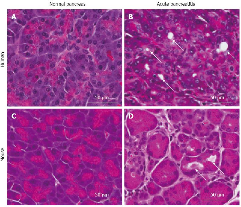
Figure 1 Comparison of histology of human and mouse pancreatic tissue.
A: Normal human pancreas; B: Human acute pancreatitis (AP); C: Normal mouse pancreas; D: Mouse AP. Arrows indicate acini with dilated lumina, as commonly seen in AP.
- Citation: Inman KS, Francis AA, Murray NR. Complex role for the immune system in initiation and progression of pancreatic cancer. World J Gastroenterol 2014; 20(32): 11160-11181
- URL: https://www.wjgnet.com/1007-9327/full/v20/i32/11160.htm
- DOI: https://dx.doi.org/10.3748/wjg.v20.i32.11160


