Published online Jan 14, 2014. doi: 10.3748/wjg.v20.i2.603
Revised: October 31, 2013
Accepted: November 18, 2013
Published online: January 14, 2014
Processing time: 89 Days and 3.6 Hours
Intestinal duplications are rare congenital anomalies that can occur anywhere in the gastrointestinal tract. They are most commonly located in the ileum and are usually detected in infancy or early childhood. Duplicated segments are usually firmly attached to and sometimes communicate with the normal gastrointestinal tract. Rarely, intestinal duplications are completely isolated, thus not associated at all with any part of the gastrointestinal tract. Such duplications do not share a common blood supply with the adjacent normal intestinal segment, unlike the usual form of duplication, but rather have a separate vascular pedicle. Reports of completely isolated duplication cysts in adults are extremely rare; we found only five such reports in the English-language medical literature. Here, we report a case of a completely isolated duplication cyst 12 cm long in an adult female. The cyst had no connection to any part of the intestinal tract and had a dedicated vascular pedicle.
Core tip: Intestinal duplications are rare congenital anomalies generally detected in infancy or early childhood. Duplicated segments are usually firmly attached to and sometimes communicate with the gastrointestinal tract. Rarely, intestinal duplications are completely isolated, thus not associated at all with the normal gastrointestinal tract. Such duplications do not share a common blood supply with the adjacent normal intestine, unlike the usual type of duplication. Reports on completely isolated duplication cysts in adults are extremely rare; we found only five in the English-language literature. Here, we report a case of a completely isolated duplication cyst mimicking an ovarian cyst in an adult female.
- Citation: Park JY, Her KH, Kim BS, Maeng YH. A completely isolated intestinal duplication cyst mimicking ovarian cyst torsion in an adult. World J Gastroenterol 2014; 20(2): 603-606
- URL: https://www.wjgnet.com/1007-9327/full/v20/i2/603.htm
- DOI: https://dx.doi.org/10.3748/wjg.v20.i2.603
Enteric duplication cysts are uncommon congenital anomalies that occur anywhere from the tongue to the anus[1]. Such cysts occur most commonly in the small bowel and about half are in the mesenteric border of the ileum[1-3]. Such cystic duplications communicate only rarely with the intestinal lumen although the cysts are attached to the intestine and may even share a common wall with the adjacent alimentary tract[2,4,5]. The duplication cysts usually share a blood supply with the adjacent normal bowel[4]. However, completely isolated duplication cysts are not attached to any part of the intestine and have their own vascular supply[4]. They are usually detected in infancy and early childhood; adult patients are extremely rare. In the present report, we describe an exceptionally rare case of a completely isolated duplication cyst in an adult female.
A 36-year-old female patient presented with a 10-d history of abdominal pain. Clinical examination revealed pain and tenderness around the periumbilical area and a semi-mobile palpable mass in the lower abdomen. Her past medical history included two Cesarean sections and colchicine treatment for Behçet’s disease, diagnosed before she attained the age of 20 years.
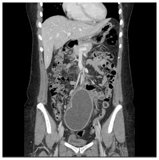
A contrast-enhanced abdominal computed tomography (CT) scan revealed a well-circumscribed and poorly attenuated lobulated mass approximately 12 cm long, located in the pelvic cavity (Figure 1). The mass exhibited peripheral enhancement and had a layered appearance. The center of the mass was of uniformly low attenuation (10 Hounsfield units) indicating the presence of a cystic cavity. The mass displaced the terminal ileum in an inferior direction. No right-side ovary was detected either ultrasonographically or on the CT scan. Ovarian cyst torsion was thus suggested both clinically and radiologically.
Upon laparotomy, a large cystic mass was located on the mesentery of the terminal ileum. The mass was not connected or attached to any part of the adjacent intestinal tract. The mass was dissected completely free of surrounding soft tissue without disturbing any normal intestinal or mesenteric structure. The mass had a dedicated vascular pedicle, which was simply ligated and removed. An enlarged lymph node was located in the vicinity of the cystic mass.
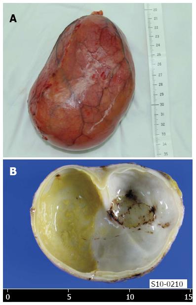
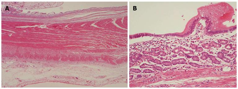
The resected specimen was of dimensions 12 cm × 8.5 cm × 6 cm and weighed 413 g. The external surface was smooth and pinkish-gray in color (Figure 2A). When the specimen was opened, the cyst was found to be uninoculated and partly septated. The cavity contained a clear mucous fluid and a quantity of dirty-yellowish sludge-like material. The cystic wall was pale-grayish and smooth, and was relatively even in thickness (up to 0.5 cm; Figure 2B). Microscopically, the cystic wall consisted of mucosa, submucosa, two layers of smooth muscle, and the serosa. The mucosal layer was largely covered by simple cuboidal-to-columnar mucous epithelium and partly by a gastric-type mucosa, including oxyntic glands (Figure 3). Islands of myenteric plexus were also present between the two layers of muscle. No epithelial dysplasia or malignancy was evident.
The postoperative course was uneventful and the patient was discharged without any complications.
Intestinal duplication cysts are rare congenital anomalies that occur in one of every 4000-5000 live births[5]. Isolated duplications occur much less frequently, in approximately 1 in 10000 live births[6]. Duplications are most commonly detected in infancy and early childhood when symptoms such as abdominal pain, obstruction, or vomiting develop[7]. The duplications occur most frequently in the small bowel and over half are ileal duplications[3]. Enteric duplications are tubular or cystic in structure and contain intestinal mucosa and muscle layers. The wall is usually attached to some part of the gastrointestinal tract and often shares a common muscle or serosal layer with the normal intestine of the region[4].
Completely isolated enteric duplication cysts are not connected or attached to any part of the intestinal tract. Such cysts have dedicated vascular pedicles, whereas the usual form of duplication cysts shares a blood supply with the adjacent bowel. Inevitably, then, the bowel must be resected upon cyst removal. Our 36-year-old female patient had a completely isolated enteric duplication cyst that was dissected without disturbing her intra-abdominal anatomy. Reports of completely isolated enteric duplication cysts in adults are extremely rare; only five cases have been described in the English-language medical literature[3,8-11] (Table 1). The cause of intestinal duplication is believed to be vascular compromise during early organogenesis. However, the precise mechanism remains to be elucidated[5].
| Ref. | Age (yr) | Gender | Clinical feature | Dimensions (cm) | Site | Mucosal type |
| Blank et al[3] | 51 | M | Incidental | 10 × 4 | Mesentery of the ileum | Villi, crypts, numerous mucous cells |
| Kim et al[8] | 28 | M | Incidental | Not mentioned | Mesentery of the Treitz ligament | Gastric |
| Nichols et al[9] | 27 | F | Abdominal fullness | 9 × 6 × 1 | Mesentery of the descending colon | Simple columnar mucinous epithelium |
| Lee et al[10] | 21 | F | Palpable mass | 3.5 × 2.5 | Mesentery in the vicinity of the round ligament | No epithelial lining |
| Kyriakos et al[11] | 20 | M | Abdominal pain and fever | 7 × 4 | Lateral region of the ascending colon | Not mentioned |
| Present case | 36 | F | Abdominal pain | 12 × 8.5 × 6 | Mesentery of the terminal ileum | Mixed |
Histopathologically, the wall of a duplication cyst is composed of a lining epithelium and thin layers of muscle[9]. The mucosa is often similar to that of the region of the bowel to which the cyst is attached[5], but some heterotopic mucosa is also commonly found[3,8,12]. In our case, the cystic mass was largely covered by a single layer of simple cuboidal-to-columnar mucous epithelium with some areas of gastric-type mucosa, including oxyntic glands. The wall contained two layers of smooth muscle and some islands of myenteric plexus were noted between the muscle layers. Thus, the wall was similar in structure to that of the normal intestine.
Enteric duplications are usually detected within the first year of life when infants present with abdominal pain, a palpable mass, or intestinal obstruction[1]. In some adult cases, however, the duplications are asymptomatic, remaining undiagnosed for years. Adenocarcinoma may arise in such adult asymptomatic cysts[3,13,14]. In the present case, our patient reported abdominal pain that we could not readily explain. Surface ulcers, distension of the cystic cavity, or impending vascular insufficiency, are among several possible causes. No dysplastic change or malignant transformation was evident.
Diagnosis of a duplication cyst is difficult even with the aid of modern imaging techniques such as CT or magnetic resonance imaging, especially when no structural connection exists between the cyst and the normal bowel. A recent report suggested that a barium meal could be used to differentiate noncommunicating from communicating cysts[7]. The cited authors reported that the alimentary tract was compressed by a noncommunicating cyst, whereas communicating cysts were themselves filled with contrast. However, it remains difficult to diagnose an enteric duplication cyst because the differential diagnosis includes all intra-abdominal cystic masses, including mesenteric and omental cysts, pancreatic pseudocysts, and ovarian cysts[1,2,8]. In the present case, ultrasonography and CT suggested the possibility of an ovarian cyst rather than a rare intestinal duplication cyst. There has been one report of torsion of an ovarian cyst mimicking an enteric duplication cyst[15]. The author said that non-enteric cysts may have double-layered features on ultrasound, which are commonly found in duplication cysts.
No consensus has been reached on treatment of asymptomatic duplication cysts. However, surgical management of asymptomatic adult cases is recommended because various potential complications, including malignancy, may arise. Exploratory laparotomy may be indicated if the preoperative diagnosis is unclear. Completely isolated duplication cysts can be treated via simple ligation and division of the pedicle; no bowel resection is required[4]. A successful laparoscopic excision of a completely isolated enteric duplication cyst was reported recently[9]. We performed simple excision of the cystic mass without disturbing the normal intraperitoneal anatomy.
A 36-year-old female patient presented with a 10-d history of abdominal pain and a palpable mass in the lower abdomen.
Ultrasonography and computed tomography (CT) scan was performed for differential diagnosis between ovarian cyst and other intra-abdominal cystic lesions.
Her complete blood count, blood chemistry, and urinalysis were within normal limits except mild anemia (Hb, 11.3g/dL; Hct, 33.9%).
A contrast-enhanced abdominal CT scan revealed a well-circumscribed and poorly attenuated lobulated mass having peripheral enhancement and a cystic cavity in the pelvis.
Microscopically, the cystic wall consisted of mucosa, submucosa, two layers of smooth muscle, and the serosa.
A simple cystectomy was performed.
A completely isolated intestinal duplication cyst should be included in the differential diagnosis of an intra-abdominal cystic lesion.
The article presents an extremely rare but important case so that the readers could use the information in the practice. However, the pathogenesis of the disease was not fully investigated in this report.
P- Reviewers: Abenavoli L, Montecucco F S- Editor: Ma YJ L- Editor: A E- Editor: Wang CH
| 1. | Gümüş M, Kapan M, Gümüş H, Onder A, Girgin S. Unusual noncommunicating isolated enteric duplication cyst in adults. Gastroenterol Res Pract. 2011;2011:323919. [RCA] [PubMed] [DOI] [Full Text] [Full Text (PDF)] [Cited by in Crossref: 18] [Cited by in RCA: 27] [Article Influence: 1.9] [Reference Citation Analysis (1)] |
| 2. | Macpherson RI. Gastrointestinal tract duplications: clinical, pathologic, etiologic, and radiologic considerations. Radiographics. 1993;13:1063-1080. [RCA] [PubMed] [DOI] [Full Text] [Cited by in Crossref: 316] [Cited by in RCA: 284] [Article Influence: 8.9] [Reference Citation Analysis (0)] |
| 3. | Blank G, Königsrainer A, Sipos B, Ladurner R. Adenocarcinoma arising in a cystic duplication of the small bowel: case report and review of literature. World J Surg Oncol. 2012;10:55. [RCA] [PubMed] [DOI] [Full Text] [Full Text (PDF)] [Cited by in Crossref: 47] [Cited by in RCA: 56] [Article Influence: 4.3] [Reference Citation Analysis (0)] |
| 4. | Sinha A, Ojha S, Sarin YK. Completely isolated, noncontiguous duplication cyst. Eur J Pediatr Surg. 2006;16:127-129. [RCA] [PubMed] [DOI] [Full Text] [Cited by in Crossref: 25] [Cited by in RCA: 29] [Article Influence: 1.5] [Reference Citation Analysis (0)] |
| 5. | Gilbert-Barness E. Potter’s pathology of the fetus, infant and child. 2nd ed. Amsterdam, Paesi Bassi: Elsevier Inc 2007; 1169-1170. |
| 6. | Simsek A, Zeybek N, Yagci G, Kaymakcioglu N, Tas H, Saglam M, Cetiner S. Enteric and rectal duplications and duplication cysts in the adult. ANZ J Surg. 2005;75:174-176. [RCA] [PubMed] [DOI] [Full Text] [Cited by in Crossref: 11] [Cited by in RCA: 15] [Article Influence: 0.8] [Reference Citation Analysis (0)] |
| 7. | Shah A, Du J, Sun Y, Cao D. Dynamic change of intestinal duplication in an adult patient: a case report and literature review. Case Rep Med. 2012;2012:297585. [PubMed] |
| 8. | Kim SK, Lim HK, Lee SJ, Park CK. Completely isolated enteric duplication cyst: case report. Abdom Imaging. 2003;28:12-14. [RCA] [PubMed] [DOI] [Full Text] [Cited by in Crossref: 32] [Cited by in RCA: 40] [Article Influence: 1.8] [Reference Citation Analysis (0)] |
| 9. | Nichols KC, Pollema T, Moncure M. Laparoscopically excised completely isolated enteric duplication cyst in adult female: a case report. Surg Laparosc Endosc Percutan Tech. 2011;21:e173-e175. [PubMed] |
| 10. | Lee JU, KIM JO, KIM SJ, SUL HJ. Completely isolated enteric duplication cyst presenting as an inguinal hernia. Kor J Pathol. 2010;44:204-206. [RCA] [DOI] [Full Text] [Cited by in Crossref: 4] [Cited by in RCA: 5] [Article Influence: 0.3] [Reference Citation Analysis (0)] |
| 11. | Kyriakos N, Andreas C, Elena S, Charalampos A, Chrisanthos G. Infected completely isolated enteric duplication cyst management with percutaneous drainage and surgical excision after retreat of infection: a case report. Case Rep Surg. 2013;2013:108126. [RCA] [PubMed] [DOI] [Full Text] [Full Text (PDF)] [Cited by in Crossref: 5] [Cited by in RCA: 10] [Article Influence: 0.8] [Reference Citation Analysis (0)] |
| 12. | Upadhyay N, Gomez D, Button MF, Verbeke CS, Menon KV. Retroperitoneal enteric duplication cyst presenting as a pancreatic cystic lesion. A case report. JOP. 2006;7:492-495. [PubMed] |
| 13. | Jung KH, Jang SM, Joo YW, Oh YH, Park YW, Paik HG, Choi JH. Adenocarcinoma arising in a duplication of the cecum. Korean J Intern Med. 2012;27:103-106. [PubMed] |
| 14. | Hsu H, Gueng MK, Tseng YH, Wu CC, Liu PH, Chen CC. Adenocarcinoma arising from colonic duplication cyst with metastasis to omentum: A case report. J Clin Ultrasound. 2011;39:41-43. [PubMed] |
| 15. | Godfrey H, Abernethy L, Boothroyd A. Torsion of an ovarian cyst mimicking enteric duplication cyst on transabdominal ultrasound: two cases. Pediatr Radiol. 1998;28:171-173. [PubMed] |


