Published online Jan 14, 2014. doi: 10.3748/wjg.v20.i2.593
Revised: October 30, 2013
Accepted: October 30, 2013
Published online: January 14, 2014
Processing time: 128 Days and 19.1 Hours
We report a rare case of a 68-year-old male with metachronous pancreatic metastasis that was resected 2 years after salvage esophagectomy for local recurrence of esophageal squamous cell carcinoma (ESCC). Two years and 8 mo ago, he had undergone definitive chemoradiotherapy for the lower thoracic ESCC and achieved a complete response. Chemoradiotherapy used the protocol of the Japan Clinical Oncology Group trial 9906. Approximately 8 mo later, he developed a local recurrence of the ESCC and underwent thoracoscopic salvage esophagectomy followed by reconstruction with a conduit colon graft via a subcutaneous route. Recently, a tumor of the pancreatic body was found on routine follow-up computed tomography (CT). The tumor diameter was 15 mm on CT, and the maximum standardized uptake value of the lesion was 5.49 at 18F-2-fluoro-2-deoxy-D-glucose positron-emission tomography, strongly suggesting pancreatic cancer. In addition, all tumor markers were within the reference intervals. Therefore, distal pancreatectomy was performed with the resultant histological diagnosis being confirmed as pancreatic metastasis of the ESCC. He was treated with adjuvant chemotherapy, and there has been no evidence of recurrence 9 mo after the surgery. Resection of pancreatic metastasis offers a good prognosis and should be considered for solitary ESCC metastasis.
Core tip: This report a 68-year-old male presenting with a pancreatic tumor following esophageal squamous cell carcinoma (ESCC) treated with curative chemoradiotherapy and thoracoscopic salvage esophagectomy approximately 2 years previously. He was treated with distal pancreatectomy and histologically diagnosed as a rare case of metachronous pancreatic metastasis of ESCC. Resection of pancreatic metastasis offers a good prognosis and should be considered for solitary ESCC metastasis. Both this case and a review of the relevant literature support this view.
- Citation: Okamoto H, Hara Y, Chin M, Hagiwara M, Onodera Y, Horii S, Shirahata Y, Kamei T, Hashizume E, Ohuchi N. An extremely rare case of pancreatic metastasis of esophageal squamous cell carcinoma. World J Gastroenterol 2014; 20(2): 593-597
- URL: https://www.wjgnet.com/1007-9327/full/v20/i2/593.htm
- DOI: https://dx.doi.org/10.3748/wjg.v20.i2.593
Pancreatic metastasis of esophageal carcinoma is rare. The disease occurs reportedly at a frequency of 0.12% in esophageal carcinomas and 0.68% in metastatic tumors of esophageal carcinoma[1]. Another study has reported the rate of pancreatic metastasis of esophageal carcinoma to be 3.9% of that of esophageal carcinomas according to pathological examination of autopsy samples[2]. Furthermore, it has been observed in 0%-4.9% of pancreatic metastatic malignancies[3-7]. To the best of our knowledge, there are few English literature reports of pancreatic metastasis of esophageal squamous cell carcinoma (ESCC). Here, we describe a case of metachronous pancreatic metastasis of ESCC with a brief review of the literature.
A 68-year-old male was found to have a tumor of the pancreatic body on routine enhanced computed tomography (CT), as follow-up for the treatment of esophageal carcinoma. Two years and eight months previously, he had been diagnosed and treated for lower thoracic ESCC (clinical T1bN0M0, stage I; according to the seventh edition of the Union for International Cancer Control system). At that time, he underwent definitive chemoradiotherapy with a complete remission. The chemoradiotherapy protocol was consistent with that of the Japan Clinical Oncology Group trial 9906, and comprised two cycles of intravenous cisplatin infusions with continuous 5-fluorouracil infusion and concurrent irradiation with a total dose of 60 Gy[8]. Approximately 8 mo later, he developed local recurrence of the ESCC (pathological T1bN0M0, stage I), and underwent thoracoscopic salvage esophagectomy followed by reconstruction with a conduit colon graft via a subcutaneous route. Thoracoscopic salvage esophagectomy was performed with two-field lymph node dissection[9].
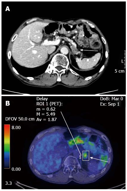
The pancreatic body tumor was evaluated prior to surgery by CT and 18F-2-fluoro-2-deoxy-D-glucose positron-emission tomography (FDG-PET). The tumor was low density and its diameter was 15 mm on CT examination, and the maximum standardized uptake value (SUVmax) of the lesion was 5.01 in the early phase and 5.49 in the late phase at FDG-PET (Figure 1). These results strongly suggested pancreatic cancer. In addition, all tumor markers were within the reference intervals, including SCC, CEA, CA19-9, SPan-1, DUPAN-2, and NCC-ST-439.
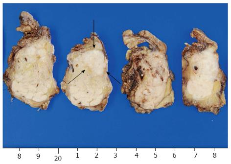
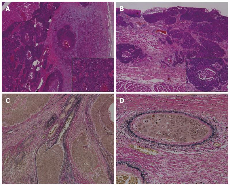
Distal pancreatectomy and splenectomy were performed to treat the pancreatic body tumor. Macroscopic findings of the specimen revealed a 15 mm × 11 mm tumor in the pancreatic body (Figure 2). Pathological examination revealed that the tumor was a squamous cell carcinoma with morphologic features similar to the previous ESCC; it was therefore diagnosed as a pancreatic metastasis of the ESCC (Figure 3). He was discharged uneventfully on the 11th postoperative day and later received one dose of adjuvant chemotherapy (intravenous cisplatin infusions with continuous 5-fluorouracil infusion). However, because of renal impairment the adjuvant chemotherapy was stopped and was followed up on an outpatient basis without adjuvant therapy. There have been no signs of recurrence 9 mo after surgical resection of the pancreatic metastasis.
To the best of our knowledge, only 3 cases of pancreatic metastasis of ESCC have been reported in the English literature till date[10-12]. Park et al[11] reported the case of 58-year-old male who underwent an esophagectomy for early ESCC followed by distal pancreatectomy for pancreatic metastasis with radiofrequency ablation for hepatocellular carcinoma. Adjuvant chemotherapy was given for 4 mo after the surgery in this case, without any evidence of recurrence. Sawada et al[12] reported the case of 73-year-old male with advanced ESCC who developed pancreatic metastasis that caused hepatic portal venous gas; he received supportive care only. Esfehani et al[10] reported a 59-year-old female with a pancreatic metastasis of ESCC who had undergone surgical treatment and adjuvant chemoradiotherapy 4 years earlier. In this case, she was treated with distal pancreatectomy and adjuvant chemotherapy for the pancreatic metastasis and showed no recurrence within the 6-mo follow-up. This is the only known case of metachronous pancreatic metastasis of ESCC reported in the literature[10]. Our report is the first case of metachronous pancreatic metastasis of ESCC following local recurrence treated with salvage esophagectomy and definitive chemoradiotherapy.
The differential diagnosis of pure squamous cell carcinoma of the pancreas is also a possibility. However, this is very rare, and it has been argued that before diagnosis, metastasis from another site should be excluded[13]. Most of the squamous differentiation that occurs in pancreatic carcinoma, exists as a component of adenosquamous carcinoma. Therefore, the World Health Organization classification of tumors of the pancreas does not specify it as a distinct entity. Al-Shehri et al[14] reported in his review that the development of pancreatic squamous cell carcinoma was explained by 5 postulated theories as follows:. (1) malignant change in a primitive cell capable of differentiating into either squamous or glandular carcinoma; (2) squamous change in a pre-existing adenocarcinoma; (3) malignant transformation in a squamous metaplasia of the ductal epithelium; (4) malignant change in an aberrant squamous cell; and (5) the theory of tumor collision. In this case, the histological morphology similar to the primary ESCC together with the history of ESCC were the basis for the diagnosis of pancreatic metastasis of ESCC. Vascular invasion was observed in the specimens of both the previously resected esophagus and the pancreatic metastasis. This fact supports the hematogenous distant metastasis of the ESCC. Although we initially diagnosed the patient with pancreatic cancer at first, primarily on the basis of the results of imaging, the tumor was ultimately diagnosed as pancreatic metastasis from the ESCC.
Finally, we need to consider whether resection of the ESCC pancreatic metastasis was the appropriate treatment option. Several investigators have reported favorable prognosis with resection of pancreatic metastasis[4,6,7,15]. However, the primary cancers in these reports consisted of renal cell cancer, colon cancer, melanoma, sarcoma, lung cancer, and breast cancer with limited consideration of esophageal cancer. Because of the rarity of the presentation, the optimal treatment regimen remains unknown. Reddy et al[6] stated that patients might benefit from pancreatic metastasectomy given the following: a primary cancer type that was associated with good outcome, control of the primary cancer site, demonstration of isolated metastasis, resectability of the metastasis, and patient fitness to tolerate pancreatectomy. We previously reported that following salvage esophagectomy, the outcome was better in patients with recurrent disease after complete response to definitive chemoradiotherapy than in those with persistent disease[16]. In addition, it was reported that resection of solitary ESCC metastasis results in a longer survival[17]. Therefore, it is therefore likely that this case will have a good prognosis.
In conclusion, we report a rare case of pancreatic metastasis of ESCC and provide a brief review of the literature. From these results, we argue that surgical excision should be considered a valid treatment strategy for solitary pancreatic metastasis in ESCC. However, further investigation and an accumulation of case reports are needed to confirm this finding.
The authors thank Dr. Akiko Nishida, Department of Pathology, Nihonkai General Hospital, for providing pathological findings.
Metachronous pancreatic metastasis that was resected 2 years after salvage esophagectomy for local recurrence of esophageal squamous cell carcinoma (ESCC).
This report performed pancreatectomy in order to clarify whether the tumor was primary pancreatic cancer or pancreatic metastasis.
Laboratory testing such as tumor makers could not contribute to the diagnosis.
Computed tomography (CT) and PET-CT had suggested that the tumor was pancreatic cancer, but it was pathologically pancreatic metastasis of ESCC.
Pathological examination revealed that the tumor was a squamous cell carcinoma with morphologic features similar to the previous ESCC; it was therefore diagnosed as a pancreatic metastasis of the ESCC.
Pancreatectomy and adjuvant chemotherapy (intravenous cisplatin infusions with continuous 5-fluorouracil infusion).
Surgical excision should be considered a valid treatment strategy for solitary pancreatic metastasis in ESCC, but further investigation and an accumulation of case reports are needed to confirm this finding.
This report is the first case of metachronous pancreatic metastasis of ESCC following local recurrence treated with salvage esophagectomy and definitive chemoradiotherapy and revealed favorable prognosis with resection of pancreatic metastasis. However, because few cases of pancreatic metastasis of ESCC have been reported it could not insist on many things. Further investigation and an accumulation of case reports are needed.
P- Reviewers: Li H, Natsugoe S, Wang SJ S- Editor: Qi Y L- Editor: A E- Editor: Wang CH
| 1. | Quint LE, Hepburn LM, Francis IR, Whyte RI, Orringer MB. Incidence and distribution of distant metastases from newly diagnosed esophageal carcinoma. Cancer. 1995;76:1120-1125. [PubMed] |
| 2. | Chan KW, Chan EY, Chan CW. Carcinoma of the esophagus. An autopsy study of 231 cases. Pathology. 1986;18:400-405. [RCA] [PubMed] [DOI] [Full Text] [Cited by in Crossref: 37] [Cited by in RCA: 45] [Article Influence: 1.2] [Reference Citation Analysis (0)] |
| 3. | Adsay NV, Andea A, Basturk O, Kilinc N, Nassar H, Cheng JD. Secondary tumors of the pancreas: an analysis of a surgical and autopsy database and review of the literature. Virchows Arch. 2004;444:527-535. [RCA] [PubMed] [DOI] [Full Text] [Cited by in Crossref: 248] [Cited by in RCA: 239] [Article Influence: 11.4] [Reference Citation Analysis (0)] |
| 4. | Masetti M, Zanini N, Martuzzi F, Fabbri C, Mastrangelo L, Landolfo G, Fornelli A, Burzi M, Vezzelli E, Jovine E. Analysis of prognostic factors in metastatic tumors of the pancreas: a single-center experience and review of the literature. Pancreas. 2010;39:135-143. [RCA] [PubMed] [DOI] [Full Text] [Cited by in Crossref: 54] [Cited by in RCA: 61] [Article Influence: 4.1] [Reference Citation Analysis (0)] |
| 5. | Nakamura E, Shimizu M, Itoh T, Manabe T. Secondary tumors of the pancreas: clinicopathological study of 103 autopsy cases of Japanese patients. Pathol Int. 2001;51:686-690. [RCA] [PubMed] [DOI] [Full Text] [Cited by in Crossref: 114] [Cited by in RCA: 101] [Article Influence: 4.2] [Reference Citation Analysis (0)] |
| 6. | Reddy S, Wolfgang CL. The role of surgery in the management of isolated metastases to the pancreas. Lancet Oncol. 2009;10:287-293. [RCA] [PubMed] [DOI] [Full Text] [Cited by in Crossref: 163] [Cited by in RCA: 167] [Article Influence: 10.4] [Reference Citation Analysis (0)] |
| 7. | Varker KA, Muscarella P, Wall K, Ellison C, Bloomston M. Pancreatectomy for non-pancreatic malignancies results in improved survival after R0 resection. World J Surg Oncol. 2007;5:145. [RCA] [PubMed] [DOI] [Full Text] [Full Text (PDF)] [Cited by in Crossref: 25] [Cited by in RCA: 28] [Article Influence: 1.6] [Reference Citation Analysis (0)] |
| 8. | Kato K, Muro K, Minashi K, Ohtsu A, Ishikura S, Boku N, Takiuchi H, Komatsu Y, Miyata Y, Fukuda H. Phase II study of chemoradiotherapy with 5-fluorouracil and cisplatin for Stage II-III esophageal squamous cell carcinoma: JCOG trial (JCOG 9906). Int J Radiat Oncol Biol Phys. 2011;81:684-690. [RCA] [PubMed] [DOI] [Full Text] [Cited by in Crossref: 236] [Cited by in RCA: 268] [Article Influence: 17.9] [Reference Citation Analysis (0)] |
| 9. | Akaishi T, Kaneda I, Higuchi N, Kuriya Y, Kuramoto J, Toyoda T, Wakabayashi A. Thoracoscopic en bloc total esophagectomy with radical mediastinal lymphadenectomy. J Thorac Cardiovasc Surg. 1996;112:1533-1540; discussion 1540-1541. [PubMed] |
| 10. | Esfehani MH, Mahmoodzadeh H, Alibakhshi A, Safavi F. Esophageal squamous cell carcinoma with pancreatic metastasis: a case report. Acta Med Iran. 2011;49:760-762. [PubMed] |
| 11. | Park C, Jang JY, Kim YH, Hwang EJ, Na KY, Kim KY, Park JH, Chang YW. A case of esophageal squamous cell carcinoma with pancreatic metastasis. Clin Endosc. 2013;46:197-200. [RCA] [PubMed] [DOI] [Full Text] [Full Text (PDF)] [Cited by in Crossref: 9] [Cited by in RCA: 11] [Article Influence: 0.9] [Reference Citation Analysis (0)] |
| 12. | Sawada T, Adachi Y, Noda M, Akino K, Kikuchi T, Mita H, Ishii Y, Endo T. Hepatic portal venous gas in pancreatic solitary metastasis from an esophageal squamous cell carcinoma. Hepatobiliary Pancreat Dis Int. 2013;12:103-105. [RCA] [PubMed] [DOI] [Full Text] [Cited by in Crossref: 7] [Cited by in RCA: 7] [Article Influence: 0.6] [Reference Citation Analysis (0)] |
| 13. | Fukushima N, Hruban RH, Y . Kato, Klimstra DS, Kloppel G, Shimizu M, Terris B. Ductal adenocarcinoma variants and mixed neoplasms of the pancreas. WHO Classification of Tumours of the Digestive System. Lyons: IARC 2010; 292-295. |
| 14. | Al-Shehri A, Silverman S, King KM. Squamous cell carcinoma of the pancreas. Curr Oncol. 2008;15:293-297. [PubMed] |
| 15. | Ballarin R, Spaggiari M, Cautero N, De Ruvo N, Montalti R, Longo C, Pecchi A, Giacobazzi P, De Marco G, D’Amico G. Pancreatic metastases from renal cell carcinoma: the state of the art. World J Gastroenterol. 2011;17:4747-4756. [RCA] [PubMed] [DOI] [Full Text] [Full Text (PDF)] [Cited by in CrossRef: 114] [Cited by in RCA: 130] [Article Influence: 9.3] [Reference Citation Analysis (0)] |
| 16. | Okamoto H, Fujishima F, Nakamura Y, Zuguchi M, Miyata G, Kamei T, Nakano T, Katsura K, Abe S, Taniyama Y. Murine double minute 2 and its association with chemoradioresistance of esophageal squamous cell carcinoma. Anticancer Res. 2013;33:1463-1471. [PubMed] |
| 17. | Cho MM, Kobayashi K, Aoki T, Nishioka K, Yoshida K, Hatano N, Hirose H, Moon JH, Matsumoto T, Uemura Y. Surgical resection of solitary adrenal metastasis from esophageal carcinoma following esophagectomy. Dis Esophagus. 2007;20:79-81. [RCA] [PubMed] [DOI] [Full Text] [Cited by in Crossref: 7] [Cited by in RCA: 10] [Article Influence: 0.6] [Reference Citation Analysis (0)] |


