Published online Feb 7, 2013. doi: 10.3748/wjg.v19.i5.781
Revised: December 15, 2012
Accepted: December 22, 2012
Published online: February 7, 2013
Processing time: 71 Days and 12.8 Hours
Lymphangioma, a congenital malformation of the lymphatic system, is usually found in children, and generally occurs in the neck and mediastinum. It is rarely found in the spleen. The clinical features of splenic lymphangioma typically include abdominal pain, nausea, and abdominal distention. Frequently, however, this condition is asymptomatic and is incidentally detected by abdominal ultrasonography or by an abdominal computed tomography (CT) scan. In this paper, we retrospectively describe a case of incidentally detected splenic lymphangioma in a 30-year-old woman with special abdominal contrast material-enhanced CT findings, which was accurately diagnosed by histopathology. The clinical and physical examinations related to the mass were negative. A few cases of splenic lymphangioma have been reported previously; however, the presentation of the mass and the enhancement pattern in the contrast medium-enhanced CT images were quite extraordinary. These findings had misled our abdominal radiologists to consider it as other neoplastic diseases of the spleen.
- Citation: Yang F, Chen WX. Splenic lymphangioma that manifested as a solid-cystic mass: A case report. World J Gastroenterol 2013; 19(5): 781-783
- URL: https://www.wjgnet.com/1007-9327/full/v19/i5/781.htm
- DOI: https://dx.doi.org/10.3748/wjg.v19.i5.781
Splenic lymphangiomas are usually benign tumors that predominantly affect children, whereas only a few cases have been reported in adults[1-3]. Previous case reports revealed no particular findings in the image analysis, which uniformly indicated solitary cysts, or multiple cysts without contrast enhancement or only with slight enhancement of the thin septa[1,3-5], except for one case of a solid mass with diffuse and prolonged enhancement[2]. Here we present a histopathologically proven case in which a solid-cystic mass of the spleen in the computed tomography (CT) images showed no enhancement of the cysts and prominent enhancement of the solid components. To the best of our knowledge, these special contrast-enhanced CT findings are relatively rare.
A 30-year year-old woman was admitted to our hospital with a splenic mass detected by abdominal ultrasonography, which was performed for recurrent epigastric pain at another hospital. The epigastric pain was later attributed to gall bladder lithiasis. The patient’s past and familial medical histories indicated no abnormalities. There were no positive symptoms related to the splenic mass. Physical examinations detected no mass or splenomegaly in the left upper quadrant of the abdomen. Peripheral blood count, coagulation studies, and liver and kidney function tests were all within normal limits.
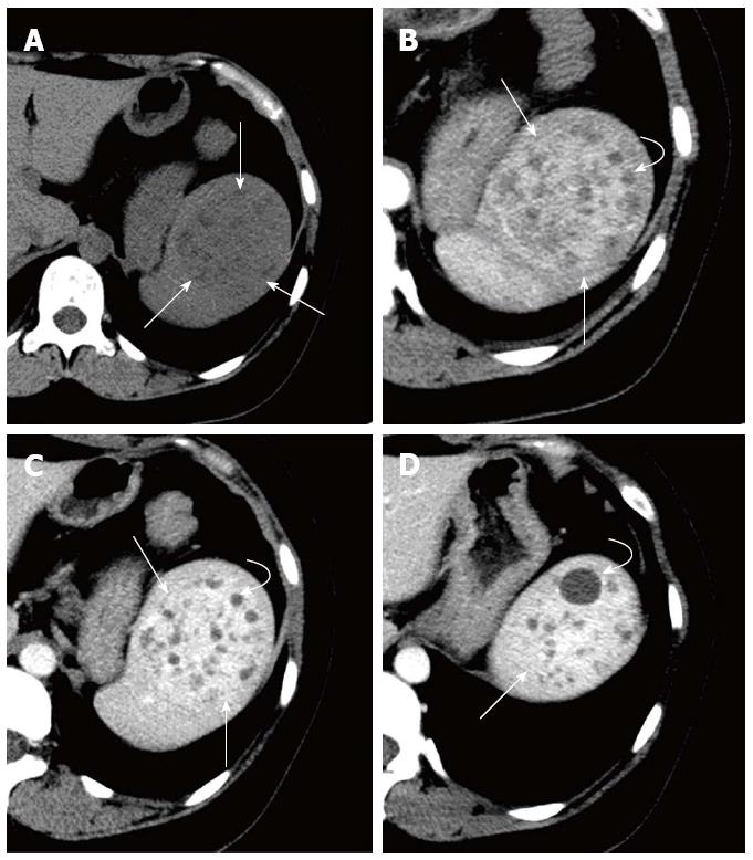
CT showed a heterogeneous solid-cystic mass with a well-defined margin located in the normal sized spleen in the plain images. Within the mass, there were multiple small cysts of variable size. The cysts showed sharp borders with no enhancement at either the arterial or parenchymal phase, whereas the solid components presented with persistently progressive enhancement (Figure 1). At this point, our radiologists suspected a splenic tumor.
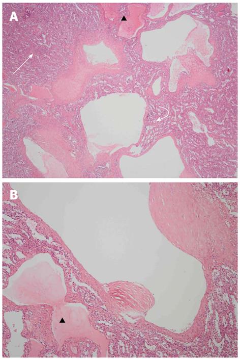
Splenectomy was eventually undertaken for the splenic mass. During the operation, a solid mass was found, which had no clear boundary with the peripheral splenic parenchyma. The final histopathological result was splenic cavernous lymphangioma. The findings revealed multiple lymphatic vesicles of variable size, and the blood sinuses in the red pulp within the mass that were dilated compared with the peripheral red pulp. The vesicles were lined by a single layer of flat endothelial cells, and some vesicles were filled with pink eosinophilic lymphatic fluid (Figure 2).
Lymphangiomas are generally considered to be congenital malformations of the lymphatic system, and they occur mostly in the neck, mediastinum and retroperitoneum[4]; they are seldom found in the spleen. Histologically, lymphangiomas are classified into three subtypes according to the congenital dilated lymphatic channels: capillary (super-microcystic), cavernous (microcystic), or cystic (macrocystic)[6]. Our case was ultimately diagnosed as the cavernous type. Cavernous lymphangiomas are composed of numerous irregular cavernous spaces containing amorphous eosinophilic fluid and few lymphocytes, lined by a single layer of flat endothelial cells[7]. Splenic lymphangiomas are mostly asymptomatic; therefore, the final diagnosis should be based on a combination of clinical, radiological, and histopathological findings.
In most cases, splenic lymphangiomas present with thin-walled cystic masses without enhancement or with only slight enhancement of the thin septa in the CT studies. We detailed a histologically confirmed case in which the mass manifested as a solid-cystic lesion. The multiple cysts showed no enhancement, as in previous cases[1,3-5]; however, the solid components showed obvious progressive enhancement. Histopathologically, however, there were no real solid components, and the solid, prominently enhanced portions in the CT images simply represented the dilated blood sinuses in the red pulp within the mass. The presence of reduced blood velocity and collecting blood in the dilated blood sinuses may have resulted in more contrast medium per voxel compared with the peripheral normal sinuses, and may have eventually led to obviously progressive enhancement.
Thus, lymphangioma presenting with this kind of solid-cystic mass in the image studies may be misdiagnosed. The differential diagnoses of splenic lymphangiomas often include other solid and cystic lesions of the spleen, such as hemangioma, chronic infection, lymphoma and metastasis. Hemangiomas may sometimes display solid and cystic portions[8]. Contrast-enhanced CT scans may show delayed enhancement within the solid portion. Punctate calcification can be seen in the central solid portion, and curvilinear calcification may be seen in the cystic areas[8]. Parasitic infection with Echinococcus granulosus is the main cause of cystic proliferations of the spleen[9]. The differential factors are the patients’ history, calcification in the cystic walls, a cyst with daughter cysts or concomitant cystic lesions in the liver or other organs[10].
In conclusion, dynamic contrast-enhanced CT is a useful method for evaluating splenic lesions. We suggest that lymphangioma should be considered in the differential diagnosis of a splenic mass that manifests as a solid-cystic lesion in the image studies. In our case, the dilated blood sinuses in the red pulp within the mass might have contributed to the indication of solid components with evident enhancement.
We thank Wen-Yan Zhang (Department of Pathology, West China Hospital of Sichuan University) for providing the histopathological resources.
P- Reviewers Triantopoulou C, Albiin N S- Editor Gou SX L- Editor Stewart GJ E- Editor Zhang DN
| 1. | Vezzoli M, Ottini E, Montagna M, La Fianza A, Paulli M, Rosso R, Mazzone A. Lymphangioma of the spleen in an elderly patient. Haematologica. 2000;85:314-317. [PubMed] |
| 2. | Chang WC, Liou CH, Kao HW, Hsu CC, Chen CY, Yu CY. Solitary lymphangioma of the spleen: dynamic MR findings with pathological correlation. Br J Radiol. 2007;80:e4-e6. [RCA] [PubMed] [DOI] [Full Text] [Cited by in Crossref: 22] [Cited by in RCA: 20] [Article Influence: 1.1] [Reference Citation Analysis (0)] |
| 3. | Beltrán MA, Barría C, Pujado B, Oliva J, Contreras MA, Wilson CS, Cruces KS. [Gigantic cystic splenic lymphangioma. Report of one case]. Rev Med Chil. 2009;137:1597-1601. [RCA] [PubMed] [DOI] [Full Text] [Cited by in Crossref: 2] [Cited by in RCA: 3] [Article Influence: 0.2] [Reference Citation Analysis (0)] |
| 4. | Abbott RM, Levy AD, Aguilera NS, Gorospe L, Thompson WM. From the archives of the AFIP: primary vascular neoplasms of the spleen: radiologic-pathologic correlation. Radiographics. 2004;24:1137-1163. [RCA] [PubMed] [DOI] [Full Text] [Cited by in Crossref: 281] [Cited by in RCA: 238] [Article Influence: 11.3] [Reference Citation Analysis (0)] |
| 5. | Solomou EG, Patriarheas GV, Mpadra FA, Karamouzis MV, Dimopoulos I. Asymptomatic adult cystic lymphangioma of the spleen: case report and review of the literature. Magn Reson Imaging. 2003;21:81-84. [RCA] [PubMed] [DOI] [Full Text] [Cited by in Crossref: 37] [Cited by in RCA: 40] [Article Influence: 1.8] [Reference Citation Analysis (0)] |
| 6. | Lee WS, Kim YH, Chee HK, Lee SA, Kim JD, Kim DC. Cavernous lymphangioma arising in the chest wall 19 years after excision of a cystic hygroma. Korean J Thorac Cardiovasc Surg. 2011;44:380-382. [RCA] [PubMed] [DOI] [Full Text] [Full Text (PDF)] [Cited by in Crossref: 6] [Cited by in RCA: 9] [Article Influence: 0.6] [Reference Citation Analysis (0)] |
| 7. | Ogun GO, Oyetunde O, Akang EE. Cavernous lymphangioma of the breast. World J Surg Oncol. 2007;5:69. [RCA] [PubMed] [DOI] [Full Text] [Full Text (PDF)] [Cited by in Crossref: 18] [Cited by in RCA: 19] [Article Influence: 1.1] [Reference Citation Analysis (0)] |
| 8. | Urrutia M, Mergo PJ, Ros LH, Torres GM, Ros PR. Cystic masses of the spleen: radiologic-pathologic correlation. Radiographics. 1996;16:107-129. [PubMed] |
| 9. | Adas G, Karatepe O, Altiok M, Battal M, Bender O, Ozcan D, Karahan S. Diagnostic problems with parasitic and non-parasitic splenic cysts. BMC Surg. 2009;9:9. [RCA] [PubMed] [DOI] [Full Text] [Full Text (PDF)] [Cited by in Crossref: 21] [Cited by in RCA: 27] [Article Influence: 1.7] [Reference Citation Analysis (0)] |
| 10. | Polat P, Kantarci M, Alper F, Suma S, Koruyucu MB, Okur A. Hydatid disease from head to toe. Radiographics. 2003;23:475-494; quiz 536-537. [RCA] [PubMed] [DOI] [Full Text] [Cited by in Crossref: 345] [Cited by in RCA: 342] [Article Influence: 15.5] [Reference Citation Analysis (0)] |


