Case Report Open Access
Copyright ©2013 Baishideng Publishing Group Co., Limited. All rights reserved.
World J Gastroenterol. Oct 28, 2013; 19(40): 6939-6942
Published online Oct 28, 2013. doi: 10.3748/wjg.v19.i40.6939
Isolated gastric variceal bleeding caused by splenic lymphoma-associated splenic vein occlusion
Bao-Chung Chen, Yu-Lueng Shih, Wei-Kuo Chang, Tsai-Yuan Hsieh, Division of Gastroenterology, Department of Internal Medicine, Tri-Service General Hospital, National Defense Medical Center, Taipei 114, Taiwan
Hong-Hau Wang, Department of Radiology, Tri-Service General Hospital, National Defense Medical Center, Taipei 114, Taiwan
Yu-Chieh Lin, Department of Pathology, Tri-Service General Hospital, National Defense Medical Center, Taipei 114, Taiwan
Author contributions: Chen BC designed the report; Hsieh TY was the attending physician for the patient; Shih YL performed the ultrasound-guided aspiration biopsy; Lin YC performed the pathological examinations; Wang HH performed the imaging diagnosis; Chang WK assisted with editing and proof-reading; Hsieh TY organized the report; and Chen BC wrote the paper.
Correspondence to: Tsai-Yuan Hsieh, MD, PhD, Division of Gastroenterology, Department of Internal Medicine, Tri-Service General Hospital, National Defense Medical Center, No.325, Sec. 2, Chenggong Rd., Neihu Dist., Taipei 114, Taiwan. tyh8121@gmail.com
Telephone: +886-2-87923311 Fax: +886-2-87927139
Received: May 27, 2013
Revised: August 12, 2013
Accepted: August 16, 2013
Published online: October 28, 2013
Processing time: 170 Days and 5.2 Hours

Abstract

Isolated gastric varices (IGV) can occur in patients with left-sided portal hypertension resulting from splenic vein occlusion caused by thrombosis or stenosis. In left-sided portal hypertension, blood flows retrogradely through the short and posterior gastric veins and the gastroepiploic veins, leading to the formation of an IGV. The most common causes of splenic vein occlusion are pancreatic diseases, such as pancreatic cancer, pancreatitis, or a pseudocyst. However, various other cancers, such as colon, gastric, or renal cancers, have also been known to cause splenic vein occlusion. Our patient presented with a rare case of IGV bleeding induced by splenic lymphoma-associated splenic vein occlusion. Splenectomy, splenic artery embolization, and stenting of the splenic vein are the current treatment choices. Chemotherapy, however, is an alternative effective treatment for splenic vein occlusion caused by chemotherapy-sensitive tumors. Our patient responded well to chemotherapy with a cyclophosphamide, hydroxydaunorubicin, oncovin, and prednisolone regimen, and the splenic vein occlusion resolved after the lymphoma regressed.

Key Words: Isolated gastric varices; Splenic vein; Lymphoma; Occlusion; Hematemesis

Core tip: Isolated gastric varices occur in patients with splenic vein occlusion caused by thrombosis, stenosis, or cancer, such as pancreatic, colon, gastric, or renal cancers. Here, we describe a rare case of an isolated gastric varices in an elderly woman who presented with tarry stool and bloody vomitus. The diagnostic tests revealed a splenic B-cell lymphoma, which caused splenic vein occlusion that resulted in isolated gastric variceal bleeding. Splenectomy, splenic artery embolization, and stenting of the splenic vein are the current treatment choices. In this patient, chemotherapy was an alternative treatment, and the splenic vein occlusion resolved after the lymphoma regressed.


INTRODUCTION

Hematemesis is a medical emergency, and in most cases, it is caused by esophageal varices or peptic ulcers. However, there are other rare, but important and treatable causes of hematemesis, such as isolated gastric varices. We report one such case and review the literature, focusing on the unusual entities presenting as isolated gastric variceal bleeding.

CASE REPORT

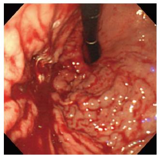
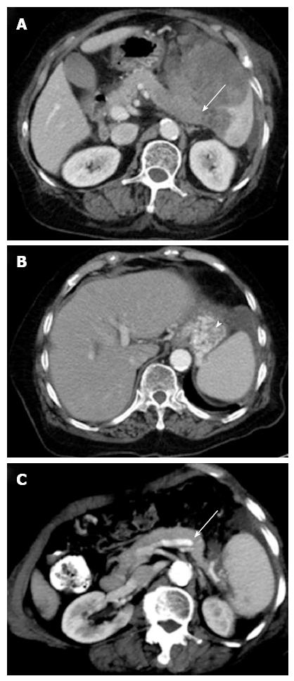
A 77-year-old woman was admitted to our hospital due to two episodes of bloody vomitus and three episodes of tarry stool. She did not have a significant medical or surgical history or any known allergies, nor was she taking any medication. The physical examination revealed pale conjunctiva without any jaundice, lymphadenopathy, or pedal edema. An abdominal palpation demonstrated an enlarged spleen, palpable below the left costal margin, and a digital rectal examination showed melena. The remainder of the patient’s systemic examination results was unremarkable. The blood pressure was 97/55 mmHg, the pulse rate was 96/min, the hemoglobin level was 8.9 g/dL, the platelet count was 118000/μL, and the stool occult blood test was positive. The patient’s biochemistry tests, including those for liver and renal function, were within the normal limits, apart from a high serum lactate dehydrogenase level (1112 U/L). Esophagogastroduodenoscopy demonstrated an isolated gastric varices (IGV) at the cardia and high body of the stomach, with active bleeding (Figure 1); thus, sclerotherapy with cyanoacrylate was successfully performed. Abdominal computed tomography (CT) showed a large mass in the enlarged spleen, with near total occlusion of the splenic vein (Figure 2A). This splenic vein occlusion led to the development of a gastric varices at the fundus (Figure 2B), and the portal and superior mesenteric veins were patent. The laboratory findings indicated the patient’s carcinoembryonic antigen and carbohydrate antigen 19-9 levels were normal.

Figure 1
Figure 1 Engorged varices at the cardia and high body of the stomach, with active bleeding.
Figure 2
Figure 2 Imaging findings. A: Axial contrast-enhanced abdominal computed tomography (CT) at the level of the splenic hilum shows a large low-attenuation mass in the enlarged spleen. The mass involves the splenic vein with near total occlusion (arrow); B: The development of a gastric varice (arrowhead) at the fundus; C: The 6-mo follow-up CT shows that the splenic mass has almost completely resolved, allowing the splenic vein (arrow) to return to patency.

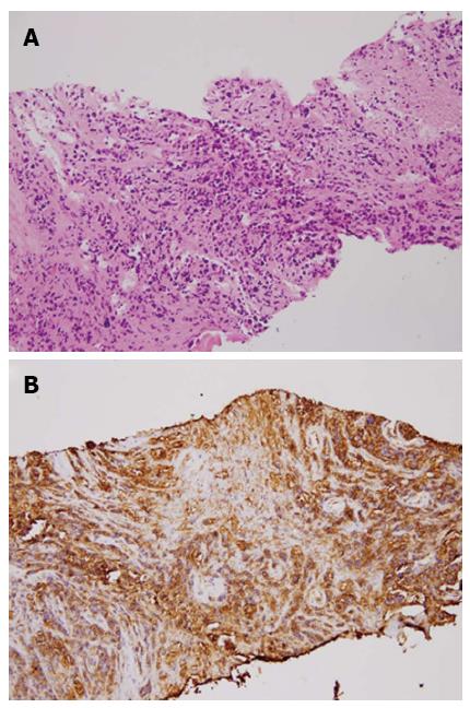
The patient underwent ultrasound-guided aspiration biopsy, and histopathology confirmed a high-grade B-cell lymphoma (Figure 3). Thereafter, the patient received chemotherapy with a cyclophosphamide, hydroxydaunorubicin, oncovin, and prednisolone (CHOP) regimen, and the splenic vein occlusion resolved after the lymphoma regressed (Figure 2C).

Figure 3
Figure 3 Pathologic findings (hematoxylin/eosin staining) and immunohistochemical staining. A: The predominant fibrous tissue with necrotic foci and scattered atypical cells characterized by pleomorphic and hyperchromatic nuclei with discohesive arrangement. B: The B-lymphocyte marker (CD20) was strongly positive in the atypical cells.
DISCUSSION

IGVs are observed in up to 5% of patients with cirrhosis and in up to 10% of patients with non-cirrhotic portal hypertension[1]. Such varices can occur in patients with segmental or left-sided portal hypertension (LSPH), and the incidence has increased over the past three decades because of increased awareness of the entity and advances in diagnostic approaches[2,3]. When a segment of the portal venous bed is obstructed, varices can develop in the area to decompress the blocked segment. In cases of segmental portal hypertension, the blood drains in a retrograde manner through the short and posterior gastric veins and the gastroepiploic veins. This process results in blood flow and a pressure increase, which causes dilation of the submucosal structures, leading to the formation of a gastric varice[4].

LSPH primarily occurs as a result of splenic vein obstruction, most commonly resulting from pancreatic disorders[5]. Because the splenic vein is posterior to the pancreas and in direct contact with it, any type of pancreatic disease, including cancer, pancreatitis, abscess formation, or a pseudocyst, can affect the splenic vein[6-8]. In a study by Moosa et al, pancreatitis, diagnosed by either biopsy or surgery, was part of the LSPH etiology in 87 (60%) of 144 cases, while pancreatic malignancy was detected in only 13 (9%) patients[9,10]. Various disorders other than pancreatic diseases, such as partial gastrectomy, metastatic carcinoma, retroperitoneal fibrosis, or protein S deficiency, have a wide spectrum of mechanisms involved in causing splenic vein occlusion[5,11-13]. Among the metastatic carcinomas, oat cell carcinoma[14], colon cancer[5], gastric cancer[5], renal cancer[12], and rare retroperitoneal liposarcoma[9] have been reported to cause splenic vein occlusion. Primary pancreatic lymphoma, with splenic vein thrombosis and IGV bleeding, has been reported as well[15]. Our patient had a rare case of splenic B-cell lymphoma that caused splenic vein occlusion and IGV bleeding.

In general, IGVs are asymptomatic and identified incidentally. In symptomatic cases, the most common clinical manifestation is gastrointestinal bleeding from ruptured gastric varices; statistically, 45%-72% of patients with LSPH present with the above-mentioned symptom[3]. Splenomegaly and abdominal pain are observed in 71% and 25%-38% of patients with LSPH, respectively[5,9], whereas ascites seldom develop[16]. Various radiological examinations can assist in making an IGV diagnosis, including upper gastrointestinal endoscopy, angiography, ultrasonography, contrast-enhanced CT portography, and magnetic resonance imaging. In one study, gastric varices could be accurately diagnosed and localized in nearly 90% of cases by endoscopy[17]. Endoscopic ultrasound (EUS) appears to be a more accurate test than transabdominal ultrasonography, which is useful for evaluating the pancreatic parenchyma and splenic vasculature[18]. Contrast-enhanced CT portography can quickly elucidate the portal venous system, but it requires a large amount of iodinated contrast material. Magnetic resonance angiography with gadopentetate dimeglumine is a promising noninvasive test that is being increasingly used to diagnosis patency or thrombosis of the portal venous system[19].

The treatment of IGV bleeding should be directed toward the splenic side of the portal circulation because the pressure increases only on that side[14]. Several choices such as conservative therapy (e.g., pharmacotherapy, balloon tamponade, endoscopic therapy with sclerotherapy or band ligation) and interventional radiologic techniques, are available for bleeding control. Japanese endoscopists have reported using a combination technique of endoscopic variceal ligation injection sclerotherapy to treat acute gastric varice bleeding; when using this procedure, the rebleeding rate was low (0%-8%), the gastric varice eradication rate was 85% at up to 2 years post-treatment, and hemostasis was achieved in 100% of patients[20]. Transcatheter splenic artery embolization has also been attempted with varying degrees of success, but it has not become a preferred approach. If a patient with active bleeding is unresponsive to conservative management, then splenectomy is indicated. A large splenectomy series that included patients with isolated splenic vein thrombosis showed that none of the patients had recurrent bleeding during the 11-mo mean follow-up period after splenectomy[9].

The current case was a rare incidence of IGV bleeding induced by splenic lymphoma-associated splenic vein occlusion. In this patient, chemotherapy was an alternative treatment for splenic vein occlusion caused by chemotherapy-sensitive tumors. Our patient responded well to chemotherapy, and the splenic vein occlusion resolved after the lymphoma regressed.

Footnotes

P- Reviewers Chen CY, Garcia-Roca R, Samarasam I S- Editor Qi Y L- Editor A E- Editor Zhang DN

References
1.  Sarin SK, Jain AK, Lamba GS, Gupta R, Chowdhary A. Isolated gastric varices: prevalence, clinical relevance and natural history. Dig Surg. 2003;20:42-47.  [PubMed]  [DOI]  [Cited in This Article: ]  [Cited by in Crossref: 27]  [Cited by in F6Publishing: 29]  [Article Influence: 1.4]  [Reference Citation Analysis (0)]
2.  Ryan BM, Stockbrugger RW, Ryan JM. A pathophysiologic, gastroenterologic, and radiologic approach to the management of gastric varices. Gastroenterology. 2004;126:1175-1189.  [PubMed]  [DOI]  [Cited in This Article: ]  [Cited by in Crossref: 227]  [Cited by in F6Publishing: 212]  [Article Influence: 10.6]  [Reference Citation Analysis (0)]
3.  Köklü S, Coban S, Yüksel O, Arhan M. Left-sided portal hypertension. Dig Dis Sci. 2007;52:1141-1149.  [PubMed]  [DOI]  [Cited in This Article: ]  [Cited by in Crossref: 101]  [Cited by in F6Publishing: 126]  [Article Influence: 7.4]  [Reference Citation Analysis (0)]
4.  Madsen MS, Petersen TH, Sommer H. Segmental portal hypertension. Ann Surg. 1986;204:72-77.  [PubMed]  [DOI]  [Cited in This Article: ]  [Cited by in Crossref: 109]  [Cited by in F6Publishing: 105]  [Article Influence: 2.8]  [Reference Citation Analysis (18)]
5.  Köklü S, Yüksel O, Arhan M, Coban S, Başar O, Yolcu OF, Uçar E, Ibiş M, Ertugrul I, Sahin B. Report of 24 left-sided portal hypertension cases: a single-center prospective cohort study. Dig Dis Sci. 2005;50:976-982.  [PubMed]  [DOI]  [Cited in This Article: ]  [Cited by in Crossref: 37]  [Cited by in F6Publishing: 46]  [Article Influence: 2.4]  [Reference Citation Analysis (0)]
6.  Little AG, Moossa AR. Gastrointestinal hemorrhage from left-sided portal hypertension. An unappreciated complication of pancreatitis. Am J Surg. 1981;141:153-158.  [PubMed]  [DOI]  [Cited in This Article: ]  [Cited by in Crossref: 86]  [Cited by in F6Publishing: 86]  [Article Influence: 2.0]  [Reference Citation Analysis (0)]
7.  Manenti A. Splenic vein obstruction secondary to pancreatic carcinoma. Acta Chir Belg. 1981;80:245-248.  [PubMed]  [DOI]  [Cited in This Article: ]
8.  Keith RG, Mustard RA, Saibil EA. Gastric variceal bleeding due to occlusion of splenic vein in pancreatic disease. Can J Surg. 1982;25:301-304.  [PubMed]  [DOI]  [Cited in This Article: ]
9.  Moossa AR, Gadd MA. Isolated splenic vein thrombosis. World J Surg. 1985;9:384-390.  [PubMed]  [DOI]  [Cited in This Article: ]  [Cited by in Crossref: 82]  [Cited by in F6Publishing: 81]  [Article Influence: 2.1]  [Reference Citation Analysis (0)]
10.  Ismail FW, Mumtaz K, Chawla T, Jafri W. Gastric variceal bleed in a patient without liver cirrhosis: an unusual cause of haematemesis. Singapore Med J. 2007;48:e171-e173.  [PubMed]  [DOI]  [Cited in This Article: ]
11.  Honda Y, Ueda M, Kyoi M, Mizunuma T, Takeda R. An unusual case of portasystemic encephalopathy caused by splenic vein occlusion following gastrectomy. Am J Gastroenterol. 1978;69:590-593.  [PubMed]  [DOI]  [Cited in This Article: ]
12.  Koehler RE. Case: splenic vein obstruction due to metastatic hypernephroma. Gastrointest Radiol. 1981;6:365-370.  [PubMed]  [DOI]  [Cited in This Article: ]
13.  Lavender S, Lloyd-Davies RW, Thomas ML. Retroperitoneal fibrosis causing localized portal hypertension. Br Med J. 1970;3:627-628.  [PubMed]  [DOI]  [Cited in This Article: ]  [Cited by in Crossref: 23]  [Cited by in F6Publishing: 43]  [Article Influence: 0.8]  [Reference Citation Analysis (0)]
14.  Evans GR, Yellin AE, Weaver FA, Stain SC. Sinistral (left-sided) portal hypertension. Am Surg. 1990;56:758-763.  [PubMed]  [DOI]  [Cited in This Article: ]
15.  Vida Pérez L, González Galilea A, Fraga Rivas E. [Bleeding from gastric varices as the initial manifestation of primary pancreatic lymphoma]. Gastroenterol Hepatol. 2010;33:165-170.  [PubMed]  [DOI]  [Cited in This Article: ]  [Cited by in Crossref: 3]  [Cited by in F6Publishing: 4]  [Article Influence: 0.3]  [Reference Citation Analysis (0)]
16.  Witte CL, Chung YC, Witte MH, Sterile OF, Cole WR. Observations on the origin of ascites from experimental extrahepatic portal congestion. Ann Surg. 1969;170:1002-1015.  [PubMed]  [DOI]  [Cited in This Article: ]  [Cited by in Crossref: 25]  [Cited by in F6Publishing: 21]  [Article Influence: 0.4]  [Reference Citation Analysis (0)]
17.  Mathur SK, Dalvi AN, Someshwar V, Supe AN, Ramakantan R. Endoscopic and radiological appraisal of gastric varices. Br J Surg. 1990;77:432-435.  [PubMed]  [DOI]  [Cited in This Article: ]  [Cited by in Crossref: 39]  [Cited by in F6Publishing: 41]  [Article Influence: 1.2]  [Reference Citation Analysis (0)]
18.  Lewis JD, Faigel DO, Morris JB, Siegelman ES, Kochman ML. Splenic vein thrombosis secondary to focal pancreatitis diagnosed by endoscopic ultrasonography. J Clin Gastroenterol. 1998;26:54-56.  [PubMed]  [DOI]  [Cited in This Article: ]  [Cited by in Crossref: 10]  [Cited by in F6Publishing: 13]  [Article Influence: 0.5]  [Reference Citation Analysis (0)]
19.  Erden A, Erden I, Yağmurlu B, Karayalçin S, Yurdaydin C, Karayalçin K. Portal venous system: evaluation with contrast-enhanced 3D MR portography. Clin Imaging. 2003;27:101-105.  [PubMed]  [DOI]  [Cited in This Article: ]  [Cited by in Crossref: 6]  [Cited by in F6Publishing: 9]  [Article Influence: 0.4]  [Reference Citation Analysis (0)]
20.  Yoshida H, Onda M, Tajiri T, Mamada Y, Taniai N, Mineta S, Yoshioka M, Hirakata A, Yamashita K. New techniques: combined endoscopic injection sclerotherapy and ligation for acute bleeding from gastric varices. Hepatogastroenterology. 2002;49:932-934.  [PubMed]  [DOI]  [Cited in This Article: ]