Case Report Open Access
Copyright ©2013 Baishideng Publishing Group Co., Limited. All rights reserved.
World J Gastroenterol. Jun 14, 2013; 19(22): 3505-3507
Published online Jun 14, 2013. doi: 10.3748/wjg.v19.i22.3505
Mini-loop ligation of a bleeding duodenal Dieulafoy's lesion
Marija Gomerčić Palčić, Neven Ljubičić, Division of Gastroenterology and Hepatology, Department of Internal Medicine, Medical and Dental Faculty, “Sestre Milosrdnice” University Hospital Center, University of Zagreb, 10000 Zagreb, Croatia
Author contributions: Gomerčić Palčić M collected the data and design and wrote the paper; Ljubičić N analyzed the data, drafted the article and approved the version to be published.
Correspondence to: Marija Gomerčić Palčić, MD, Division of Gastroenterology and Hepatology, Department of Internal Medicine, Medical and Dental Faculty, “Sestre Milosrdnice” University Hospital Center, University of Zagreb, Vinogradska 29, 10000, Zagreb, Croatia. marijagomercic@yahoo.com
Telephone: +385-1-3787448 Fax: +385-1-3787448
Received: January 15, 2013
Revised: March 4, 2013
Accepted: March 15, 2013
Published online: June 14, 2013
Processing time: 150 Days and 17.5 Hours

Abstract

Two percent of gastrointestinal hemorrhages are caused by Dieulafoy’s lesions, which are located in duodenum in only 15% of cases. There are no recommendations regarding the prime endoscopic treatment technique for this condition. A 61-year-old woman presented with melena without signs of hemodynamic instability. During an urgent upper endoscopy, blood oozing from the normal mucosa of the duodenum was seen and this was classified as a Dieulafoy’s lesion. A mini-loop was opened at the rim of a transparent ligation chamber, at the end of the endoscope, and after aspiration of the lesion, closed and detached. Complete hemostasis was achieved without early or postponed complications. In every day clinical practice, mini-loop ligation is rarely used because of possible complications, such as site ulceration, organ perforation, re-bleeding and possible inexperience of the operator. To the best of our knowledge this is the first case of successful treatment of bleeding duodenal Dieulafoy’s lesion by mini-loop ligation.

Key Words: Dieulafoy’s lesion; Duodenum; Endoscopy; Mini-loop; Hemostasis

Core tip: This is a case of 61-year-old woman who presented with upper gastrointestinal hemorrhage caused by duodenal Dieulafoy’s lesion that presented with blood oozing from the normal mucosa on upper endoscopy. Complete hemostasis was achieved using a mini-loop ligation without a mucosal lesion, such as ulceration, on two month follow-up endoscopy. Thus, mini-loop ligation is an effective, easy to use and safe method for the treatment of Dieulafoy’s lesion. However, case reports with longer follow-up are needed for a definitive statement because of the substantial risk of re-bleeding from a residual aberrant artery.


INTRODUCTION

Dieulafoy’s lesion (DL), a condition first reported in 1884, and named in 1898, refers to dilated submucosal arterial malformation (width of 1-3 mm) that protrudes through overlying epithelium erosion accompanied with normal surrounding mucosa[1]. It can be found in any segment of the gastrointestinal (GI) tract with a preference for the lesser curvature of the stomach, 6 cm distally from the esophagogastric junction[2]. The incidence of DL is unknown because it is usually an asymptomatic condition that is often undiagnosed; however, its most common complication, GI hemorrhage, accounts for 2%-3% of all GI hemorrhage. A combination of abundant arterial bleeding with an inapproachable site of the lesion implies urgent and reliable treatment; the available endoscopic hemostatic methods are thermal, mechanical and injection[3,4]. To the best of our knowledge mini-loop ligation has never been reported as a treatment option in a case of DL.

CASE REPORT

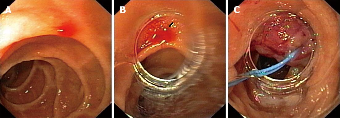
A 61-year-old woman was admitted to our emergency unit because of melenic stool without abdominal pain, nausea or vomiting. Her previous history indicated that she did not have any serious health problems, just arterial hypertension and hyperlipoproteinemia, which were adequately regulated with chronic therapy. There was no history of alcohol abuse or excess intake of non-steroidal anti-inflammatory drugs (NSAIDs). She was anemic, but showed no signs of hemodynamic instability. Although a nasogastric tube aspirate was clear after insertion, a rectal examination showed evidence of melena. Laboratory tests revealed low levels of erythrocytes 2.8 × 10⁹/L and hemoglobin 79 g/L with normal coagulation parameters. After signing an informed consent, the patient underwent an urgent upper endoscopy (GIF Q160, Olympus Optical Co., Japan). Blood oozing from a pin-point defect with normal surrounding mucosa of the duodenum was visualized and that was suggestive of DL (Figure 1A). A detachable nylon ring was opened at the rim of a transparent ligation chamber attached to the tip of the endoscope (Figure 1B). The lesion was aspirated into the chamber and subsequently the mini-loop was closed and detached, achieving complete hemostasis (Figure 1C). A blood transfusion was administered later, together with pantoprazole in the infusion over a 48 h period. A repeated endoscopy showed that the mini-loop was located at the site of the lesion with no visible sign of acute hemorrhage. The patient was discharged from the hospital on the fourth day with a satisfactory complete blood count (erythrocytes 3.9 × 10⁹/L and hemoglobin 98 g/L). On follow up examination two months later, no pathology of a duodenal mucosa was observed, such as ulceration on the site, and values of her complete blood count were normal.

Figure 1
Figure 1 Sixty-one-year-old woman was admitted to our emergency unit because of melenic stool without abdominal pain, nausea or vomiting. A: Bleeding duodenal Dieulafoy's lesion; B: Visible bleeding Dieulafoy's lesion through the rim of a transparent ligation chamber attached to the tip of the endoscope; C: The mini-loop is closed and detached, achieving complete hemostasis.
DISCUSSION

DL of the duodenum is an uncommon cause of upper GI bleeding, currently experiencing an increase of its frequency because operators’ increased awareness of this condition. The duodenum is the second commonest site of DLs, accounting for 15% of patients, second only to the lesser curvature of the stomach, which accounts for around 70%[3,5]. Although the incidence of DL is unknown, it accounts for 2%-3% of all GI hemorrhages. Clinical presentations of this condition are extremely wide-ranging: from symptom free, and therefore, frequently undiagnosed cases, to signs of severe and often fatal GI hemorrhage. These lesions usually appear among the elderly, predominantly in men, and are associated with multiple co-morbidities such as chronic renal and heart failure. Other risk factors include the usage of NSAIDs, and antiplatelet and anticoagulant therapy. The patient we treated was an elderly woman with a history of arterial hypertension and hyperlipoproteinemia.

Upper endoscopy, the gold standard, is diagnostic in 90% of the cases; other available diagnostic techniques are angiography and endoscopic ultrasound (EUS). Selective catheter-directed embolization, especially in patients at high risk for surgery, is usually a salvage method in cases of unsuccessful endoscopic treatment, particularly if the lesion cannot be visualized because of excessive hemorrhage[6]. EUS is used to help detect the site of the submucosal vessel, especially in cases of spouting bleeding and to confirm ablation of a DL after therapy.

The endoscopic appearance of a DL is typically arterial spurting or oozing streaming from a minimal defect of the normal surrounding mucosa, or, less commonly, a protruding vessel without signs of active bleeding or with an adherent clot. In this case, blood oozing out of the normal mucosa was visualized without the possibility to distinguishing the underlying vessel. Once the diagnosis is established, several endoscopic techniques can be used, depending on the site of the lesion, severity of symptoms and previous operator experience. Available endoscopic procedures are electrocoagulation, heat probe coagulation, argon plasma coagulation, local epinephrine injection, sclerotherapy, banding and hemoclip. There is no consensus as to which is the most appropriate method. Based on the available data, endoscopic mechanical haemostatic methods seem to be superior to thermal or injection treatment methods[7,8]. However, the most commonly used methods are injection therapy and mechanical endoscopic therapy with hemoclips. In our case, a mechanical hemostatic method was used; the bleeding vessel with the surrounding mucosa was aspirated into the transparent ligation chamber and a preloaded nylon ring, called a mini-loop, was closed and detached with consequent complete hemostasis. A search of the published literature using PubMed with the phrase “Dieulafoy’s lesion” resulted in 211 articles, of which the first was dated to early 1978 reporting two cases of DL of the jejunum[9]. First report of a duodenal DL was published in 1993[10] and to the best of our knowledge, there have been no reports of mini-loop ligation as a choice of endoscopic treatment for DL. Despite the fact that the first choice therapy for the bleeding duodenal DLs among most endoscopists includes injection of epinephrine and electrocoagulation[11], mechanical techniques are used more frequently and endoscopic band ligation (EBL) has been demonstrated to be an effective treatment for bleeding esophageal, stomach, duodenal and rectal DLs[12,13]. Chung et al[14] and Yanar et al[15] demonstrated higher efficacy of mechanical hemostatic therapy in initial hemostasis and lowering re-bleeding rate with hemoclip and band ligation compared to injection therapy in patients with DL. On the other hand, EBL has been shown to be as effective as injection therapy with epinephrine, with or without thermal therapy, with a significantly longer length of hospitalization among patients treated with injection therapy or combined therapy with epinephrine and thermal therapy[16]. In a study that compared two mechanical methods, EBL and endoscopic hemoclip placement (EHP), no difference was detected in efficacy or safety in the management of bleeding gastric DLs and only one patient from each group experienced re-bleeding[17]. A larger study (66 patients) showed a lower recurrent bleeding rate in patients treated with EBL compared with EHP (3.1% vs 14.7%)[18]. A combined therapy, such as EBL with EUS, promises a new era in diagnostic efficacy in terms of establishing accurate diagnosis, guiding EBL and confirming success of the treatment[19].

Therapeutic endoscopy is the first line of treatment of DL. However, the choice of technique is not specified. In our case, endoscopic mini-loop ligation proved to be an effective, easy to use and safe method for the treatment of DL. Mini-loop ligation may become a method of choice in resolving bleeding DL; however, case reports with longer follow-up periods are needed for a definitive statement because of the substantial risk of re-bleeding from a residual aberrant artery.

Footnotes

P- Reviewers Chamberlain SM, Kouraklis G, Loffroy R, Wilcox CM S- Editor Gou SX L- Editor Stewart G E- Editor Xiong L

References
1.  Dieulafoy G. Exulceratio simplex: Leçons 1-3. Clinique medicale de l’Hotel Dieu de Paris. Paris: Masson et Cie 1898; 1-38.  [PubMed]  [DOI]  [Cited in This Article: ]
2.  Ibrarullah M, Wagholikar GD. Dieulafoy’s lesion of duodenum: a case report. BMC Gastroenterol. 2003;3:2.  [PubMed]  [DOI]  [Cited in This Article: ]  [Cited by in Crossref: 10]  [Cited by in F6Publishing: 14]  [Article Influence: 0.7]  [Reference Citation Analysis (0)]
3.  Baxter M, Aly EH. Dieulafoy’s lesion: current trends in diagnosis and management. Ann R Coll Surg Engl. 2010;92:548-554.  [PubMed]  [DOI]  [Cited in This Article: ]  [Cited by in Crossref: 190]  [Cited by in F6Publishing: 161]  [Article Influence: 11.5]  [Reference Citation Analysis (0)]
4.  Ljubicic N. Efficacy of endoscopic clipping and long-term follow-up of bleeding Dieulafoy’s lesions in the upper gastrointestinal tract. Hepatogastroenterology. 2006;53:224-227.  [PubMed]  [DOI]  [Cited in This Article: ]
5.  Schmulewitz N, Baillie J. Dieulafoy lesions: a review of 6 years of experience at a tertiary referral center. Am J Gastroenterol. 2001;96:1688-1694.  [PubMed]  [DOI]  [Cited in This Article: ]  [Cited by in Crossref: 82]  [Cited by in F6Publishing: 104]  [Article Influence: 4.5]  [Reference Citation Analysis (0)]
6.  Loffroy R, Guiu B. Role of transcatheter arterial embolization for massive bleeding from gastroduodenal ulcers. World J Gastroenterol. 2009;15:5889-5897.  [PubMed]  [DOI]  [Cited in This Article: ]  [Cited by in CrossRef: 59]  [Cited by in F6Publishing: 59]  [Article Influence: 3.9]  [Reference Citation Analysis (0)]
7.  Matsui S, Kamisako T, Kudo M, Inoue R. Endoscopic band ligation for control of nonvariceal upper GI hemorrhage: comparison with bipolar electrocoagulation. Gastrointest Endosc. 2002;55:214-218.  [PubMed]  [DOI]  [Cited in This Article: ]  [Cited by in Crossref: 64]  [Cited by in F6Publishing: 67]  [Article Influence: 3.0]  [Reference Citation Analysis (0)]
8.  Ljubicić N. Endoscopic detachable mini-loop ligation for treatment of gastroduodenal angiodysplasia: case study of 11 patients with long-term follow-up. Gastrointest Endosc. 2004;59:420-423.  [PubMed]  [DOI]  [Cited in This Article: ]  [Cited by in F6Publishing: 7]  [Reference Citation Analysis (0)]
9.  Matuchansky C, Babin P, Abadie JC, Payen J, Gasquet C, Barbier J. Jejunal bleeding from a solitary large submucosal artery. Report of two cases. Gastroenterology. 1978;75:110-113.  [PubMed]  [DOI]  [Cited in This Article: ]
10.  Pollack R, Lipsky H, Goldberg RI. Duodenal Dieulafoy’s lesion. Gastrointest Endosc. 1993;39:820-822.  [PubMed]  [DOI]  [Cited in This Article: ]
11.  Goldenberg SP, DeLuca VA, Marignani P. Endoscopic treatment of Dieulafoy’s lesion of the duodenum. Am J Gastroenterol. 1990;85:452-454.  [PubMed]  [DOI]  [Cited in This Article: ]
12.  Soetikno RM, Piper J, Montes H, Ukomadu C, Carr-Locke DL. Use of endoscopic band ligation to treat a Dieulafoy’s lesion of the esophagus. Endoscopy. 2000;32:S15.  [PubMed]  [DOI]  [Cited in This Article: ]
13.  Yoshikumi Y, Mashima H, Suzuki J, Yamaji Y, Okamoto M, Ogura K, Kawabe T, Omata M. A case of rectal Dieulafoy’s ulcer and successful endoscopic band ligation. Can J Gastroenterol. 2006;20:287-290.  [PubMed]  [DOI]  [Cited in This Article: ]
14.  Chung IK, Kim EJ, Lee MS, Kim HS, Park SH, Lee MH, Kim SJ, Cho MS. Bleeding Dieulafoy’s lesions and the choice of endoscopic method: comparing the hemostatic efficacy of mechanical and injection methods. Gastrointest Endosc. 2000;52:721-724.  [PubMed]  [DOI]  [Cited in This Article: ]  [Cited by in Crossref: 121]  [Cited by in F6Publishing: 124]  [Article Influence: 5.2]  [Reference Citation Analysis (0)]
15.  Yanar H, Dolay K, Ertekin C, Taviloglu K, Ozcinar B, Guloglu R, Barbaros U. An infrequent cause of upper gastrointestinal tract bleeding: “Dieulafoy’s lesion”. Hepatogastroenterology. 2007;54:1013-1017.  [PubMed]  [DOI]  [Cited in This Article: ]
16.  Mumtaz R, Shaukat M, Ramirez FC. Outcomes of endoscopic treatment of gastroduodenal Dieulafoy’s lesion with rubber band ligation and thermal/injection therapy. J Clin Gastroenterol. 2003;36:310-314.  [PubMed]  [DOI]  [Cited in This Article: ]  [Cited by in Crossref: 47]  [Cited by in F6Publishing: 57]  [Article Influence: 2.7]  [Reference Citation Analysis (0)]
17.  Park CH, Joo YE, Kim HS, Choi SK, Rew JS, Kim SJ. A prospective, randomized trial of endoscopic band ligation versus endoscopic hemoclip placement for bleeding gastric Dieulafoy’s lesions. Endoscopy. 2004;36:677-681.  [PubMed]  [DOI]  [Cited in This Article: ]  [Cited by in Crossref: 69]  [Cited by in F6Publishing: 78]  [Article Influence: 3.9]  [Reference Citation Analysis (0)]
18.  Ahn DW, Lee SH, Park YS, Shin CM, Hwang JH, Kim JW, Jeong SH, Kim N, Lee DH. Hemostatic efficacy and clinical outcome of endoscopic treatment of Dieulafoy’s lesions: comparison of endoscopic hemoclip placement and endoscopic band ligation. Gastrointest Endosc. 2012;75:32-38.  [PubMed]  [DOI]  [Cited in This Article: ]  [Cited by in Crossref: 26]  [Cited by in F6Publishing: 29]  [Article Influence: 2.4]  [Reference Citation Analysis (0)]
19.  Folvik G, Nesje LB, Berstad A, Odegaard S. Endosonography-guided endoscopic band ligation of Dieulafoy’s malformation: a case report. Endoscopy. 2001;33:636-638.  [PubMed]  [DOI]  [Cited in This Article: ]  [Cited by in Crossref: 18]  [Cited by in F6Publishing: 22]  [Article Influence: 1.0]  [Reference Citation Analysis (0)]