Published online Oct 21, 2010. doi: 10.3748/wjg.v16.i39.4922
Revised: May 28, 2010
Accepted: June 5, 2010
Published online: October 21, 2010
AIM: To investigate the effect of aqueous extract from Mangifera indica L. (MIE) on dextran sulfate sodium (DSS)-induced colitis in rats.
METHODS: MIE (150 mg/kg) was administered in two different protocols: (1) rectally, over 7 d at the same time as DSS administration; and (2) once daily over 14 d (by oral gavage, 7 d before starting DSS, and rectally for 7 d during DSS administration). General observations of clinical signs were performed. Anti-inflammatory activity of MIE was assessed by myeloperoxidase (MPO) activity. Colonic lipid peroxidation was determined by measuring the levels of thiobarbituric acid reactive substances (TBARS). Reduced glutathione (GSH) levels, expression of inflammatory related mediators [inducible isoforms of nitric oxide synthase (iNOS) and cyclooxygenase (COX)-2, respectively] and cytokines [tumor necrosis factor (TNF)-α and TNF receptors 1 and 2] in colonic tissue were also assessed. Interleukin (IL)-6 and TNF-α serum levels were also measured.
RESULTS: The results demonstrated that MIE has anti-inflammatory properties by improvement of clinical signs, reduction of ulceration and reduced MPO activity when administered before DSS. In addition, administration of MIE for 14 d resulted in an increase in GSH and reduction of TBARS levels and iNOS, COX-2, TNF-α and TNF R-2 expression in colonic tissue, and a decrease in IL-6 and TNF-α serum levels.
CONCLUSION: MIE has anti-inflammatory activity in a DSS-induced rat colitis model and preventive administration (prior to DSS) seems to be a more effective protocol.
-
Citation: Márquez L, Pérez-Nievas BG, Gárate I, García-Bueno B, Madrigal JL, Menchén L, Garrido G, Leza JC. Anti-inflammatory effects of
Mangifera indica L. extract in a model of colitis. World J Gastroenterol 2010; 16(39): 4922-4931 - URL: https://www.wjgnet.com/1007-9327/full/v16/i39/4922.htm
- DOI: https://dx.doi.org/10.3748/wjg.v16.i39.4922
Ulcerative colitis (UC) is a chronic, idiopathic inflammatory bowel disease that is characterized by bloody diarrhea, colonic mucosal ulceration and, in severe cases, systemic symptoms. An abnormal immune response against antigens of the colonic microbiota in genetically predisposed individuals is suggested to be involved in the etiology of UC[1]. Several authors have proposed that such intestinal conditions are mediated by the activation of lymphocytes and non-lymphoid cells such as macrophages and neutrophils. Once a large number of neutrophils and macrophages are activated, these cells enter the injured mucosa of the large intestine, which leads to over-production of oxygen free radicals that can cause injury to target cells in inflamed tissue[2]. Many animal models have been designed to study pathogenic events during colitis development. The symptoms and colonic histopathology of the rodent colitis model induced by dextran sulfate sodium (DSS) salt resemble more human UC than other chemically induced colitis, and has become a research model for the pathogenesis of UC and for the development of new drugs[3].
In spite of several pharmacological treatments for UC, new therapies must be developed to increase the number and duration of remissions. In this regard, traditional medicine worldwide is nowadays being re-evaluated by extensive research on different plants and their therapeutic principles. Many plants produce antioxidant compounds to control the oxidative stress caused by sunbeams and oxygen, and represent a source of new compounds with antioxidant activity[4]. An aqueous stem bark extract from Mangifera indica (M. indica) L. (Anacardiaceae family) has been traditionally used as a nutritional supplement. The composition is a defined mixture of polyphenols, flavonoids, triterpenoids, steroids, phytosterols, fatty acids and microelements (mainly zinc, copper and selenium)[5]. The extract has been described as an antioxidant with anti-inflammatory and immunomodulatory activities in several experimental settings[6-8]. In addition, some experimental models have demonstrated that M. indica extract (MIE) improves its effects when it is given on various days before the induction of damage[9]. For example, when administered orally 1 h before lipopolysaccharide (LPS), MIE inhibited LPS-induced tumor necrosis factor (TNF)-α production in mice dose-dependently with ED50 64.5 mg/kg. However, the extract inhibited the TNF serum levels but with ED50 37.4 mg/kg when it was administered orally during 7 d before LPS challenge. The increasing evidence related to the positive effects of natural compounds with antioxidant and anti-inflammatory properties on UC prompted us to investigate whether MIE could protect colonic mucosa of rats from damage induced by oral administration of DSS, using two different treatment protocols, and to elucidate the possible mechanism(s) involved.
Twenty-eight male outbred Wistar Hannover rats (HsdRccHan:Wist, from Harlan Spain), initially weighing 190-200 g, were housed five per cage and maintained in an animal holding room controlled at a constant temperature of 24 ± 2°C, with a relative humidity of 70% ± 5% and a 12-h light/dark cycle. Animals were fed a standard pellet chow with free access to fresh tap water. All experimental protocols followed the guidelines of the Animal Welfare Committee of the Universidad Complutense according to European legislation (2003/65/EC). Chemicals were from Sigma (Spain) or as indicated.
M. indica L. was collected from a cultivated field located in the region of Pinar del Rio, Cuba. Voucher specimens of the plant (Code: 41722) were deposited at Herbarium of the Academy of Sciences, Institute of Ecology and Systematics, Ministry of Science, Technology and Environment, La Habana, Cuba. Stem bark extract was concentrated by evaporation and spray-dried to obtain a fine brown powder, which is used as the standardized active ingredient of MIE formulations. It melts at 210-215°C with decomposition. The chemical composition of MIE has been characterized by chromatographic (planar, liquid and gas) methods, mass spectrometry, nuclear magnetic resonance (NMR), and UV-V spectrophotometry (fully described in[5]). The elemental inorganic composition has been determined by inductively coupled plasma spectrometry[6]. Extracts were prepared by suspending powder in 0.5% carboxymethylcellulose for oral administration and in melted suppository vehicle (Witepsol H15; Sasol, Witten, Germany) for rectal administration.
The experiment lasted for 21 d. The rats were randomly divided into four groups (Table 1). Control, A and B groups received vehicle orally during 2 wk. Group C received MIE (150 mg/kg) orally once daily. At day 15, oral administration was stopped and colitis was induced by 4% DSS (MP Biomedicals) in drinking water during 7 d for groups A, B and C. The control group received water. At the same time, groups B and C were co-treated rectally with extract at an equal dose while the controls and group A received vehicle rectally.
| Group | Treatment regimens | n | ||
| Week 1 | Week 2 | Week 3 | ||
| Control | Vehicle po | Vehicle po | DSS no + vehicle | 4 |
| A | Vehicle po | Vehicle po | DSS yes + vehicle | 8 |
| B | Vehicle po | Vehicle po | DSS yes + MIE 150 mg/kg rectal | 8 |
| C | MIE 150 mg/kg po | MIE 150 mg/kg po | DSS yes + MIE 150 mg/kg rectal | 8 |
Macroscopic assessments, including weight changes, visible fecal blood and stool consistency were determined. The severity of diarrhea was evaluated according to the following score: no diarrhea = 0; mild diarrhea = 2; severe watery diarrhea = 3; and severe watery diarrhea with blood = 4[10]. Seven days after DSS (or 21 d from the onset of the study), animals were sacrificed after terminal anesthesia with sodium pentobarbital, and the entire colon was removed. The colon length was measured and colon samples were collected for biochemical determinations and histological assessment.
Each removed colon was washed in saline solution and cut longitudinally. Distal fractions were immediately embedded in Tissue-Teck OCT (Sakura), frozen and cut in transverse sections (7 μm) in a microtome cryostat. Samples were mounted on glass slides, cleaned and stained with hematoxylin and eosin for histological evaluation. Each slide was coded and analyzed in a blinded fashion by two investigators who assigned to each sample a histological score based on mucosal injury, with particular attention paid to alterations of the colonic crypts and the presence of inflammation in the colon. Colonic epithelial damage was assessed as: grade 0, normal; grade 1, slight damage and a few inflammatory cells infiltrated in a small area of mucosa; grade 2, moderate damage in two or more areas of the mucosa, with slight bleeding of the submucosa and mild inflammatory infiltrate; and grade 3, severe damage of the mucosa that extended into the muscular mucosa, with loss of the epithelium, and a large inflammatory infiltrate[1].
Immediately after removal, colon samples were minced on ice and homogenized (glass/glass) in 0.5% hexadecyltrimethylammonium bromide, 0.5% Nonidet P40 (Boehringer, Mannheim, Germany) in 20 mmol/L phosphate buffer, pH 6.0. The homogenates were then centrifuged for 20 min at 12 000 g. Tissue levels of myeloperoxidase (MPO) were determined in supernatants using hydrogen peroxide as a substrate for the enzyme. A unit of MPO activity was defined as that which converted 1 μmol hydrogen peroxide to water in 1 min at 40°C[11].
Lipid peroxidation was measured by the thiobarbituric acid test for malondialdehyde (MDA) following a previously described method[12] with some modifications. Colonic samples were homogenized (glass/glass) in 10 vol 50 mmol/L phosphate buffer and deproteinized with 40 % trichloroacetic acid and 5 mol/L HCl, followed by addition of 2 % (w/v) thiobarbituric acid in 0.5 mol/L NaOH. The reaction mixture was heated in a water bath at 90°C for 15 min and centrifuged at 12 000 g for 20 min. The pink chromogen was measured at 532 nm in a Beckman DU-7500 spectrophotometer. The results were expressed as nmol/mg protein.
Reduced glutathione (GSH) levels were determined in accordance with a procedure described by Kamencic et al[13]. Frozen colonic samples were homogenized (glass/glass) in 20 vol cold 50 mmol/L Tris buffer, pH 7.4. Homogenates were centrifuged at 12 000 g for 20 min and the supernatants were collected. The samples were then treated with monochlorobimane (mCB) 100 μmol/L and glutathione-s-transferase 1 U/mL, and were incubated at room temperature for 30 min. The GSH-mCB adducts were measured in a Labsystems Fluoroskan reader with excitation at 380 nm and emission measured at 470 nm. Concentration of GSH in samples were calculated by standard curve of GSH and expressed as μg/mg protein.
To determine the levels of inducible nitric oxide synthase (iNOS), inducible cyclooxygenase (COX)-2, TNF-α and its receptors TNF-R1, and TNF-R2, tissues were homogenized at 4°C in 5 vol buffer that contained 320 mmol/L sucrose, 1 mmol/L, DL-dithiothreitol, 10 μg/mL leupeptin, 10 μg/mL soybean trypsin inhibitor, 2 μg/mL aprotinin and 50 nmol/L Tris brought to pH 7.0, and supernatants after centrifugation at 12 000 g for 20 min were used. The supernatants were diluted (Laemmli) and heated at 90°C for 10 min. After loading (20 μg protein), proteins were sized-separated in 10% or 14% (for TNF-α analysis) SDS-PAGE (90 mV). The gels were processed against the antigens and after blotting onto a polyvinylidene difluoride membrane (Millipore, Bedford, MA, USA), they were incubated with specific goat polyclonal anti-rat COX-2 (1:1000), rabbit polyclonal anti-rat iNOS (1:1000), polyclonal rabbit anti-rat TNF-α (1:1000), polyclonal rabbit anti-rat TNF-R1 (1:500) and polyclonal rabbit anti-rat TNF-R2 (1:500) antibodies (all from Santa Cruz Biotechonology, Santa Cruz, CA, USA, except anti-rat TNF-α that was purchased from PeproTech EC). The correspondent peroxidase secondary antibody was used and proteins recognized by the antibody were visualized on X-ray film by chemiluminescence following the manufacturer’s instructions (Amersham Ibérica, Madrid, Spain). Autoradiographs were quantified by densitometry (Software Total Lab Dynamics Ltd, Phoretix, Newcastle, UK), and several time expositions were analyzed to ensure the linearity of the band intensities.
ABC-ELISAs of double antibodies sandwich were adopted for determination of the two cytokines (kits were obtained from R&D Corporation).
All results are presented as mean ± SE. Data were analyzed using the Graph Pad Prism 4 statistical software. One-way analysis of variance followed by Newman-Keuls test were used for statistical evaluation of the parametric data. Non-parametric data were analyzed by Kruskal-Wallis one-way analysis followed by Dunn’s test. P < 0.05 was considered as statistically significant.
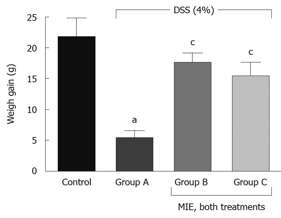
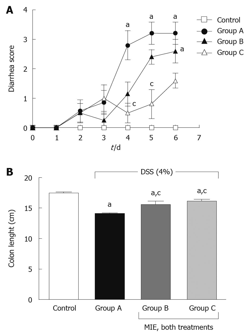
None of the animals in the four experimental groups died throughout the experiment. The intake of drinking water in the three groups administered with DSS (A-C) decreased significantly from the beginning compared with that in the control group (data not shown). The weight gain of rats in the DSS group (A) was significantly lower than in the control group. Administration of MIE in the both pre/co-treated (C) and co-treated only (B) groups prevented this effect (Figure 1). On the other hand, all groups with DSS exhibited an increase in diarrhea and rectal bleeding from day 4 post-DSS until the end of experiment. However, in the case of group C (pre/co-treated group), diarrhea score was found to be less severe than in the DSS group (A) at days 4 and 5 post-DSS. Group B did not show any significant differences compared to the group that received DSS alone (Figure 2A). Colon length is a useful assessment of colitis and it is considered as a marker of inflammation. As shown in Figure 2B, 7 d after DSS administration, there was a significant shortening of the colon length in the group given DSS only (group A: 14.1 ± 0.1 cm) compared with the control group (17.4 ± 0.2 cm). In both pre-and co-treated groups (C and B), MIE significantly improved this inflammatory marker.
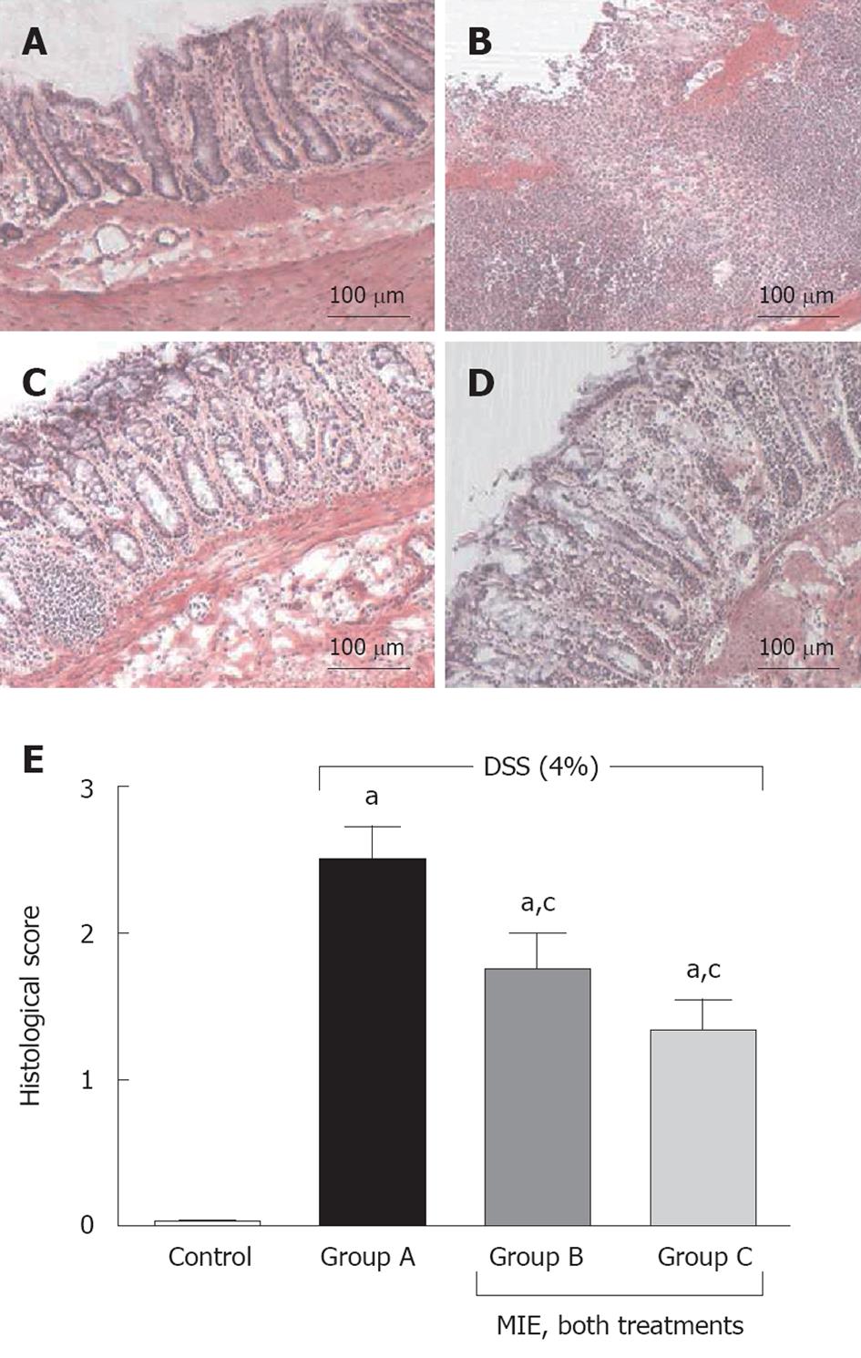
The occurrence of UC was corroborated on the basis of histological damage and inflammatory infiltrate as shown in Figure 3. Figure 3D summarizes the microscopical damage scores from DSS rats and DSS rats treated with MIE. The control group exhibited normal mucosal morphology. Rats that received DSS and vehicle (group A) showed extensive mucosal damage with a large number of inflammatory cells, obtaining as a result, the highest score in the microscopic analysis. MIE in both treatment protocols (groups B and C) decreased the grade and number of ulcerations and diminished the inflammatory infiltrate.
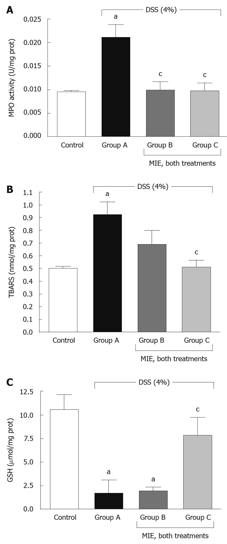
DSS colitis was also characterized by increased MPO activity in colonic tissue, an indicator of polymorphonuclear leukocyte accumulation. The DSS group (A) showed a significant elevation of MPO levels in colonic tissue (21.1 ± 2.7 mU/mg), P < 0.05 vs the control group. The increase observed in the DSS group was clearly diminished by both treatments with MIE as shown in Figure 4A.
The effect of MIE on lipid peroxidation - an indicator of cell membrane damage as a result of oxidative toxicity - in rats treated with 4% DSS is shown in Figure 4B. In the DSS-induced colitis rats, the level of TBARS was significantly increased (0.71 ± 0.07 nmol/mg) when compared with the control group (0.41 ± 0.05 nmol/mg). Although previous administration of MIE resulted in a reduction in TBARS level (group C, 0.4 ± 0.04 nmol/mg), co-treatment with MIE (group B) did not decrease TBARS level (0.57 ± 0.16 nmol/mg) compared with that in the DSS-treated rats.
GSH is one of the most important endogenous antioxidants. Figure 4C shows a significant decrease of GSH in group A (1.68 ± 1.4 μg/mg) compared to the control group (10.55 ± 1.6 μg/mg). In this case, there were no significant differences between the DSS group and group B (co-treated but not pre-treated with 1.91 ± 0.4 μg/mg MIE). However, the administration of MIE prior to 4% DSS resulted in an increase in GSH level (7.80 ± 1.91 μg/mg) compared with that in the 4% DSS treatment group.
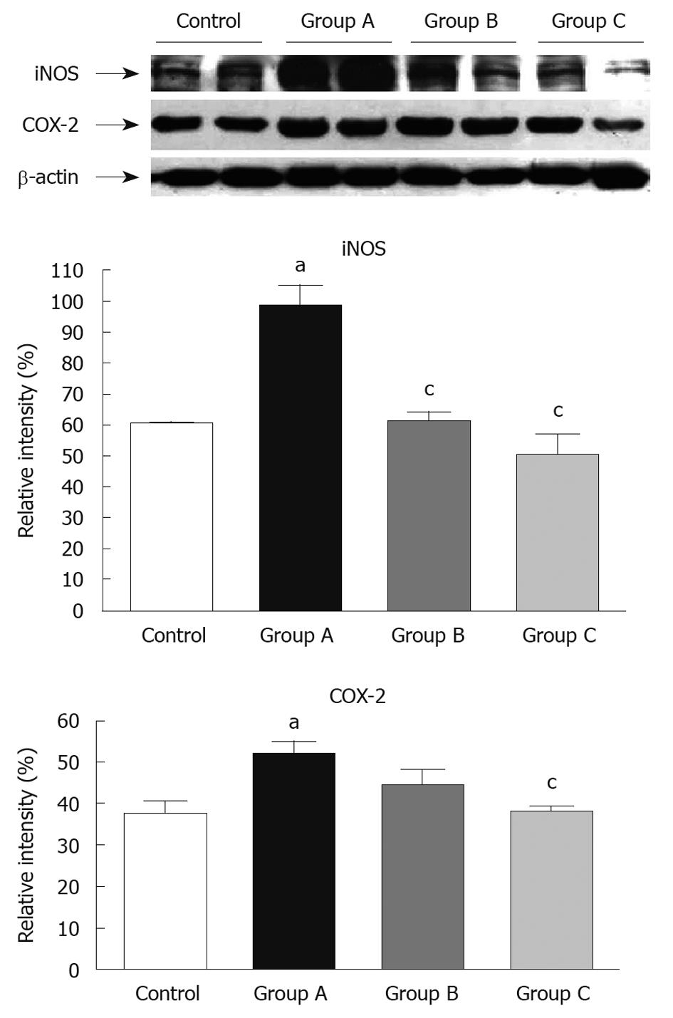
When rats were treated with 4% DSS, the levels of inflammation-related proteins (iNOS and COX-2) in colonic tissue were significantly increased (Figure 5). In the case of iNOS, both treatments with MIE resulted in a decrease of expression. However, for COX-2 expression, attenuation of band intensities was observed only in group C (pre-treated group).
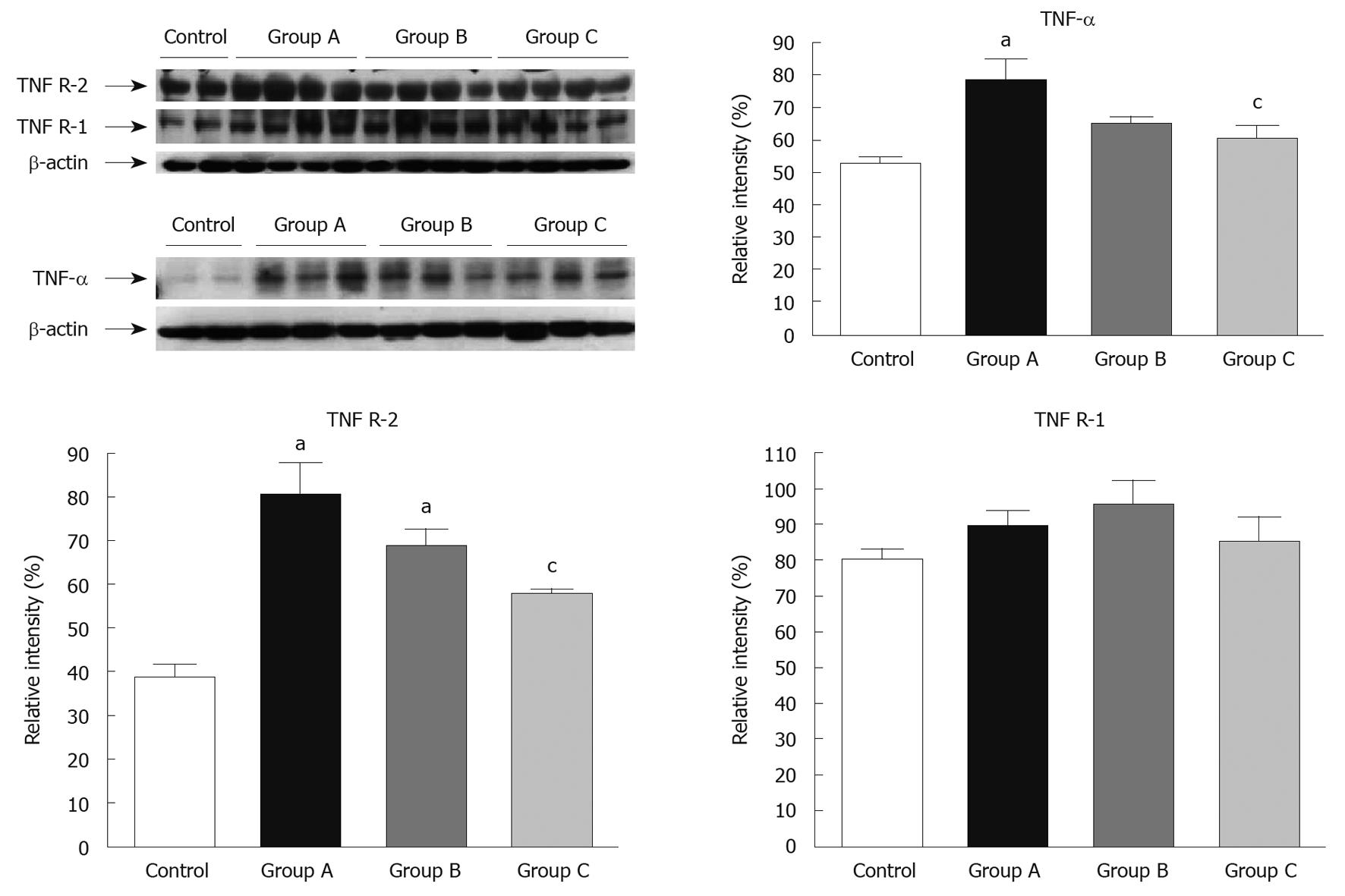
Administration of 4% DSS induced a significant increase in TNF-α (Figure 6, lanes 3-5) and TNF R-2 in inflamed tissue (Figure 6, lanes 3-6). Treatment with MIE 15 d before 4% DSS resulted in a gradual weakness of band intensities for TNF-α and TNF R-2. Relative band intensities of increased TNF-α and TNF R-2 expression caused by DSS were reduced by prior treatment with MIE (group C) in 17.8 and 22.8% respectively vs DSS. There were no significant differences between relative intensities in group B compared with the group treated with DSS alone. Expression of TNF R-1 was not affected by DSS supplementation.
Based on the effects of MIE given before colitis induction (group C) on tissue cytokine expression, we tested the systemic levels of cytokines. Administration of 4% DSS produced an increase in TNF-α serum levels (46.2%), whereas interleukin (IL)-6 serum levels showed a tendency to elevation in group A (treated with DSS alone) but this was not statistically significant (18.1%). Treatment with MIE, before DSS intake, clearly decreased TNF-α levels by 44.3% and reduced IL-6 serum levels (down to control serum levels) by 58.8% (Table 2).
UC is a chronic, relapsing disease that causes inflammation and ulcerations of the colonic mucosa with a variable extent and severity. The etiology of UC remains essentially unknown but the results from many studies in humans and animal models suggest that it is related to an abnormal immune response in the gastrointestinal tract, possibly associated with genetic and environmental - mainly microbial - factors[14]. Aminosalicylates, glucocorticoids and immunosuppressive drugs have been mainly used for the treatment and maintenance of remission of UC, but the side effects or toxicity of these drugs represents a major clinical problem[15]. For these reasons, natural medicine has become an alternative therapy in addition to the conventional therapies that are used to treat UC[16].
In the present study, we demonstrated that MIE has an anti-inflammatory effect on colonic injury provoked by oral supplementation with DSS in rats, mainly when it is administered before the induction of damage. DSS-induced colitis is a well-established model that is phenotypically similar to UC in humans[17]. Oral administration of DSS for several days, leads to colonic epithelial lesions and acute inflammation characterized by the presence of neutrophils and macrophages within damaged segments. The reason for the deleterious effects of DSS is not well understood, however, epithelial cell permeability and macrophage activation have been proposed as potential mechanisms. We administered M. indica extract in two different protocols to evaluate the role of pretreatment with this product. The decrease in colitis induced by MIE was accompanied by a lower weight loss of rats and a partial restoration of colon length, which is an indirect assessment of colon inflammation. However, a decrease in the occurrence of diarrhea was only observed when MIE was administered before DSS. Confirming clinical results, microscopic analysis established a protective action of MIE, which was measured as a decrease in ulceration, conservation of epithelial crypts, and a reduction in infiltrated cells. These effects were more evident in the pretreated group.
The infiltration of leukocytes into the mucosa contributes significantly to the tissue necrosis and mucosal dysfunction, as they represent a major source of reactive oxygen species (ROS)[18]. MPO is an enzyme that is found predominantly in neutrophils, and a good marker of inflammation and tissue injury. Therefore, the decrease of MPO activity can be explained through the reduction of neutrophil accumulation in inflamed tissue[19]. In addition, oxygen radicals and NO can interact and exert a cytotoxic effect by causing lipid peroxidation, which results in the formation of MDA[20]. Our results showed that MIE in both treatment protocols inhibited MPO activity, whereas the decrease in MDA production was only observed when animals received MIE before DSS administration. A decrease of MPO activity with MIE treatment in different experimental models of inflammation (ear and paw edema) has been described[21]. In addition, several studies have established the high antioxidant capacity of the extract by blocking oxygen radical formation[22,23]. The mechanism involved is associated with the antioxidant activity reported for mangiferin, which has a low redox potential that proves its ROS scavenger ability[24]. Therefore, the antioxidant capacity of the extract, administered prior to colitis development, probably leads to a decrease of lipid peroxidation and MPO activity. However, the results presented here indicated that co-treatment with MIE was not sufficient to reduce MDA levels. This could be related to the necessary oxidative pre-conditioning that has been described for many antioxidants[25]. We hypothesize that MIE could be useful in the prevention of relapse in patients with quiescent UC.
Furthermore, the increased generation of highly toxic ROS in UC exceeds the limited intestinal antioxidant defense system, thereby contributing to intestinal oxidative injury. Glutathione, as the most abundant cellular antioxidant system in animal cells, plays an essential role in modulating cell responses to redox changes[26]. GSH deficiency predisposes animals to organ failure and death after an otherwise nonlethal period of hypotension[27,28]. GSH deficiency is associated with severe injury such as inflammation and sepsis, therefore, treatment strategies that maintain GSH stores might decrease the incidence of organ failure. Our findings demonstrated that MIE administered before colitis induction produced a significant increase in GSH levels, which were probably associated with the radical scavenger capacity of the extract and the protection of thiol groups described by numerous polyphenols[29]. Polyphenols are the main constituent of MIE (around 50%)[5].
Moreover, pathological invasion of inflammatory cells into the mucosa produces increased concentrations of inflammatory cytokines such as interleukins, TNF-α and interferon-γ[30]. Pro-inflammatory cytokines induce the expression of genes associated with inflammation, such as iNOS, and stimulate iNOS activity, which increases the production of the free radical NO[31]. Studies in knockout mice have demonstrated that iNOS plays an important role in the pathogenesis of colitis[32], and the role of iNOS in the pathogenesis of human UC has been previously suggested[33]. In the present study, MIE inhibited iNOS expression, as described in other inflammatory experimental settings[34].
In addition to iNOS, DSS-induced expression of COX-2 was also inhibited by prior administration of MIE. Previous studies in endotoxin-stimulated macrophages also have demonstrated that MIE inhibits COX-2 protein and mRNA levels, but at doses higher than those required for iNOS inhibition, which suggests that longer treatments or higher doses of MIE than those needed for inhibition of COX-2[34] are necessary. This might explain the lack of effect when the extract was administered only in the co-treatment regimen. The synthesis and activity of iNOS and COX-2 are induced by almost the same pro-inflammatory stimuli and are associated with inflammatory conditions. Therefore, it is possible that inhibition of iNOS and COX-2 induced by prior treatment with MIE could provide the most potent anti-inflammatory effect.
On the other hand, TNF-α has been described as a key molecule in UC pathogenesis, and a monoclonal antibody against this molecule, such as infliximab, has proven to be effective in the treatment of moderate to severe UC[35]. This cytokine, by interaction with its receptors I and II, recruits leukocytes to inflammatory sites, stimulates monocytes and vascular endothelial cells to express cytokines, induces the cascade effects for other cytokines, and finally results in inflammatory lesions in tissues[36,37]. Our results demonstrated that prior administration of MIE inhibits DSS-induced increased TNF-α and TNF R-II expression. TNF R-I is expressed constitutively, whereas TNF R II is induced by diverse stimuli and plays a key role in the local inflammatory response[38]. Previous in vivo and in vitro studies have appointed MIE as a potent TNF-α inhibitor[9] and some polyphenols structurally related to those present in MIE inhibit lymphocyte proliferation and cytokine production[39,40]. Moreover, the reduction of TNF R-II receptor expression seems to enhance the inhibitory action of the extract on the TNF-α signaling system.
The reduction of inflammatory enzymes iNOS and COX-2, TNF-α and TNF R-II expression induced by MIE can be correlated with its antioxidant properties. The effects of antioxidant agents have been ascribed by some authors to inhibition of activation of the nuclear transcription factor nuclear factor (NF)-κB, which is activated by ROS with the subsequent induction and expression of various cytokines (such as TNF-α) and enzymes (i.e. iNOS and COX-2)[41,42] that are involved in the induction and development of UC. Although in vitro studies have demonstrated that MIE inhibits NF-κB in macrophages[43], further research is necessary to demonstrate that MIE exerts an inhibitory effect on NF-κB signaling pathways.
In addition, TNF-α and IL-6 serum levels were determined in our study. Administration of DSS produced an increase in systemic TNF-α levels, which was reversed by prior administration of MIE. This fact is probably associated with the molecular changes found in the local inflammatory focus. Although several studies have establish an increase in IL-6 serum levels after DSS supplementation[3,44], our results demonstrated a non-significant tendency to increase IL-6 levels in serum. Nevertheless, prior administration of MIE produced a significant decrease in this cytokine. A previous study has demonstrated the ability of MIE to modulate macrophage function through inhibition of chemotaxis and phagocytosis[43]. Macrophages are one of the main sources of cytokines (i.e IL-6 and TNF-α), therefore, a possible modulation of macrophage activity by MIE could influence the decrease in cytokine production. This result suggests an important role for MIE as a modulator of the immune system and should be taken into account for future investigations.
In conclusion, the results showed that MIE administered in co-treatment regimens is able to prevent body weight loss and colon shortness, as well as modulate MPO activity and reduce iNOS expression levels. However, when MIE is administered before DSS damage, its protective effects are broader and enhanced, as demonstrated by a decrease in diarrhea and lipid peroxidation; an increase in GSH levels; a decrease in iNOS, COX-2, TNF-α and TNF R-II expression levels, as well as a reduction in TNF-α and IL-6 serum levels.
Ulcerative colitis (UC) is a chronic inflammatory bowel disease (IBD) that is characterized by bloody diarrhea, colonic mucosal ulceration and, in severe cases, systemic symptoms. An exaggerated immune response against antigens of the colonic microbiota in genetically predisposed individuals is suggested to be involved in the etiology of UC. Current available treatment includes anti-inflammatory and immunosuppressive agents, all of which have many adverse reactions after long-term treatment to prevent remissions of the disease.
New therapies must be developed to increase the number and duration of remissions. In this vein, traditional medicine around the world is now being re-evaluated by extensive research on different plants and their therapeutic principles. Many plants produce antioxidant compounds to control oxidative stress. An aqueous stem bark extract from Mangifera indica L. (MIE, Anacardiaceae family), has been traditionally used as a nutritional supplement. The composition is a defined mixture of polyphenols, flavonoids, triterpenoids, steroids, phytosterols, fatty acids and microelements (mainly zinc, copper and selenium). The extract has been described as an antioxidant with anti-inflammatory and immunomodulatory activities in several experimental settings.
MIE has an anti-inflammatory effect on colonic injury in a rat model of UC, mainly when it is administered before the induction of damage. The decrease in colitis induced by MIE was accompanied by a lower weight loss of rats and a partial restoration of colon length, which is an indirect assessment of colon inflammation. Furthermore, a decrease was also observed in occurrence of diarrhea, which is the main clinical finding. By confirming the clinical results, microscopic analysis established protective activity of MIE, as measured by a decrease in ulceration, conservation of epithelial cells, and a reduction in infiltrating cells. Finally, MIE modulated most of the inflammatory mediators in colitis: inducible nitric oxide synthase (NOS), inducible cyclooxygenase (COX), and consequent lipid peroxidation. MIE inhibited two of the main inflammatory cytokines, tumor necrosis factor (TNF)-α and interleukin (IL)-6.
MIE is able to prevent body weight loss and colon shortness, as well as decrease some of the intra- and intercellular mechanisms of inflammatory damage in the colon, in an animal model of UC. In this way, this study might represent a future strategy for therapeutic intervention in the preventive management of patients with UC.
NOS and COX are two enzymatic sources of inflammatory mediators, and their activation leads to an increase in reactive oxygen species, which can damage cells. Peroxidation of lipid components of the cell membranes is the result of this damage. Cytokines are a family of pleiotropic intercellular proteins, mainly in immunological cells, and most of them are pro-inflammatory, such as TNF-α and IL-6.
This is a novel and interesting study that demonstrates the anti-inflammatory effects of MIE on colonic mucosa in a DSS colitis model in rats. The results are important and potentially relevant for designing therapy in IBD. The results have are well presented and support the authors’ conclusions. However, the addition of another colitis model would improve the paper.
Peer reviewer: Didier Merlin, PhD, Associate Professor, Department of Medicine Division of Digestive Diseases, Emory University, 615 Michael Street, Atlanta, GA 30322, United States
S- Editor Wang YR L- Editor Kerr C E- Editor Lin YP
| 1. | Murakami A, Hayashi R, Tanaka T, Kwon KH, Ohigashi H, Safitri R. Suppression of dextran sodium sulfate-induced colitis in mice by zerumbone, a subtropical ginger sesquiterpene, and nimesulide: separately and in combination. Biochem Pharmacol. 2003;66:1253-1261. |
| 2. | Oh PS, Lim KT. Plant originated glycoprotein has anti-oxidative and anti-inflammatory effects on dextran sulfate sodium-induced colitis in mouse. J Biomed Sci. 2006;13:549-560. |
| 3. | Zheng P, Niu FL, Liu WZ, Shi Y, Lu LG. Anti-inflammatory mechanism of oxymatrine in dextran sulfate sodium-induced colitis of rats. World J Gastroenterol. 2005;11:4912-4915. |
| 4. | Hernández P, Delgado R, Walczak H. Mangifera indica L. extract protects T cells from activation-induced cell death. Int Immunopharmacol. 2006;6:1496-1505. |
| 5. | Núñez Sellés AJ, Vélez Castro HT, Agüero-Agüero J, González-González J, Naddeo F, De Simone F, Rastrelli L. Isolation and quantitative analysis of phenolic antioxidants, free sugars, and polyols from mango (Mangifera indica L.) stem bark aqueous decoction used in Cuba as a nutritional supplement. J Agric Food Chem. 2002;50:762-766. |
| 6. | Sellés AJ, Rodríguez MD, Balseiro ER, Gonzalez LN, Nicolais V, Rastrelli L. Comparison of major and trace element concentrations in 16 varieties of Cuban mango stem bark (Mangifera indica L.). J Agric Food Chem. 2007;55:2176-2181. |
| 7. | Martínez G, Delgado R, Pérez G, Garrido G, Núñez Sellés AJ, León OS. Evaluation of the in vitro antioxidant activity of Mangifera indica L. extract (Vimang). Phytother Res. 2000;14:424-427. |
| 8. | Garrido G, González D, Delporte C, Backhouse N, Quintero G, Núñez-Sellés AJ, Morales MA. Analgesic and anti-inflammatory effects of Mangifera indica L. extract (Vimang). Phytother Res. 2001;15:18-21. |
| 9. | Garrido G, Delgado R, Lemus Y, Rodríguez J, García D, Núñez-Sellés AJ. Protection against septic shock and suppression of tumor necrosis factor alpha and nitric oxide production on macrophages and microglia by a standard aqueous extract of Mangifera indica L. (VIMANG). Role of mangiferin isolated from the extract. Pharmacol Res. 2004;50:165-172. |
| 10. | Kim TW, Seo JN, Suh YH, Park HJ, Kim JH, Kim JY, Oh KI. Involvement of lymphocytes in dextran sulfate sodium-induced experimental colitis. World J Gastroenterol. 2006;12:302-305. |
| 11. | Ponferrada A, Caso JR, Alou L, Colón A, Sevillano D, Moro MA, Lizasoain I, Menchén P, Gómez-Lus ML, Lorenzo P. The role of PPARgamma on restoration of colonic homeostasis after experimental stress-induced inflammation and dysfunction. Gastroenterology. 2007;132:1791-1803. |
| 12. | Das NP, Ratty AK. Studies on the effects of the narcotic alkaloids, cocaine, morphine, and codeine on nonenzymatic lipid peroxidation in rat brain mitochondria. Biochem Med Metab Biol. 1987;37:258-264. |
| 13. | Kamencic H, Lyon A, Paterson PG, Juurlink BH. Monochlorobimane fluorometric method to measure tissue glutathione. Anal Biochem. 2000;286:35-37. |
| 14. | Chung HL, Yue GG, To KF, Su YL, Huang Y, Ko WH. Effect of Scutellariae Radix extract on experimental dextran-sulfate sodium-induced colitis in rats. World J Gastroenterol. 2007;13:5605-5611. |
| 15. | Sands BE. Therapy of inflammatory bowel disease. Gastroenterology. 2000;118:S68-S82. |
| 16. | Langmead L, Dawson C, Hawkins C, Banna N, Loo S, Rampton DS. Antioxidant effects of herbal therapies used by patients with inflammatory bowel disease: an in vitro study. Aliment Pharmacol Ther. 2002;16:197-205. |
| 17. | Björck S, Jennische E, Dahlström A, Ahlman H. Influence of topical rectal application of drugs on dextran sulfate-induced colitis in rats. Dig Dis Sci. 1997;42:824-832. |
| 18. | Yoshida N, Yoshikawa T, Yamaguchi T, Naito Y, Tanigawa T, Murase H, Kondo M. A novel water-soluble vitamin E derivative protects against experimental colitis in rats. Antioxid Redox Signal. 1999;1:555-562. |
| 19. | Babbs CF. Oxygen radicals in ulcerative colitis. Free Radic Biol Med. 1992;13:169-181. |
| 20. | Guzik TJ, Korbut R, Adamek-Guzik T. Nitric oxide and superoxide in inflammation and immune regulation. J Physiol Pharmacol. 2003;54:469-487. |
| 21. | Garrido G, González D, Lemus Y, García D, Lodeiro L, Quintero G, Delporte C, Núñez-Sellés AJ, Delgado R. In vivo and in vitro anti-inflammatory activity of Mangifera indica L. extract (VIMANG). Pharmacol Res. 2004;50:143-149. |
| 22. | Pardo Andreu G, Delgado R, Velho J, Inada NM, Curti C, Vercesi AE. Mangifera indica L. extract (Vimang) inhibits Fe2+-citrate-induced lipoperoxidation in isolated rat liver mitochondria. Pharmacol Res. 2005;51:427-435. |
| 23. | Sánchez GM, Re L, Giuliani A, Núñez-Sellés AJ, Davison GP, León-Fernández OS. Protective effects of Mangifera indica L. extract, mangiferin and selected antioxidants against TPA-induced biomolecules oxidation and peritoneal macrophage activation in mice. Pharmacol Res. 2000;42:565-573. |
| 24. | Mishra B, Indira PK, Sudheerkumar M, Unnikrishhnan MK, Mohan H. Pulse radiolysis studies of mangiferin: A C-glycosyl xanthone isolated from Mangifera indica. Rad Physics Chem. 2006;75:70-77. |
| 25. | Galvez J, de la Cruz JP, Zarzuelo A, Sanchez de la Cuesta F. Flavonoid inhibition of enzymic and nonenzymic lipid peroxidation in rat liver differs from its influence on the glutathione-related enzymes. Pharmacology. 1995;51:127-133. |
| 26. | Oz HS, Chen TS, McClain CJ, de Villiers WJ. Antioxidants as novel therapy in a murine model of colitis. J Nutr Biochem. 2005;16:297-304. |
| 27. | Robinson MK, Rounds JD, Hong RW, Jacobs DO, Wilmore DW. Glutathione deficiency increases organ dysfunction after hemorrhagic shock. Surgery. 1992;112:140-147; discussion 148-149. |
| 28. | Koch TR, Yuan LX, Fink JG, Petro A, Opara EC. Induction of enlarged intestinal lymphoid aggregates during acute glutathione depletion in a murine model. Dig Dis Sci. 2000;45:2115-2121. |
| 29. | Srinivasan P, Sabitha KE, Shyamaladevi CS. Therapeutic efficacy of green tea polyphenols on cellular thiols in 4-Nitroquinoline 1-oxide-induced oral carcinogenesis. Chem Biol Interact. 2004;149:81-87. |
| 30. | Sandborn WJ, Targan SR. Biologic therapy of inflammatory bowel disease. Gastroenterology. 2002;122:1592-1608. |
| 32. | Krieglstein CF, Cerwinka WH, Laroux FS, Salter JW, Russell JM, Schuermann G, Grisham MB, Ross CR, Granger DN. Regulation of murine intestinal inflammation by reactive metabolites of oxygen and nitrogen: divergent roles of superoxide and nitric oxide. J Exp Med. 2001;194:1207-1218. |
| 33. | Menchén L, Colón AL, Madrigal JL, Beltrán L, Botella S, Lizasoain I, Leza JC, Moro MA, Menchén P, Cos E. Activity of inducible and neuronal nitric oxide synthases in colonic mucosa predicts progression of ulcerative colitis. Am J Gastroenterol. 2004;99:1756-1764. |
| 34. | Leiro J, García D, Arranz JA, Delgado R, Sanmartín ML, Orallo F. An Anacardiaceae preparation reduces the expression of inflammation-related genes in murine macrophages. Int Immunopharmacol. 2004;4:991-1003. |
| 35. | Ferrante M, Vermeire S, Katsanos KH, Noman M, Van Assche G, Schnitzler F, Arijs I, De Hertogh G, Hoffman I, Geboes JK. Predictors of early response to infliximab in patients with ulcerative colitis. Inflamm Bowel Dis. 2007;13:123-128. |
| 36. | Myers KJ, Murthy S, Flanigan A, Witchell DR, Butler M, Murray S, Siwkowski A, Goodfellow D, Madsen K, Baker B. Antisense oligonucleotide blockade of tumor necrosis factor-alpha in two murine models of colitis. J Pharmacol Exp Ther. 2003;304:411-424. |
| 37. | Murthy S, Flanigan A, Coppola D, Buelow R. RDP58, a locally active TNF inhibitor, is effective in the dextran sulphate mouse model of chronic colitis. Inflamm Res. 2002;51:522-531. |
| 38. | Grell M, Douni E, Wajant H, Löhden M, Clauss M, Maxeiner B, Georgopoulos S, Lesslauer W, Kollias G, Pfizenmaier K. The transmembrane form of tumor necrosis factor is the prime activating ligand of the 80 kDa tumor necrosis factor receptor. Cell. 1995;83:793-802. |
| 39. | Hsu MF, Raung SL, Tsao LT, Lin CN, Wang JP. Examination of the inhibitory effect of norathyriol in formylmethionyl-leucyl-phenylalanine-induced respiratory burst in rat neutrophils. Free Radic Biol Med. 1997;23:1035-1045. |
| 40. | Middleton E Jr, Kandaswami C. Effects of flavonoids on immune and inflammatory cell functions. Biochem Pharmacol. 1992;43:1167-1179. |
| 41. | Bremner P, Heinrich M. Natural products as targeted modulators of the nuclear factor-kappaB pathway. J Pharm Pharmacol. 2002;54:453-472. |
| 42. | Taylor BS, de Vera ME, Ganster RW, Wang Q, Shapiro RA, Morris SM Jr, Billiar TR, Geller DA. Multiple NF-kappaB enhancer elements regulate cytokine induction of the human inducible nitric oxide synthase gene. J Biol Chem. 1998;273:15148-15156. |
| 43. | García D, Delgado R, Ubeira FM, Leiro J. Modulation of rat macrophage function by the Mangifera indica L. extracts Vimang and mangiferin. Int Immunopharmacol. 2002;2:797-806. |
| 44. | Araki Y, Andoh A, Fujiyama Y. The free radical scavenger edaravone suppresses experimental dextran sulfate sodium-induced colitis in rats. Int J Mol Med. 2003;12:125-129. |


