Published online Apr 21, 2010. doi: 10.3748/wjg.v16.i15.1854
Revised: January 18, 2010
Accepted: January 25, 2010
Published online: April 21, 2010
AIM: To develop a molecular therapy for pancreatic cancer, the insulin-like growth factor-I (IGF-I) signaling pathway was analyzed.
METHODS: Pancreatic cancer cell lines (MIA-Paca2, NOR-P1, PANC-1, PK-45H, PK-1, PK-59 and KP-4) were cultured in media with 10 mL/L fetal bovine serum. Western blotting analysis was performed to clarify the expression of IGF-I receptor (IGF-IR). Picropodophyllin (PPP), a specific inhibitor of IGF-IR, LY294002, a specific inhibitor of phosphatidylinositol 3 kinase (PI3K), and PD98059, a specific inhibitor of mitogen-activated protein kinase, were added to the media. After 72 h, a 3-(4,5-dimethylthiazol-2-yl)-5-(3-carboxymethoxyphenyl)-2-(4-sulfophenyl)-2H-tetrazolium inner salt (MTS) assay was performed to analyze cell proliferation. A wound assay was performed to analyze cell motility with hematoxylin and eosin (HE) staining 48 h after addition of each inhibitor.
RESULTS: All cell lines clearly expressed not only IGF-IR but also phosphorylated IGF-IR. PPP significantly suppressed proliferation of MIA-Paca2, NOR-P1, PANC-1, PK-45H, PK-1, PK-59 and KP-4 cells to 36.9% ± 2.4% (mean ± SD), 30.9% ± 5.5%, 23.8% ± 3.9%, 37.1% ± 5.3%, 10.4% ± 4.5%, 52.5% ± 4.5% and 22.6% ± 0.4%, at 2 μmol/L, respectively (P < 0.05). LY294002 significantly suppressed proliferation of MIA-Paca2, NOR-P1, PANC-1, PK-45H, PK-1, PK-59 and KP-4 cells to 44.4% ± 7.6%, 32.9% ± 8.2%, 53.9% ± 8.0%, 52.8% ± 4.0%, 32.3% ± 4.2%, 51.8% ± 4.5%, and 30.6% ± 9.4%, at 50 μmol/L, respectively (P < 0.05). PD98059 did not significantly suppress cell proliferation. PPP at 2 μmol/L suppressed motility of MIA-Paca2, NOR-P1, PANC-1, PK-45H, PK-1, PK-59 and KP-4 cells to 3.0% ± 0.2%, 0%, 0%, 2.0% ± 0.1%, 5.0% ± 0.2%, 3.0% ± 0.1%, and 5.0% ± 0.2%, respectively (P < 0.05). LY294002 at 50 μmol/L suppressed motility of MIA-Paca2, NOR-P1, PANC-1, PK-45H, PK-1, PK-59 and KP-4 to 3.0% ± 0.2%, 0%, 3.0% ± 0.2%, 0%, 0%, 0% and 3% ± 0.1%, respectively (P < 0.05). PD980509 at 20 μmol/L did not suppress motility. Cells were observed by microscopy to analyze the morphological changes induced by the inhibitors. Cells in medium treated with 2 μmol/L PPP or 50 μmol/L LY294002 had pyknotic nuclei, whereas those in medium with 20 μmol/L PD98059 did not show apoptosis.
CONCLUSION: IGF-IR and PI3K are good candidates for molecular therapy of pancreatic cancer.
- Citation: Tomizawa M, Shinozaki F, Sugiyama T, Yamamoto S, Sueishi M, Yoshida T. Insulin-like growth factor-I receptor in proliferation and motility of pancreatic cancer. World J Gastroenterol 2010; 16(15): 1854-1858
- URL: https://www.wjgnet.com/1007-9327/full/v16/i15/1854.htm
- DOI: https://dx.doi.org/10.3748/wjg.v16.i15.1854
Molecular alterations of pancreatic cancer need to be clarified to develop molecular therapy. Growth factors transmit signals to stimulate tumor growth and enhance metastasis. Insulin-like growth factor (IGF)-I signaling plays an important role in the growth and development of many tissues[1]. IGF-I signaling is also thought to be involved in tumorigenesis. Upon ligand binding, the tyrosine kinase of IGF-I receptor (IGF-IR) is activated, and results in autophosphorylation of tyrosine[2]. This initiates a phosphorylation cascade to the phosphatidylinositol-3 kinase (PI3K) and mitogen-activated protein kinase (MAPK) pathways.
IGF-I is upregulated in human pancreatic cancer tissues but is not expressed in surrounding non-cancerous tissue[3]. Serum level of IGF-I is elevated in pancreatic cancer patients[4]. Histological analysis has shown that IGF-IR is positive in the membrane of cells of pancreatic cancer tissues but is not expressed in surrounding non-tumorous tissues[3,5]. These facts imply that IGF-I acts as a growth factor for pancreatic cancer. IGF-IR is phosphorylated solely in pancreatic cancer tissues, and not in surrounding non-tumorous tissues[6]. The present study suggests that IGF-IR transmits signals to downstream pathways. This hypothesis is supported by the facts that IGF-I stimulates DNA synthesis of PANC-1 and MIA Paca2 pancreatic cancer cell lines, and motility of HPAF-II, another pancreatic cancer cell line[7,8].
IGF-IR is not involved in metabolism, therefore, it was expected that inhibition of IGF-IR and its downstream pathway would not cause any adverse effects[1]. We therefore focused on the IGF-IR signaling pathway. Pancreatic cancer tissues express growth factors other than IGF-I[9]. We used fetal bovine serum (FBS) as a growth model of pancreatic cancer cells instead of addition of IGF-I in serum-free medium, because pancreatic cancer tissues and serum from patients contain various growth factors.
Pancreatic cancer cell lines, PANC-1, NOR-P1, PK-45H, PK-1, PK-59, and MIA-Paca2 were purchased from RIKEN Cell Bank (Tsukuba, Japan)[10]. NOR-P1 was cultured in Dulbecco’s Minimum Essential Medium (DMEM): F12 medium and MIA-Paca2 was cultured in DMEM, and the others in Roswell Park Memorial Institute (RPMI)-1640 (Sigma, St. Louis, MO, USA). All the media were purchased from Sigma, and supplemented with 100 mL/L FBS (Trace Scientific, Melbourne, Australia). All the cell lines were cultured in 50 mL/L carbon dioxide at 37°C in a humidified chamber. LY294002, a specific inhibitor of PI3K, and PD98059, a specific inhibitor of MAPK, were purchased from Wako Pure Chemicals (Osaka, Japan), and picropodophyllin (PPP), a specific inhibitor of IGF-IR, was from Calbiochem (Darmstadt, Germany)[11].
Protein was isolated from cells after 72 h culture. Twenty micrograms of protein was subjected to sodium dodecyl sulfate polyacrylamide gel electrophoresis (SDS-PAGE), and transferred to a nylon filter. Primary antibodies were polyclonal rabbit anti-IGF-IR antibody, anti-phosphorylated IGF-IR (Cell Signaling Technology, Danvers, MA, USA), and mouse monoclonal anti-tubulin-α antibody (Lab Vision, Fremont, CA, USA). Second antibodies were horseradish peroxidase (HRP)-linked anti-rabbit antibody (Amersham Bioscience, Tokyo, Japan) and HRP-linked anti-mouse antibody (Amersham Bioscience). Dilutions were 1:500 for primary antibodies, and 1:1000 for second antibodies. The filter was reprobed with anti-tubulin-α antibody. The specific antigen-antibody complexes were visualized by enhanced chemiluminescence (GE Healthcare Bio-Sciences Corp, Piscataway, NJ, USA).
Cells were trypsinized, harvested, and spread onto 96-well flat-bottom plates (Asahi Techno Glass, Tokyo, Japan) at a density of 1000 cells per well. Following 24 h of culture under RPMI-1640, DMEM, or DMEM: F12 with 100 mL/L FBS, medium was changed to RPMI-1640, DMEM, or DMEM: F12 without FBS, respectively, to quench the FBS effects. After 24 h of culture, the medium was changed to RPMI-1640, DMEM, or DMEM:F12 with 100 mL/L FBS, in addition to LY294002, PD98059, or PPP. After 72 h, a 3-(4,5-dimethylthiazol-2-yl)-5-(3-carboxymethoxyphenyl)-2-(4-sulfophenyl)-2H-tetrazolium inner salt (MTS) assay was performed according to the manufacturer’s instructions (Promega Corporation, Tokyo, Japan). MTS was bioreduced by cells into a colored formazan product that reduced absorbance at 490 nm. Absorbance was analyzed with a multiple plate reader at a wavelength of 490 nm with a BIO-RAD iMark microplate reader (Bio-RAD, Hercules, CA, USA).
Wound assays were performed according to a previously described procedure[12]. Briefly, cells were spread onto four-well chambers (Becton Dickinson, Franklin Lakes, NJ, USA). Cells were then cut with a sterile razor, and stained with hematoxylin and eosin (HE). Five images were taken by a light microscope (Olympus, Tokyo, Japan). For each experiment, the number of cells that migrated more than 150 μm per 100 μm cut surface was counted.
Cell proliferation and wound assay were analyzed statistically by one-factor analysis of variance. Statistical analysis was performed with JMP5.0J (SAS Institute Japan, Tokyo, Japan). P < 0.05 was accepted as statistically significant.
To reveal involvement of IGF-IR in proliferation of pancreatic cancer cell lines stimulated with FBS, protein was isolated and subjected to Western blotting analysis. All cell lines clearly expressed not only IGF-IR but also phosphorylated IGF-IR, which suggested that IGF-IR played a role in proliferation with FBS (Figure 1). This result prompted us to analyze the effects of IGF-IR inhibitors.
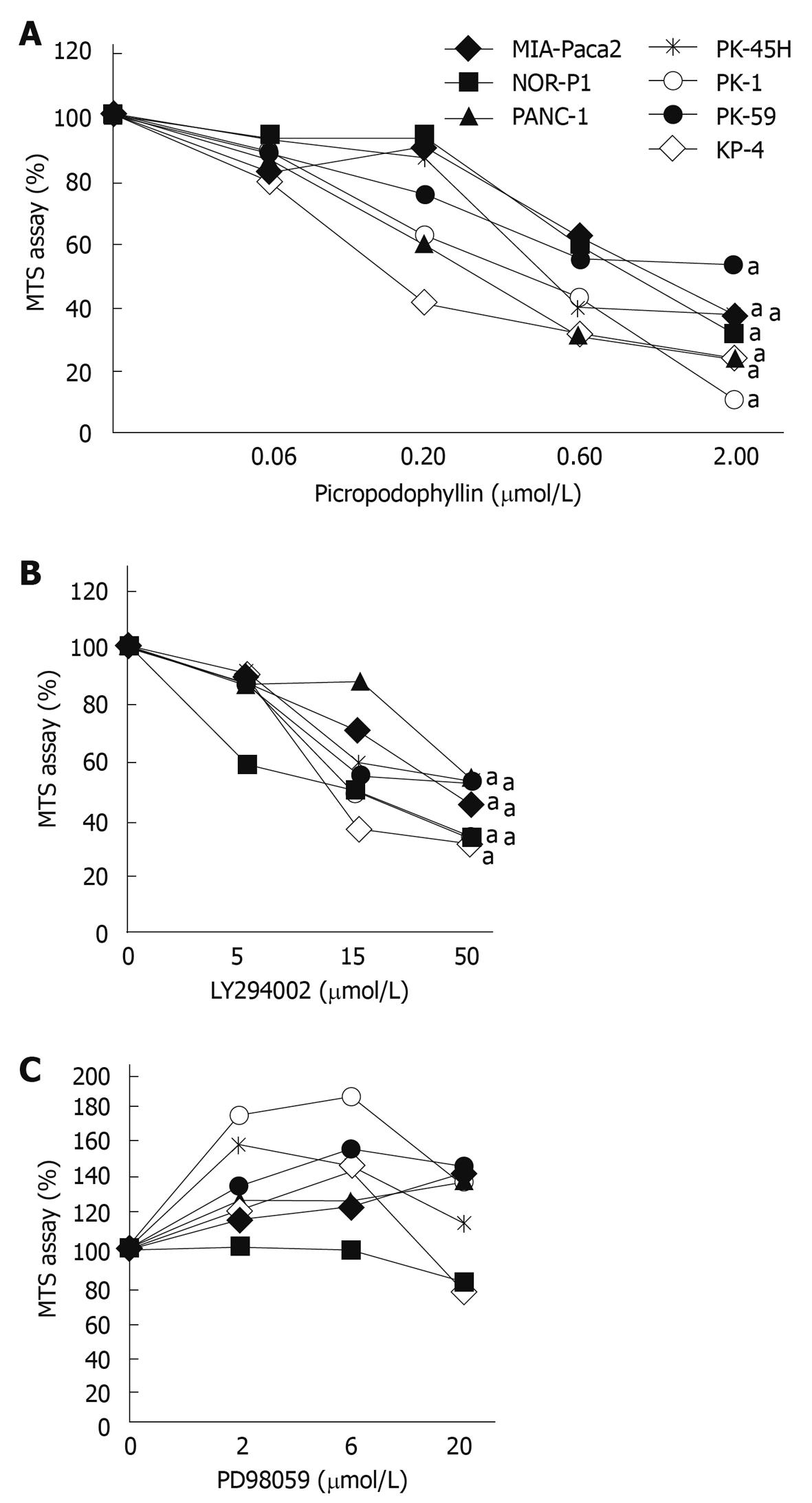
The MTS assay was performed to clarify whether PPP, LY294002, or PD98059 suppressed proliferation of pancreatic cancer cell lines. PPP suppressed proliferation of all the cell lines examined (Figure 2A). At 2 μmol/L, MIA-Paca2, NOR-P1, PANC-1, PK-45H, PK-1, PK-59 and KP-4 proliferation was reduced to 36.9% ± 2.4% (mean ± SD), 30.9% ± 5.5%, 23.8% ± 3.9%, 37.1% ± 5.3%, 10.4% ± 4.5%, 52.5% ± 4.5% and 22.6% ± 0.4%, respectively (P < 0.05, n = 3). Next, we analyzed the downstream pathway of IGF-IR. LY294002 suppressed proliferation of all the cell lines examined (Figure 2B). At 50 μmol/L, proliferation of MIA-Paca2, NOR-P1, PANC-1, PK-45H, PK-1, PK-59 and KP-4 cells was reduced significantly to 44.4% ± 7.6%, 32.9% ± 8.2%, 53.9% ± 8.0%, 52.8% ± 4.0%, 32.3% ± 4.2%, 51.8% ± 4.5%, and 30.6% ± 9.4%, respectively (P < 0.05, n = 3). PD98059 did not suppress cell proliferation (Figure 2C). Although NOR-P1 and KP-4 cells showed a marginal decrease in proliferation in the presence of 20 μmol/L PD98059, we could not analyze higher concentrations because PD98059 precipitated in the medium. Analysis with 50 μmol/L PD98059 failed since PD98059 crystallized and precipitated. The other inhibitor of MAPK was not analyzed.
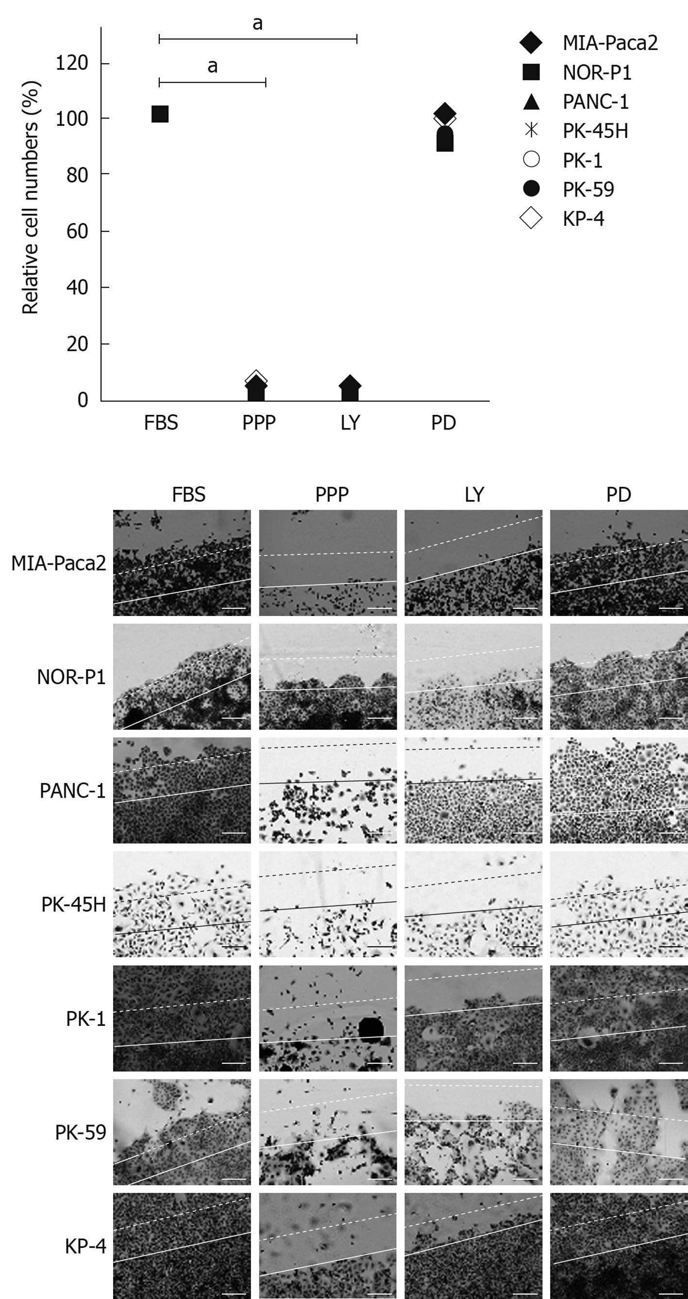
Suppression of motility is the initial step in the inhibition of metastasis. We analyzed changes in motility with inhibitors, by the wound assay (Figure 3). PPP at 2 μmol/L suppressed motility of MIA-Paca2, NOR-P1, PANC-1, PK-45H, PK-1, PK-59 and KP-4 cells to 3.0% ± 0.2%, 0%, 0%, 2.0% ± 0.1%, 5.0% ± 0.2%, 3.0% ± 0.1%, and 5.0% ± 0.2%, respectively (P < 0.05, n = 3). LY294002 at 50 μmol/L suppressed motility of MIA-Paca2, NOR-P1, PANC-1, PK-45H, PK-1, PK-59 and KP-4 cells to 3.0% ± 0.2%, 0%, 3.0% ± 0.2%, 0%, 0%, 0% and 3% ± 0.1%, respectively (P < 0.05, n = 3). PD980509 at 20 μmol/L did not suppress motility, although NOR-P1 cells showed a marginal, non-significant decrease.
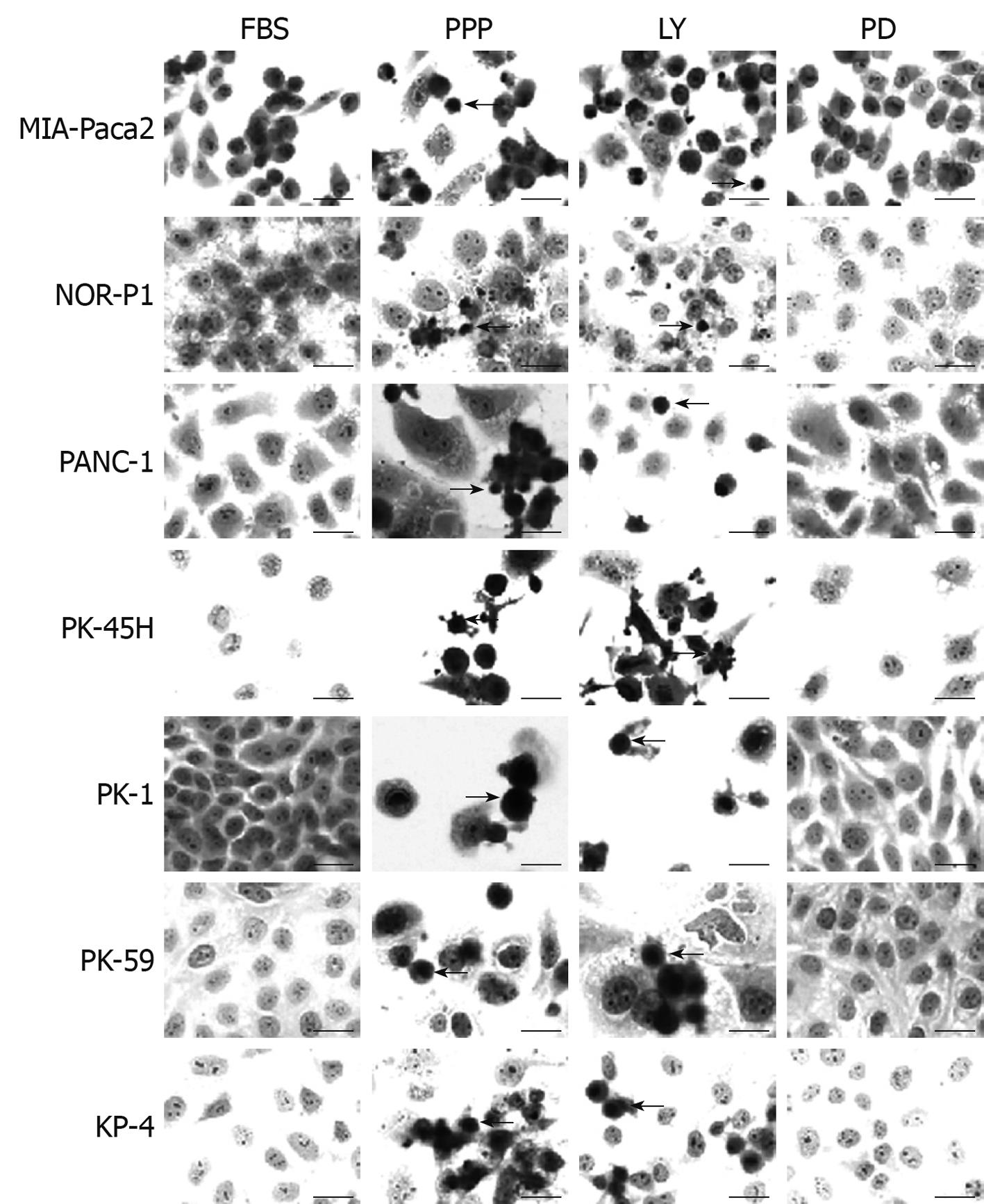
Cells were observed under the microscope to analyze the morphological changes induced by inhibitors. Cells in medium with 2 μmol/L PPP or 50 μmol/L LY294002 had pyknotic nuclei, which suggested that they were apoptotic (Figure 4). Cells in medium with 20 μmol/L PD98059 did not show signs of apoptosis (Figure 4).
IGF-I stimulates thymidine incorporation of pancreatic cancer cell lines, such as MIA-Paca2, as strongly as FBS, which suggests that it is an autocrine growth factor[13]. It was expected that inhibition of IGF-IR activity would lead to pancreatic cancer regression. Antisense oligonucleotide to IGF-IR suppresses proliferation of ASPC-1, a pancreatic cancer cell line[3]. Antisense oligonucleotide to K-ras synergistically enhances the suppression of pancreatic cancer cell lines by antisense oligonucleotide to IGF-IR[14]. Antisense oligonucleotide is unstable in blood, therefore, small molecules that inhibit the tyrosine kinase of IGF-IR are desirable. NVP-AEW541, an IGF-IR inhibitor, suppresses growth of HPAF-II cells, a pancreatic cancer cell line[15]. However, it is possible that NVP-AEW541 does not affect other cell lines. PPP is the first inhibitor to discriminate IGF-IR and insulin receptor[16]. In our experiments, PPP suppressed proliferation of all pancreatic cancer cell lines. Previous studies and the present one indicate that IGF-IR is a good candidate for molecular therapy of pancreatic cancer.
The downstream pathway of the IGF-I receptor is intriguing since shutting down the pathway could reduce cancer. Antibody to IGF-IR suppresses phosphorylation of Akt, a downstream molecule of PI3K and MAPK[13]. LY294002 significantly suppresses proliferation of PANC-1 and BxPC3 cells, and viability of KP-4 and PANC-1 cells[7,17]. Our results showed that LY294002 suppressed proliferation of all the pancreatic cancer cell lines examined. In addition to proliferation, our data clearly demonstrated that LY294002 suppressed cell migration. We conclude that PI3K is a suitable target for molecular therapy of pancreatic cancer.
MAPK is another downstream pathway of IGF-IR. MIA-PaCa2 treated with 20 μmol/L PD98059 in 100 mL/L FBS showed downregulation of phosphorylated extracellular signal-regulated kinase 1/2[18]. Although it was expected that PD98059 would suppress proliferation of pancreatic cancer cells, 20 μmol/L PD98059 did not suppress proliferation and motility of pancreatic cancer cells in our experiments. PD98059 at 20 μmol/L does not suppress DNA synthesis stimulated by IGF-I in the culture medium, although phosphorylation of MAPK is suppressed[19]. Curiously, PD98059 stimulates cell growth at concentrations of 0.1 μmol/L to 0.1 pmol/L[20]. SB203580, another inhibitor of MAPK, stimulates growth of PANC-1 cells in serum-free conditions[21]. In our experiments, PD98059 marginally stimulated growth of PANC-1 cells with 10 mL/L FBS. These results suggest that inhibition of MAPK does not always suppress growth of pancreatic cancer cell lines with unknown mechanisms.
It may be concluded that IGF-IR and PI3K are good candidates for molecular therapy of pancreatic cancer.
Insulin-like growth factor-I receptor (IGF-IR) is expressed in pancreatic cancer tissues but not in surrounding non-cancerous tissues. Although it was speculated that IGF-I might play a role in proliferation of pancreatic cancer, the detailed mechanism is not known.
IGF-IR is not involved in metabolism, therefore, it is expected that inhibition of IGF-IR and its downstream pathway should not cause adverse effects when used as a target for molecular therapy of pancreatic cancer.
IGF-IR was phosphorylated in cultured pancreatic cancer cell lines, which suggested that IGF-IR and its downstream pathway were activated. Each inhibitor of IGF-IR or phosphatidylinositol-3 kinase (PI3K) suppressed not only cell proliferation but also motility.
By revealing that IGF-IR was activated, this study indicated that IGF-IR might be a good candidate for molecular therapy of pancreatic cancer.
IGF-I is a growth factor that is involved in cell proliferation and differentiation. Stimulation of IGF-I is transmitted via IGF-IR to downstream pathways, such as PI3K and mitogen-activated protein kinase.
The authors have demonstrated that IGF-IR, phosphorylated and total, is involved in the proliferation of pancreatic cell lines. In Figure 1, we see that all cell lines expressed phosphorylated and total IGF-IR. Their experiments have shown that inhibition of IGF-IR activity results in a decrease in proliferation and motility of pancreatic cancer cell lines. These findings, along with the inhibition of PI3K, are interesting and show promise.
Peer reviewer: Gianfranco D Alpini, PhD, Professor, VA Research Scholar Award Recipient, Professor, Medicine and Systems Biology and Translation Medicine, Dr, Nicholas C. Hightower Centennial Chair of Gastroenterology, Central Texas Veterans Health Care System, The Texas A & M University System Health Science Center College of Medicine, Medical Research Building, 702 SW H.K. Dodgen Loop, Temple, TX 76504, United States
S- Editor Tian L L- Editor Kerr C E- Editor Lin YP
| 1. | LeRoith D, Roberts CT Jr. The insulin-like growth factor system and cancer. Cancer Lett. 2003;195:127-137. |
| 2. | Samani AA, Yakar S, LeRoith D, Brodt P. The role of the IGF system in cancer growth and metastasis: overview and recent insights. Endocr Rev. 2007;28:20-47. |
| 3. | Bergmann U, Funatomi H, Yokoyama M, Beger HG, Korc M. Insulin-like growth factor I overexpression in human pancreatic cancer: evidence for autocrine and paracrine roles. Cancer Res. 1995;55:2007-2011. |
| 4. | Karna E, Surazynski A, Orłowski K, Łaszkiewicz J, Puchalski Z, Nawrat P, Pałka J. Serum and tissue level of insulin-like growth factor-I (IGF-I) and IGF-I binding proteins as an index of pancreatitis and pancreatic cancer. Int J Exp Pathol. 2002;83:239-245. |
| 5. | Ouban A, Muraca P, Yeatman T, Coppola D. Expression and distribution of insulin-like growth factor-1 receptor in human carcinomas. Hum Pathol. 2003;34:803-808. |
| 6. | Stoeltzing O, Liu W, Reinmuth N, Fan F, Parikh AA, Bucana CD, Evans DB, Semenza GL, Ellis LM. Regulation of hypoxia-inducible factor-1alpha, vascular endothelial growth factor, and angiogenesis by an insulin-like growth factor-I receptor autocrine loop in human pancreatic cancer. Am J Pathol. 2003;163:1001-1011. |
| 7. | Perugini RA, McDade TP, Vittimberga FJ Jr, Callery MP. Pancreatic cancer cell proliferation is phosphatidylinositol 3-kinase dependent. J Surg Res. 2000;90:39-44. |
| 8. | Douziech N, Calvo E, Lainé J, Morisset J. Activation of MAP kinases in growth responsive pancreatic cancer cells. Cell Signal. 1999;11:591-602. |
| 9. | Ozawa F, Friess H, Tempia-Caliera A, Kleeff J, Büchler MW. Growth factors and their receptors in pancreatic cancer. Teratog Carcinog Mutagen. 2001;21:27-44. |
| 10. | Sato N, Mizumoto K, Beppu K, Maehara N, Kusumoto M, Nabae T, Morisaki T, Katano M, Tanaka M. Establishment of a new human pancreatic cancer cell line, NOR-P1, with high angiogenic activity and metastatic potential. Cancer Lett. 2000;155:153-161. |
| 11. | Tomizawa M, Saisho H. Signaling pathway of insulin-like growth factor-II as a target of molecular therapy for hepatoblastoma. World J Gastroenterol. 2006;12:6531-6535. |
| 12. | Pennisi PA, Barr V, Nunez NP, Stannard B, Le Roith D. Reduced expression of insulin-like growth factor I receptors in MCF-7 breast cancer cells leads to a more metastatic phenotype. Cancer Res. 2002;62:6529-6537. |
| 13. | Nair PN, De Armond DT, Adamo ML, Strodel WE, Freeman JW. Aberrant expression and activation of insulin-like growth factor-1 receptor (IGF-1R) are mediated by an induction of IGF-1R promoter activity and stabilization of IGF-1R mRNA and contributes to growth factor independence and increased survival of the pancreatic cancer cell line MIA PaCa-2. Oncogene. 2001;20:8203-8214. |
| 14. | Shen YM, Yang XC, Yang C, Shen JK. Enhanced therapeutic effects for human pancreatic cancer by application K-ras and IGF-IR antisense oligodeoxynucleotides. World J Gastroenterol. 2008;14:5176-5185. |
| 15. | Moser C, Schachtschneider P, Lang SA, Gaumann A, Mori A, Zimmermann J, Schlitt HJ, Geissler EK, Stoeltzing O. Inhibition of insulin-like growth factor-I receptor (IGF-IR) using NVP-AEW541, a small molecule kinase inhibitor, reduces orthotopic pancreatic cancer growth and angiogenesis. Eur J Cancer. 2008;44:1577-1586. |
| 16. | Girnita A, Girnita L, del Prete F, Bartolazzi A, Larsson O, Axelson M. Cyclolignans as inhibitors of the insulin-like growth factor-1 receptor and malignant cell growth. Cancer Res. 2004;64:236-242. |
| 17. | Takeda A, Osaki M, Adachi K, Honjo S, Ito H. Role of the phosphatidylinositol 3'-kinase-Akt signal pathway in the proliferation of human pancreatic ductal carcinoma cell lines. Pancreas. 2004;28:353-358. |
| 18. | Boucher MJ, Morisset J, Vachon PH, Reed JC, Lainé J, Rivard N. MEK/ERK signaling pathway regulates the expression of Bcl-2, Bcl-X(L), and Mcl-1 and promotes survival of human pancreatic cancer cells. J Cell Biochem. 2000;79:355-369. |
| 19. | Freeman JW, Mattingly CA, Strodel WE. Increased tumorigenicity in the human pancreatic cell line MIA PaCa-2 is associated with an aberrant regulation of an IGF-1 autocrine loop and lack of expression of the TGF-beta type RII receptor. J Cell Physiol. 1995;165:155-163. |
| 20. | Axelson J, Lindell M, Hörlin K, Ohlsson B. Inhibition of different intracellular signal cascades in human pancreatic cancer cells. Pancreatology. 2005;5:251-258. |
| 21. | Ding XZ, Adrian TE. MEK/ERK-mediated proliferation is negatively regulated by P38 map kinase in the human pancreatic cancer cell line, PANC-1. Biochem Biophys Res Commun. 2001;282:447-453. |


