Published online Oct 14, 2009. doi: 10.3748/wjg.15.4763
Revised: August 26, 2009
Accepted: September 1, 2009
Published online: October 14, 2009
AIM: To analyze the accuracy of short-term bio-impedance as a means of measuring gastric motility.
METHODS: We evaluated differences in the short-term electrical bio-impedance signal from the gastric region in the following conditions: (1) fasting state, (2) after the administration of metoclopramide (a drug that induces an increase in gastric motility) and (3) after food ingestion in 23 healthy volunteers. We recorded the real component of the electrical impedance signal from the gastric region for 1000 s. We performed a Fast Fourier Transform (FFT) on this data and then compared the signal among the fasting, medicated, and postprandial conditions using the median of the area under the curve, the relative area under the curve and the main peak activity.
RESULTS: The median of the area under the curve of the frequency range in the region between 2-8 cycles per minute (cpm) decreased from 4.7 cpm in the fasting condition to 4.0 cpm in the medicated state (t = 3.32, P = 0.004). This concurred with the decrease seen in the relative area under the FFT curve in the region from 4 to 8 cpm from 38.3% to 26.6% (t = 2.81, P = 0.012) and the increase in area in the region from 2 to 4 cpm from 22.4% to 27.7%, respectively (t = -2.5, P = 0.022). Finally the main peak position also decreased in the region from 2 to 8 cpm. Main peak activity in the fasting state was 4.72 cpm and declined to 3.45 cpm in the medicated state (t = 2.47, P = 0.025). There was a decrease from the fasting state to the postprandial state at 3.02 cpm (t = 4.0, P = 0.0013).
CONCLUSION: Short-term electrical bio-impedance can assess gastric motility changes in individuals experiencing gastric stress by analyzing the area medians and relative areas under the FFT curve.
- Citation: Huerta-Franco MR, Vargas-Luna M, Capaccione KM, Yañez-Roldán E, Hernández-Ledezma U, Morales-Mata I, Córdova-Fraga T. Effects of metoclopramide on gastric motility measured by short-term bio-impedance. World J Gastroenterol 2009; 15(38): 4763-4769
- URL: https://www.wjgnet.com/1007-9327/full/v15/i38/4763.htm
- DOI: https://dx.doi.org/10.3748/wjg.15.4763
Numerous studies have demonstrated that gastric motility occurs at a rate of three cycles, or peristaltic waves, per minute. This consistent rate is due to myoelectrical activity, which produces ring contractions through the distal gastric wall. These peristaltic waves facilitate food digestion and gastric emptying[1-3]. In various studies, gastric emptying has been used to assess many diseases using several different measurement techniques and methods of data analysis[4-8].
Scintigraphy has become the gold standard technique for the evaluation of gastric activity despite the fact that it measures gastric emptying as the main consequence of gastric motility[3,4,9]. Also for the evaluation of the gastric motility, there are invasive methods such as manometry, which uses a pressure probe device in a specific region of the gastric tract to directly measure the amplitude and frequency of the gastric movements[10-12]. Electrogastrography could be another invasive method, if electrodes are introduced into the gastro-intestinal tract in order to record the myoelectrical activity in a specific area of the tract[13]. The bio-impedance technique can also be invasive, for example, when used to measure intraluminal impedance in esophageal function studies[14,15]. Non-invasive options include electrogastrography if recordings are performed through the skin using cutaneous electrodes[16-23], and electrical bio-impedance in the gastric region when measured using cutaneous electrodes[24-26]. As opposed to gastric electrical stimulation[27,28], the bio-impedance technique uses a low current and voltage to obtain passive gastric impedance characteristics. In these two cases, uncertainties arise in the interpretation of the signal because cutaneous recording leads to the simultaneous acquisition of signals at multiple frequencies within the gastrointestinal (GI) system. The main challenge of this technique is the decomposition of the data to isolate the frequencies originating in the gastric tract[16,25,29,30].
In looking for a balance between accuracy, reproducibility and patient comfort, electrogastrography and electrical bio-impedance are the most suitable techniques for the evaluation of gastric motility[31], despite the fact that occasionally electrical activity does not correlate directly with mechanical movements[11]. In the context of selecting one of these two techniques for gastric motility assessment, electrical bio-impedance has the advantage of being directly sensitive to movements and therefore a better option than electrogastrography for gastric motility evaluation.
In the evaluation of gastric motility, bio-impedance techniques have been shown to be sensitive to material changes, including internal conformational changes (bowel movements)[26,32] and conduction properties (stomach acidity or properties of ingested food)[33]. This method has been used in gastric emptying studies for long-term measurements. Measurements are evaluated by looking for global impedance changes, which are changes over long time periods and ignoring impedance oscillations, short-term faster oscillating changes, of the gastric region[24]. Recently, this long-term assessment method has been proposed for the evaluation of gastric secretions because it is highly sensitive to the conductive properties of ingested food and internal fluid secretions, and therefore may be a useful method for such evaluations[34].
For gastric motility, bio-impedance evaluation produces results consistent with those of other gastric motility methods, mainly the position of the main peak activity around three cycles per minute (cpm)[24,25]. However, the sensitivity to other gastrointestinal regions and the overlapping of movements from those regions produce signals in other frequency ranges that are neither due to bradygastria or tachygastria but are associated with characteristic intestinal or colon frequencies[1].
There are several benefits to the bio-impedance technique: it directly measures motility, it is non-invasive, and its use for interval measurements (short term bio-impedance) can accurately reconstruct a long period of measurement. The first benefit emphasizes accuracy while the latter two maximize patient comfort. Additionally, this approach is less expensive than other techniques and so the development of this method for clinical purposes can benefit a wider range of people worldwide.
In a previous study, we evaluated normal subjects in both the fasting and postprandial states using short-term electrical bio-impedance[32]. We compared our data with those obtained from the concurrent use of long-term bio-impedance, and found consistent results. Thus, we concluded that short-term electrical bio-impedance is an accurate evaluation method in healthy volunteers. In particular, the position of the main activity around 3 cpm was strong support for the usefulness of this method in healthy volunteers because of its unequivocal agreement with several other methods of gastric motility assessment. However, for this technique to attain clinical relevance, bio-impedance must be shown to be equivalent to validated methods in subjects experiencing gastric stress, which manifests itself through small changes in peristaltic wave frequency. Thus, we must evaluate if short-term electrical bio-impedance (5-15 min) has sufficient sensitivity to record these changes.
In this study, we use the drug metoclopramide to induce gastric stress in healthy volunteers, mimicking a state of gastric stress that would warrant clinical evaluation. By comparing the data from the area under the Fast Fourier Transform (FFT) spectra curves to those obtained from the main peak of the spectra, we assessed the potential of electrical bio-impedance as a method for the evaluation of gastric motility in systems experiencing gastric stress.
For this study we recruited twenty three volunteers. Each met the following criteria: 18-30 years of age, no prior gastrointestinal disease, no prior disease that may affect the GI system function (including diabetes, Parkinson’s disease, amyloidosis, myotonic dystrophy, polymyositis, HIV infection or cytomegalovirus infection), and they must not have been taking medicine that could interfere with gastric activity. Additionally, participants must not have had significant weight loss within the 3 mo prior to the study, and must not have been overweight (BMI ≥ 25 kg/m2). In addition to these physical criteria, subjects must have been free from psychological problems such as anxiety, stress, depression and mental disease, because these conditions could potentially affect gastric activity through endocrine and central nervous system modulation. Also, subjects were free from any other endocrine disorders and had no food allergies that interfered with the food administered in this trial.
All women who participated in this study underwent evaluation within the first 10 d (the early follicular phase) of their menstrual cycle. All subjects who participated in this study signed a consent form approved by the Human Ethics Committee of the University of Guanajuato. The study was conducted according to the Declaration of Helsinki[35].
Metoclopramide [IUPAC name: 4-amino-5-chloro-N- (2-diethylamino ethyl)-2-methoxybenzamide] is a dopamine receptor antagonist with both antiemetic and prokinetic properties. The antiemetic property treats nausea and vomiting while the prokinetic properties enhance the peristaltic waves of the stomach and intestine, increasing the rate of absorption and thus the rate of passage through the stomach. Therefore, the administration of metoclopramide in healthy volunteers speeds gastric motility and subsequently the rate of gastric emptying in a measurable way. In healthy patients, metoclopramide is absorbed rapidly and almost completely from the GI tract following oral administration. The pharmacological action of orally administered metoclopramide begins 30-60 min after the subject takes the drug and persists for 1-2 h[36]. Therefore, after performing our baseline recordings in the fasting individual, metoclopramide was administered and we took bio-impedance measurements after a 60 min resting period. This ensured that the subjects were experiencing the maximal pharmacological effects of metoclopramide while the bio-impedance measurements were taken.
Before beginning the experiment, a clinical history was taken from each of the subjects. This evaluation included the following sections: (1) General Information: name, age, gender, occupation; (2) Pathological Antecedents: clinical history of disease related to the GI tract; (3) Physiological Information: anthropometrical measurements and body mass index; (4) Life-style variables: exercise, sleep habits, substance use (smoking, alcohol, medication); (5) Observations: additional information gained from the physical examination; (6) Gynecologic-Obstetric Characteristics: for all women, we collected data regarding the onset of menarche, last menstrual period, and history of gynecological problems.
Before participation, each subject fasted for 8 h immediately prior to the experiment. In addition, they abstained from smoking, alcohol consumption, strenuous exercise, and substances containing caffeine for 24 h before testing.
A basal bio-impedance measurement of each individual in the fasting state was acquired during 16.7 min (1000 s) and served as a baseline for comparison with the experimental condition (metoclopramide). Patients subsequently ingested a 20 mg dose of metoclopramide with 150 mL of water. We waited 1 h before taking the second bio-impedance measurement so that patients would experience the peak pharmacological effects of metoclopramide during the test period. After this, another 1000 s bio-impedance measurement was recorded. We performed a third bio-impedance recording after the administration of a test meal that consisted of one cereal bar containing 124 calories (cal): 6.8 cal (1.7 g) of protein, 44.1 cal (4.9 g) of fat, and 73.1 cal (18.3 g) of carbohydrates and a fat free Yoghurt Drink containing 83 cal: 32 cal (7.8 g) of protein, zero cal (0 g) of fat, and 51 cal (12.5 g) of carbohydrates. In total, the subjects consumed 207 cal, of which 38.8 cal (9.5 g) were protein, 44.1 cal (4.9 g) were fat, and 124 cal (30.8 g) were carbohydrates.
To control the consistency of the ingested food, we divided the bars into four equal pieces and instructed the subjects to chew each piece 10 times and then immediately swallow. They were to repeat this with all four pieces of the cereal bar and when finished, immediately drink the yoghurt.
Approximately 5 min after food ingestion, subjects were measured again using the bio-impedance technique to assess gastric motility. There are two factors that affect the bio-impedance signal: the first is the dielectric properties of the food ingested, where fats lead to deadening of the signal. In our experiment, we controlled for this by administering a test meal low in fats. The second factor affecting the signal is noise produced by involuntary movements of the subjects, such as yawning, coughing and heavy breathing. Here, the use of short-term bio-impedance mitigated these effects by reducing the consecutive testing time for subjects.
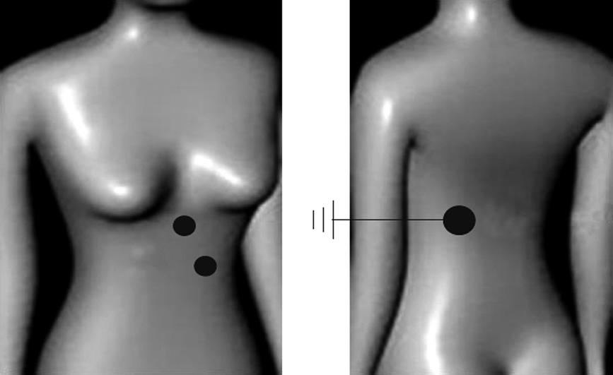
For this experiment, we used a configuration of three electrodes as shown in Figure 1. This configuration consisted of two electrodes placed on the abdomen and a ground electrode placed on the back. The first (excitation) electrode was placed using the midline as a marker, and then by following the ribcage to the level just above and to the left of the umbilicus. The second electrode was placed approximately five centimeters from the first at a 45 degree angle up and to the left of this (recording) electrode. For the ground electrode, the vertebral column was used as a reference and the electrode was placed at approximately the average height of the corresponding abdominal electrodes.
All measurements were obtained with a SOLARTRON 1260 impedance analyzer in conjunction with a SOLARTRON 1294 biological sample interface. Disposable 3M “Red Dot” monitoring electrodes were used in each patient. A BI stimulus frequency of 1 V AC was applied at 50 kHz. During the experiment, data were recorded four times per second for 1000 consecutive seconds.
Before conducting the data analysis, we preliminarily assessed the results of the anthropometrical, lifestyle, and clinical variables to ensure that our group was homogeneous in terms of these variables and that there were no unusual fluctuations in our sample.
To analyze the information from each individual over time, we used a Butterworth filter in the framework of a FFT analysis for the frequency range of 1-12/min (or cpm) (0.017-0.2 Hz). We particularly noted the data in the region between 2-4 cpm (0.033-0.066 Hz) and between 4 and 8 cpm (0.066-0.132 Hz). These analyses were carried out using MatLab 6.5 and Origin 6.0.
The data recordings were cleaned of noisy periods, sudden fluctuations and data drifting, thus the remaining data periods for analysis were in the range of 5-10 min; we discarded recordings of less than 5 min.
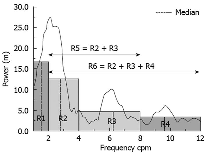
The frequency range was divided in four regions: the first region (R1) spans 1-2 cpm, and is defined as a very low frequency range and was therefore not considered to be an important region in this work. The second region (R2), spans the frequencies from 2 to 4 cpm and has generally been recognized as the main frequency range in the literature regarding gastric studies[1,24,25,32]. The third region (R3) spans 4-8 cpm, and contains the frequencies where movements from the intestines are important[1]. The fourth region (R4) spans 8-12 cpm and corresponded with the respiration frequency range of the subjects. This region may contain important data regarding the gastrointestinal system, but because respiration generates significant signal noise, we were not able to use it. Other, more expansive regions were defined as region R5, spanning 2-8 cpm and region R6, spanning 2-12 cpm (Figure 2).
For the statistical analysis, we divided the FFT signal from 1-12 cpm into the regions described above and we used both the area under the FFT curve and the median of this area (the frequency which divides the area of the frequency region into two equal parts) and the position of the main peak. Together, these measures described the characteristics of the entire frequency range from 2-8 cpm. This range was used to compare the relative activities within the high and low frequency ranges. We disregarded the activity due to respiratory movements, which contributed data at approximately 8-12 cpm.
To test the accuracy of our results, we also analyzed the position of the highest activity in the range of 2-4 cpm, which is the frequency range typically used in the assessment of gastric activity.
For data analysis, we used a MatLab platform to analyze the spectra. T-tests for dependent samples were performed to assess differences between fasting, medicated and post-prandial states using the parameters mentioned. Previously, the use of bio-impedance has been limited because of the complexity of the signal acquired from the impedance measurements. Therefore, the only parameter used was the average magnitude of resistance of the raw signal and its change over time. Our technique refines this previous method by parsing more parameters that can be independently evaluated and statistically analyzed. All statistical analyses were performed using Statistica (Stat-Soft, Inc, Tulsa OK, USA), and the threshold for significance was standardized at P = 0.05.
As mentioned above, three parameters were considered in this study: (1) The median of the area under the FFT curve for each region, (2) The ratio of the area of each region to the total area under the curve of the range from 1 to 12 cpm; and (3) the position of the highest peak in each region. The three parameters for each region were obtained from smoothed versions of the FFT curves. We took the average of all individuals evaluated in this study and used these data for further comparison.
Based on the three parameters used, we found the following.
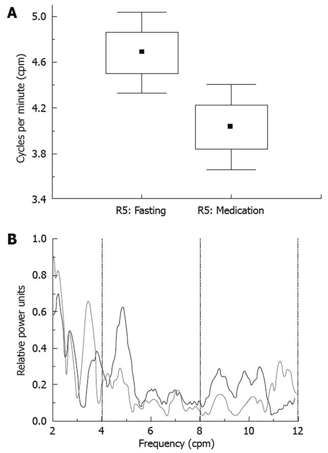
In R2, the median changed from 2.8 cpm in the fasting state to 2.75 cpm in the medicated state. However, this change was not statistically significant (P = 0.46). In R3, this parameter changed from 5.75 cpm in the fasting state to 5.9 cpm in the medicated state, however, this change was also not statistically significant (P = 0.2). As shown in Figure 3, R5 (R2 + R3) displayed a significant decrease in the median from 4.7 cpm in the fasting condition to 4.0 cpm in the medicated state (t = 3.32, P = 0.004). We did not find a statistically significant change in the median value in any region between the medicated and post-prandial states in the regions analyzed in this study. However, in the instances where the changes were not statistically significant, these changes showed a similar trend to those indicated in previous studies[32].
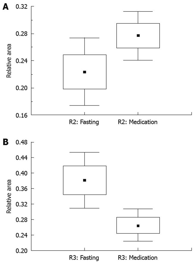
The change observed in the median of R5 may be understood as the combination of the changes seen in the area under the FFT curve of R2 and R3. For R2, when we compare the ratios of the regional area to the total area between the fasting and medicated states, we observed an increase from 22.4% to 27.7%, respectively (t = -2.5, P = 0.022). The change was in the opposite direction for R3, with a decrease from 38.3% to 26.6% (t = 2.81, P = 0.012) for the fasting and medicated states, respectively. Figure 4A and B show these changes for R2 and R3, respectively.
In contrast to our findings regarding the changes in the median of the area under the FFT curve for R5, there were no significant differences in the area of R5 between the post-prandial state and either the fasting or medicated state.
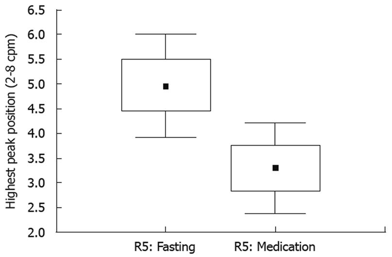
Previously, the main parameter considered in the evaluation of gastric motility was the main peak in R2 (from 2 to 4 cpm) of the frequency domain and its shift across states. As discussed above as well as in previous works, the large overlap of activity in the abdominal cavity makes determination of the main peak difficult in certain frequency regions. When the main peak activity of R5 was analyzed, we found differences in the position of the highest peak which were statistically significant. Main peak activity in the fasting state was 4.72 cpm and declined to 3.45 cpm in the medicated state (t = 2.47, P = 0.025), as shown in Figure 5. There was also a decrease from the fasting state to the post-prandial state at 3.02 cpm (t = 4.0, P = 0.0013) but this change was not statistically significant with respect to the medicated state (P = 0.28).
These results demonstrate that global changes in the gastrointestinal tract can be measured using short-term electrical bio-impedance, giving useful information on gastric motility.
The typical parameter for gastric motility evaluation is the position of the main peak in the frequency region between 2-4 cpm and should be reconsidered in the context of global behavior of the combined frequency ranges. The results described in our previous work were confirmed with the accurate measurement of gastric motility in the fasting state[32]. In addition, this study demonstrated that the bio-impedance technique is also valid for systems experiencing gastric stress, as induced by metoclopramide. In this study, we demonstrated that there was a decrease in the median of the area under the FFT curve over time in the region from 2 to 8 cpm, which was a consequence of the change in the relative areas under the curves in the regions from 2 to 4 cpm (increase) and from 4 to 8 cpm (decrease). The position of the main peak can also be used as a parameter for gastric motility assessment, and in this case the main peak would be representative of the entire 2-8 cpm region.
When analyzing the lack of significant change in the main peak position in R5 between the medicated and post-prandial states, we can understand this physiologically as a result of the continued effects of metoclopramide during the post-prandial state. Peak effects of metoclopramide occur at 60 min after administration, but can persist for up to 2.2 h[36]. Food administration and subsequent testing were well within this period, likely accounting for the lack of significant differences between the two states. The increased smooth muscle activity induced by metoclopramide administration persisted throughout the post-prandial state, causing similar bio-impedance activity in both states; this resulted in a lack of significant changes in gastric motility.
This study should be followed up with comparative studies between electrical bio-impedance and electrogastrography and other standard techniques to ensure the equivalence of this technique to other proven methods and the overall validity of electrical bio-impedance for gastric motility assessment so that it can be used in a clinical context. Frequent and easy monitoring of gastric motility in subjects with problems that involve gastric motility may be an important clinical application of this technique, for example in the clinical diagnosis, and evaluation of gastroparesis. Other applications may include pediatrics, because this technique allows easy clinical evaluation. Extended monitoring of patients experiencing gastric distress may lead to more accurate assessment of the pathological antecedents of the chief complaint. This technique could also be used in the evaluation of the effects of diverse drugs which act in the gastric motility.
The results of this study demonstrate that electrical bio-impedance can be used as an accurate and valuable clinical technique for gastric motility evaluation in individuals experiencing gastric stress.
Gastric motility assessment is an important diagnostic technique for a variety of diseases of the gastrointestinal tract. For the evaluation of gastric motility, there are invasive methods such as scintigraphy and manometry. Electrogastrography is an invasive method, if electrodes are introduced into the gastro-intestinal tract. Non-invasive options include electrogastrography, when recordings are performed through the skin using cutaneous electrodes, and electrical bio-impedance in the gastric region when cutaneous electrodes are applied. In these latter two cases, uncertainties arise in the interpretation of the signal because the cutaneous recording leads to the simultaneous acquisition of signals at multiple frequencies within the gastrointestinal system. The main challenge of this technique is the decomposition of the data to isolate the frequencies originating in the gastric tract. Presently, research is underway to develop accurate gastric motility assessment tools which optimize patient comfort and cost.
Electrical bio-impedance is a non-invasive technique that can measure gastric motility. Previously, assessment has been over long time intervals in gastric emptying studies that required immobilization of the patient for the duration of the test. Movement compromises results and the overlap of internal movements from the gastric region produce signals in other frequency ranges. Other alternatives to analyze bio-impedance data from the gastric region have been proposed in the literature, for example, discrete wavelet transform.
Here, the authors demonstrate that short-term bio-impedance can be used to assess gastric motility in healthy volunteers with gastric systems under stress. The areas and the median of the areas of specific regions under the Fast Fourier Transform (FFT) spectra were analyzed, as an alternative, to accurately depict gastric motility. Stress induced by metoclopramide mirrors a disease state by enhancing the peristaltic waves of the stomach and intestines. Thus, this work supports the clinical application of the bio-impedance technique.
In future studies, the authors hope to test short-term bio-impedance in parallel with other, previously validated methods for gastric motility assessment. If studies prove these methods to be equivalent, short-term bio-impedance could be used clinically to optimize gastric motility assessment with regard to both cost and patient comfort.
Bio-impedance is a term used to describe the response or resistance of a living organism to an externally applied electric current. It can be described by the resistance or the conductivity. Short-term bio-impedance is a term used for short time recordings of bio-impedance signal. FFT is a mathematical technique to transform data from time domain to frequency domain. It gives information about the main variation frequencies for the data. Metoclopramide, is a dopamine receptor antagonist with both antiemetic and prokinetic properties. The antiemetic property treats nausea and vomiting while the prokinetic properties enhance the peristaltic waves of the stomach and intestine, increasing the rate of absorption and thus the rate of passage through the stomach.
The manuscript entitled “Effects of metoclopramide on gastric motility measured by short-term bio-impedance” is an interesting report. They found that electrical bio-impedance can assess changes in gastric motility in individuals experiencing gastric stress. The manuscript add important information to the current knowledge.
Peer reviewer: Atsushi Nakajima, Professor, Division of Gastroenterology, Yokohama City University Graduate School of Medicine, 3-9 Fuku-ura, Kanazawa-ku, Yokohama 236-0004, Japan
S- Editor Tian L L- Editor Webster JR E- Editor Zheng XM
| 1. | Mariani G, Boni G, Barreca M, Bellini M, Fattori B, AlSharif A, Grosso M, Stasi C, Costa F, Anselmino M. Radionuclide gastroesophageal motor studies. J Nucl Med. 2004;45:1004-1028. |
| 2. | Quigley EM. Review article: gastric emptying in functional gastrointestinal disorders. Aliment Pharmacol Ther. 2004;20 Suppl 7:56-60. |
| 3. | Akkermans LM, van Isselt JW. Gastric motility and emptying studies with radionuclides in research and clinical settings. Dig Dis Sci. 1994;39:95S-96S. |
| 4. | Vantrappen G. Methods to study gastric emptying. Dig Dis Sci. 1994;39:91S-94S. |
| 5. | Mangnall YF, Kerrigan DD, Johnson AG, Read NW. Applied potential tomography. Noninvasive method for measuring gastric emptying of a solid test meal. Dig Dis Sci. 1991;36:1680-1684. |
| 6. | Ferdinandis TG, Dissanayake AS, De Silva HJ. Effects of carbohydrate meals of varying consistency on gastric myoelectrical activity. Singapore Med J. 2002;43:579-582. |
| 7. | Brzana RJ, Koch KL, Bingaman S. Gastric myoelectrical activity in patients with gastric outlet obstruction and idiopathic gastroparesis. Am J Gastroenterol. 1998;93:1803-1809. |
| 8. | Siegel JA, Urbain JL, Adler LP, Charkes ND, Maurer AH, Krevsky B, Knight LC, Fisher RS, Malmud LS. Biphasic nature of gastric emptying. Gut. 1988;29:85-89. |
| 9. | Soulsby CT, Khela M, Yazaki E, Evans DF, Hennessy E, Powell-Tuck J. Measurements of gastric emptying during continuous nasogastric infusion of liquid feed: electric impedance tomography versus gamma scintigraphy. Clin Nutr. 2006;25:671-680. |
| 10. | Stanghellini V, Ghidini C, Tosetti C, Franceschini A, Ricci Maccarini M, Corinaldesi R, Barbara L. [Comparison of methods: gastro-duodenal manometry and study of gastric emptying]. Minerva Chir. 1991;46:125-130. |
| 11. | Abid S, Lindberg G. Electrogastrography: poor correlation with antro-duodenal manometry and doubtful clinical usefulness in adults. World J Gastroenterol. 2007;13:5101-5107. |
| 12. | Wilmer A, Van Cutsem E, Andrioli A, Tack J, Coremans G, Janssens J. Ambulatory gastrojejunal manometry in severe motility-like dyspepsia: lack of correlation between dysmotility, symptoms, and gastric emptying. Gut. 1998;42:235-242. |
| 13. | Chen JD, Schirmer BD, McCallum RW. Serosal and cutaneous recordings of gastric myoelectrical activity in patients with gastroparesis. Am J Physiol. 1994;266:G90-G98. |
| 14. | Magistà AM, Indrio F, Baldassarre M, Bucci N, Menolascina A, Mautone A, Francavilla R. Multichannel intraluminal impedance to detect relationship between gastroesophageal reflux and apnoea of prematurity. Dig Liver Dis. 2007;39:216-221. |
| 15. | Tutuian R, Vela MF, Shay SS, Castell DO. Multichannel intraluminal impedance in esophageal function testing and gastroesophageal reflux monitoring. J Clin Gastroenterol. 2003;37:206-215. |
| 16. | Jonderko K, Kasicka-Jonderko A, Krusiec-Swidergoł B, Dzielicki M, Strój L, Doliński M, Doliński K, Błońska-Fajfrowska B. How reproducible is cutaneous electrogastrography? An in-depth evidence-based study. Neurogastroenterol Motil. 2005;17:800-809. |
| 17. | Wu HC, Wang KC, Chang YW, Chang FY, Young ST, Kuo TS. Power distribution analysis of cutaneous electrogastrography using discrete wavelet transform. Conf Proc IEEE Eng Med Biol Soc. 1998;6:3227-3229. |
| 18. | Lin X, Chen JZ. Abnormal gastric slow waves in patients with functional dyspepsia assessed by multichannel electrogastrography. Am J Physiol Gastrointest Liver Physiol. 2001;280:G1370-G1375. |
| 19. | Chen JD, Zou X, Lin X, Ouyang S, Liang J. Detection of gastric slow wave propagation from the cutaneous electrogastrogram. Am J Physiol. 1999;277:G424-G430. |
| 20. | Liang J, Co E, Zhang M, Pineda J, Chen JD. Development of gastric slow waves in preterm infants measured by electrogastrography. Am J Physiol. 1998;274:G503-G508. |
| 21. | Lin ZY, McCallum RW, Schirmer BD, Chen JD. Effects of pacing parameters on entrainment of gastric slow waves in patients with gastroparesis. Am J Physiol. 1998;274:G186-G191. |
| 22. | Jonderko K, Kasicka-Jonderko A, Błońska-Fajfrowska B. Does body posture affect the parameters of a cutaneous electrogastrogram? J Smooth Muscle Res. 2005;41:133-140. |
| 23. | Liang J, Chen JD. What can be measured from surface electrogastrography. Computer simulations. Dig Dis Sci. 1997;42:1331-1343. |
| 24. | McClelland GR, Sutton JA. Epigastric impedance: a non-invasive method for the assessment of gastric emptying and motility. Gut. 1985;26:607-614. |
| 25. | Huerta-Franco MR, Vargas-Luna M, Vallejo-Villalpando JM, Hernandez E, Cordova T. Utility of the short time bioelectrical impedance for the gastric motility assessment: Preliminary Results. 13th International Conference on Electrical Bioimpedance and the 8th Conference on Electrical Impedance Tomography. Graz: Springer-Verlag 2007; 820-823. |
| 26. | Kothapalli B. Origin of changes in the epigastric impedance signal as determined by a three-dimensional model. IEEE Trans Biomed Eng. 1992;39:1005-1010. |
| 27. | Zhang J, Chen JD. Systematic review: applications and future of gastric electrical stimulation. Aliment Pharmacol Ther. 2006;24:991-1002. |
| 28. | Jalilian E, Onen D, Neshev E, Mintchev MP. Implantable neural electrical stimulator for external control of gastrointestinal motility. Med Eng Phys. 2007;29:238-252. |
| 29. | Amaris MA, Sanmiguel CP, Sadowski DC, Bowes KL, Mintchev MP. Electrical activity from colon overlaps with normal gastric electrical activity in cutaneous recordings. Dig Dis Sci. 2002;47:2480-2485. |
| 30. | Buist ML, Cheng LK, Sanders KM, Pullan AJ. Multiscale modelling of human gastric electric activity: can the electrogastrogram detect functional electrical uncoupling? Exp Physiol. 2006;91:383-390. |
| 31. | Smout AJ, Jebbink HJ, Akkermans LM, Bruijs PP. Role of electrogastrography and gastric impedance measurements in evaluation of gastric emptying and motility. Dig Dis Sci. 1994;39:110S-113S. |
| 32. | Huerta-Franco R, Vargas-Luna M, Hernandez E, Capaccione K, Cordova T. Use of short-term bio-impedance for gastric motility assessment. Med Eng Phys. 2009;31:770-774. |
| 33. | Giouvanoudi A, Amaee WB, Sutton JA, Horton P, Morton R, Hall W, Morgan L, Freedman MR, Spyrou NM. Physiological interpretation of electrical impedance epigastrography measurements. Physiol Meas. 2003;24:45-55. |
| 34. | Giouvanoudi AC, Spyrou NM. Epigastric electrical impedance for the quantitative determination of the gastric acidity. Physiol Meas. 2008;29:1305-1317. |
| 36. | Food and Drug Administration: FDA Application REGLAN NDA No. 017854. Label and Approval History: Approved 07/26/2004 (NDA 17-854/S-047). Available from: URL: http://www.accessdata.fda.gov/Scripts/cder/DrugsatFDA/index.cfm?fuseaction=Search.Label_ApprovalHistory#apphist. |


