Published online Sep 28, 2008. doi: 10.3748/wjg.14.5549
Revised: June 23, 2008
Accepted: June 30, 2008
Published online: September 28, 2008
AIM: To study the relationship between nm23H1 gene genetic instability and its clinical pathological characteristics in Chinese digestive system cancer patients.
METHODS: Polymerase chain reaction-single strand conformation polymorphism (PCR-SSCP) was used to analyze the microsatellite instability (MSI) and loss of heterozygosity (LOH). Immunohistochemistry was employed to detect the expression of nm23H1.
RESULTS: The MSI was higher in TNM stageI + II than in stage III + IV of gastric, colonic and gallbladder carcinomas. The LOH was higher in TNM stage III + IV than in stageI + II of gastric, colonic and hepatocellular carcinomas. Lymphatic metastasis was also observed. The expression of nm23H1 protein was lower in TNM stage III + IV than in stageI + II of these tumors and in patients with lymphatic metastasis.The nm23H1 protein expression was higher in the LOH negative group than in the LOH positive group.
CONCLUSION: MSI and LOH may independently control the biological behaviors of digestive system cancers. MSI could serve as an early biological marker of digestive system cancers. Enhanced expression of nm23H1 protein could efficiently inhibit cancer metastasis and improve its prognosis. LOH mostly appears in late digestive system cancer.
- Citation: Yang YQ, Wu L, Chen JX, Sun JZ, Li M, Li DM, Lu HY, Su ZH, Lin XQ, Li JC. Relationship between nm23H1 genetic instability and clinical pathological characteristics in Chinese digestive system cancer patients. World J Gastroenterol 2008; 14(36): 5549-5556
- URL: https://www.wjgnet.com/1007-9327/full/v14/i36/5549.htm
- DOI: https://dx.doi.org/10.3748/wjg.14.5549
| Clinical pathological factors | Cases (n) | MSI+ (%) | LOH+ (%) | nm23H1+ (%) | nm23H1 expression intensity (mean ± SD) |
| Histological type | 40 | 8 (20.00) | 7 (17.50) | 22 (55.00) | 40.63 ± 2.95 |
| Tubular adenocarcinoma | 33 | 7 (22.21) | 6 (18.18) | 21 (63.64) | 41.56 ± 2.78 |
| High differentiation | 10 | 3 (30.00) | 1 (10.00) | 10 (100.00) | 41.83 ± 1.52 |
| Middle differentiation | 15 | 3 (20.00) | 2 (13.33) | 9 (60.00) | 40.69 ± 2.35 |
| Low differentiation | 8 | 1 (12.50) | 3 (37.50) | 2 (25.00)1 | 41.43 ± 1.89 |
| Mucoid adenocarcinoma | 7 | 1 (14.29) | 1 (14.29) | 1 (14.29)2 | 39.87 ± 2.31 |
| Serosa infiltration | |||||
| Positive | 24 | 3 (12.50) | 6 (25.00) | 10 (41.67) | 39.76 ± 2.64 |
| Negative | 16 | 5 (31.25) | 1 (6.25) | 12 (75.00)3 | 41.45 ± 2.23 |
| Lymph node metastasis | |||||
| Positive | 20 | 1 (5.00) | 6 (30.00) | 6 (30.00) | 39.14 ± 2.34 |
| Negative | 20 | 7 (35.00)4 | 1 (5.00)5 | 16 (80.00)6 | 41.75 ± 1.65 |
| TNM stage | |||||
| I+ II | 22 | 7 (31.82) | 1 (4.55) | 17 (77.27) | 41.22 ± 1.87 |
| III + IV | 18 | 1 (5.56)7 | 6 (33.33)8 | 5 (17.86)9 | 40.13 ± 2.35 |
| Group | Positive frequency of nm23H1 protein % (n/n) | |||
| Gastric cancer | Colon cancer | HCC | Gallbladder cancer | |
| MSI positive | 87.50 (7/8) | 75.00 (6/8) | 83.33 (5/6) | 63.64 (7/11) |
| MSI negative | 46.88 (15/32)1 | 45.45 (10/22) | 52.38 (22/42) | 41.67 (15/36) |
| LOH positive | 14.29 (1/7) | 33.33 (2/6) | 27.27 (3/11) | 11.11 (1/9) |
| LOH negative | 63.64 (21/33)2 | 58.33 (14/24) | 64.86 (24/37)3 | 55.26 (21/38)4 |
| Group | nm23H1 protein expression intensity (n = 10) | |||
| Gastric cancer | Colon cancer | HCC | Gallbladder cancer | |
| MSI positive | 26.34 ± 2.17 | 29.34 ± 2.14 | 29.34 ± 2.14 | 26.34 ± 2.17 |
| MSI negative | 25.78 ± 2.34 | 24.78 ± 2.06 | 24.78 ± 2.06 | 25.78 ± 2.34 |
| LOH positive | 27.64 ± 2.38 | 22.64 ± 2.38 | 22.64 ± 2.38 | 27.64 ± 2.38 |
| LOH negative | 25.88 ± 2.52 | 26.88 ± 2.52 | 26.88 ± 2.52 | 25.88 ± 2.52 |
| Clinical pathological factors | Cases | MSI+ (%) | LOH+ (%) | nm23H1+ (%) | nm23H1 expression intensity (mean ± SD) |
| Histological type | 30 | 8 (26.67) | 6 (20.00) | 16 (53.33) | 40.21 ± 3.29 |
| Tubular adenocarcinoma | 25 | 7 (28.00) | 5 (20.00) | 15 (60.00) | 40.76 ± 2.74 |
| High differentiation | 8 | 2 (25.00) | 1 (12.50) | 8 (100.00) | 41.49 ± 2.01 |
| Middle differentiation | 13 | 5 (38.46) | 2 (15.38) | 6 (46.15) | 40.41 ± 1.98 |
| Low differentiation | 4 | 0 (00.00) | 2 (50.00) | 1 (25.00)1 | 40.18 ± 2.17 |
| Mucoid adenocarcinoma | 5 | 1 (20.00) | 1 (20.00) | 1 (20.00) | 39.53 ± 2.61 |
| Serosa infiltration | |||||
| Positive | 20 | 7 (35.00) | 3 (15.00) | 12 (48.00) | 41.02 ± 2.14 |
| Negative | 10 | 1 (10.00) | 3 (30.00) | 4 (40.00) | 41.45 ± 2.23 |
| Lymph node metastasis | |||||
| Positive | 11 | 1 (9.09) | 5 (45.45) | 3 (27.27) | 39.14 ± 2.34 |
| Negative | 19 | 7 (36.84) | 1 (5.26)2 | 13 (68.42)3 | 41.75 ± 1.65 |
| TNM stage | |||||
| I+ II | 16 | 7 (43.75) | 1 (11.76) | 13 (81.25) | 41.49 ± 2.01 |
| III + IV | 14 | 1 (7.14)4 | 5 (35.71)5 | 3 (21.43)6 | 39.53 ± 2.61 |
| Clinical pathological factors | Cases | MSI+ (%) | LOH+ (%) | nm23H1+ (%) | nm23H1 expression intensity (mean ± SD) |
| Differentiation degree | 48 | 6 (12.50) | 11 (22.92) | 27 (56.25) | 25.96 ± 2.53 |
| High differentiation | 15 | 3 (20.00) | 2 (13.33) | 10 (66.67) | 27.87 ± 2.83 |
| Middle differentiation | 20 | 2 (10.00) | 5 (25.00) | 12 (60.00) | 22.37 ± 2.35 |
| Low differentiation | 13 | 1 (7.70) | 4 (30.77) | 5 (38.46) | 22.58 ± 2.56 |
| Liver infiltration | |||||
| Positive | 15 | 0 (00.00) | 8 (53.33) | 6 (40.00) | 23.84 ± 2.43 |
| Negative | 33 | 6 (18.18) | 3 (9.09) | 21 (63.64) | 25.63 ± 2.63 |
| Lymph node metastasis | |||||
| Positive | 20 | 1 (5.00) | 9 (45.00) | 4 (20.00) | 21.67 ± 2.56 |
| Negative | 28 | 5 (17.86) | 2 (7.14) | 23 (82.14)1 | 28.56 ± 2.01 |
| TNM stage | |||||
| I+ II | 29 | 5 (17.24) | 1 (3.45) | 22 (75.86) | 25.58 ± 2.33 |
| III + IV | 19 | 1 (5.26) | 10 (52.63)2 | 5 (26.32)2 | 24.27 ± 2.27 |
| Clinical pathological factors | Cases | MSI + (%) | LOH + (%) | nm23H1 + (%) | nm23H1 expression intensity (mean ± SD) |
| Differentiation degree | 47 | 11 (22.40) | 9 (19.14) | 22 (46.81) | 25.96 ± 2.73 |
| High differentiation | 17 | 9 (52.94) | 0 (00.00) | 10 (58.82) | 24.15 ± 2.49 |
| Middle differentiation | 13 | 2 (15.38) | 3 (23.08) | 7 (53.85) | 27.07 ± 2.53 |
| Low differentiation | 17 | 0 (00.00)1 | 6 (35.29)2 | 5 (29.41) | 28.03 ± 2.63 |
| Liver infiltration | |||||
| Positive | 18 | 1 (5.56) | 7 (38.89) | 6 (33.33) | 24.87 ± 2.83 |
| Negative | 29 | 10 (34.48)3 | 2 (6.90)4 | 16 (55.17) | 26.37 ± 2.45 |
| lymph node metastasis | |||||
| Positive | 20 | 1 (5.00) | 8 (40.00) | 5 (25.00) | 23.56 ± 2.19 |
| Negative | 27 | 10 (37.04)5 | 1 (3.70)6 | 17 (62.96)7 | 26.67 ± 2.33 |
| TNM stage | |||||
| I+ II | 23 | 10 (43.48) | 0 (00.00) | 15 (65.22) | 24.15 ± 2.78 |
| III + IV | 24 | 1 (4.17)8 | 9 (37.50)9 | 7 (29.17)10 | 29.84 ± 2.53 |
Digestive malignant tumor remains one of the commonly seen tumors in clinical practice, accounting for 70% of all digestive tumors. The mortality rate of gastric, colonic, liver and gallbladder tumors are the highest. Malignant tumors increase with the changing eating habit and environment changes. Since early diagnosis is always a challenge, the disease threatens the life of more and more patients. Therefore, great attention should be paid to finding new early markers and effective treatment methods for this disease.
Growing evidence suggests that accumulation of multiple alterations, such as activation of proto-oncogenes and inactivation of tumor suppressor genes, is responsible for the development and progression of digestive system cancer. Genetic instability of oncogenes, such as microsatellite instability (MSI) and loss of heterozygosity (LOH), is probably associated with mutations of genes responsible for tumor-genesis, which play an important role in tumor pathology[1-5]. Studies on MSI and LOH of digestive system cancer have been focused on genetic instability of P53[6], P16 and FHIT[7]. However, few studies are available on gene nm23H1[8-11]. nm23H1 is a metastasis-associated gene of various tumors and its coding protein has the NDPK function, which determines the biological behaviors of cell proliferation, differentiation and migrations. Inhibited expression of nm23H1 shows malignant behaviors of melanoma[12], gastric[13], colon[14] and breast carcinomas[15]. The present study was to investigate the MSI and LOH of gene nm23H1 at locus D17S396 in Chinese patients with colon, gastric, hepatocellular and gallbladder cancers, and their influence on nm23H1 protein expression.
Tissue samples were obtained from 40 gastric, 30 colon, 48 HCC and 47 gallbladder carcinoma patients. The final pathological diagnosis was based on the result of histological examination. No patient received radioactive therapy or chemotherapy prior to operation. Fresh surgical tissue samples were fixed immediately in formaldehyde solution for 12-24 h and paraffin-embedded for polymerase chain reaction-single strand conformation polymorphism (PCR-SSCP) and immunohistochemistry study.
DNA was extracted with a Qiagen’s kit according its manufacturer’s instructions. Designed primers were designed as previously described[16] and synthesized by Shanghai Shengyou Biology Company. The primer sequences are sense: 5'-TTGACCGGGGTAGAGAACTC-3', antisense: 5'-TCTCAGTACTTCCCGTGACC-3'. The PCR mixture contained 200 ng of template-DNA while the PCR reaction buffer contained 50 mmol/L KCl, 10 mmol/L Tris-HCl (pH 8.4), 1.5 mmol/L MgCl2, 0.5 μmol/L each of two fragment-specific primers, 100 μmol/L each of dATP, dGTP, dTTP and dCTP, and 2 units of Taq DNA polymerase (Shanghai Shengyou Biology Company) for a reaction volume of 50 μL. The conditions for PCR amplification were as follows: a pre-denaturation at 94°C for 5 min, then 35 cycles at 94°C for 45 s, at 62°C for 45 s, at 72°C for 45 s, and a final extension at 72°C for 10 min. The amplified fragments were run on 1% agarose gel.
SSCP analysis of fragments was performed on a mini electrophoresis unit (Bio-Rad Company, USA). Ten microlitres of the PCR product was diluted with 10 μL of sample buffer containing 90% formamide, 0.05% bromophenol blue dye and 0.05% xylene cyanol. The samples were heated at 100°C for 8 min, transferred into an ice-cold water bath for 3 min, and analyzed by 10% polyacrylamide gel electrophoresis (PAGE) in 45 mmol/L-Tris-borate (pH 8.0) of 1 mmol/L-EDTA (TBE) buffer under 13 v/cm at 10°C.
Gels were stained with silver as follows. In brief, gels were fixed in 100 mL/L alcohol for 10 min, and then oxidized in 100 mL/L nitric acid for 3 min. After washed for 1 min with double distilled water, they were stained in 2 g/L silver nitric acid for 5 min and washed for 1 min with double distilled water. Gels showed an appropriate color in 15 g/L anhydrous sodium carbonate and 4 mL/L formalin. The reaction was terminated by 7.5 mL/L glacial acetic acid, and finally, the gels were washed with double distilled water.
Immunohistochemical study was performed using the Envision method. Briefly, 5 μm thick tissue sections were deparaffinized and dehydrated. Endogenous peroxide activity was blocked with 3% hydrogen peroxide for 20 min. After three times of rinsing with 0.01 mol/L phosphate-buffered saline (PBS, pH = 7.4), the sections were incubated with 10% normal goat serum at room temperature for 10 min to block the nonspecific reaction followed by 2 h with anti-nm23H1 antibody. After rinsed in PBS for 5 min, the sections were incubated with Envision complex for 2 h at room temperature, and stained with 3,3-diaminobenzidine (DAB) after washed in PBS.
After immunohistochemistry staining, the sections were analyzed with Leica-Qwin computer imaging techniques. We selected 20 continuous high microscopical views that did not overlap. Then we tested the gray-value of background and named it as GA. The gray-value of nm23H1 positive granules was named as Ga and area density of BRCA1 positive cells as AAa. We used Excel function to compute the value of positive unit (PU) which represents the expression intensity of nm23H1 protein in gastric cancer cells. The highest gray value was 255 in Leica-Qwin system.
Statistical analysis was performed using analysis of variance (AVONA). P < 0.05 was considered statistically significant.
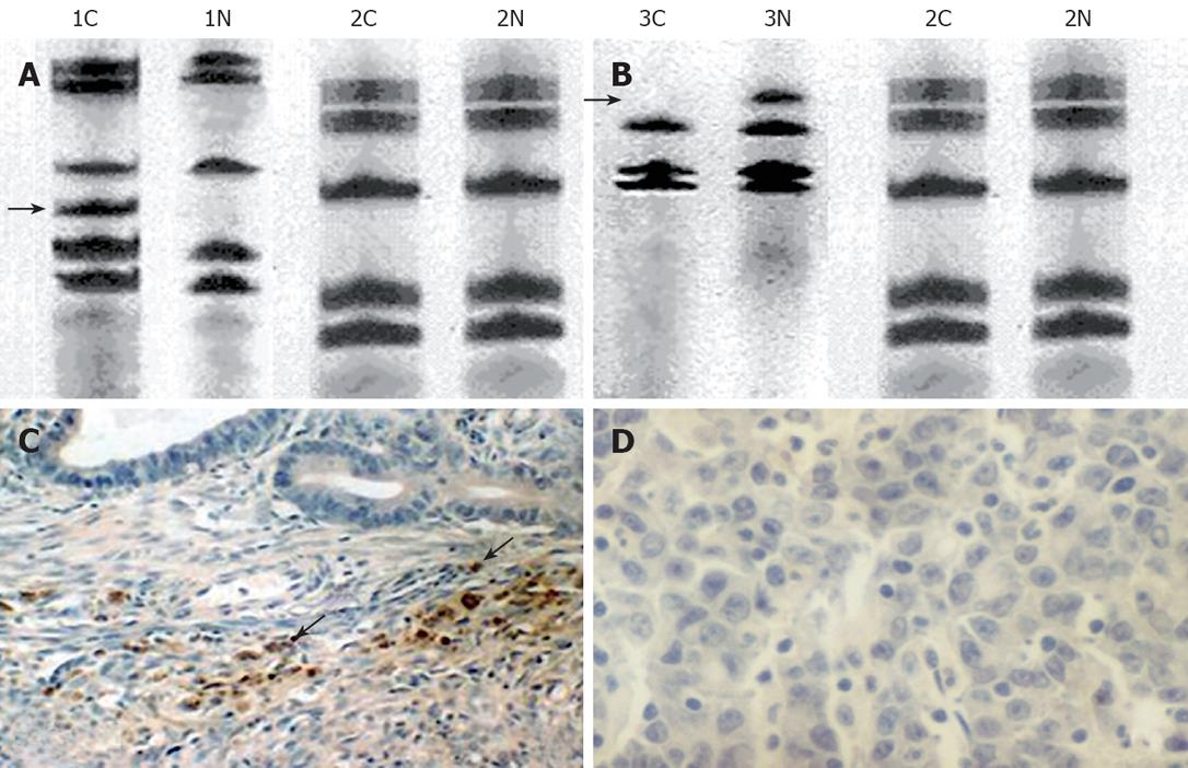
The positive rate of D17S396 MSI (Figure 1A), LOH (Figure 1B) and nm23H1 protein (Figure 1C) was 20.00%, 17.50% and 55.00%, respectively, in 40 cases of gastric cancer (Table 1).
MSI and LOH were independent of the histological type of gastric cancer, the degree of differentiation and serosa infiltration. MSI was related to the clinical TNM stage. The frequency of MSI was higher in stageI+ II than in stages III + IV of gastric cancer (31.82% vs 5.56%, P = 0.04). In contrast, LOH was higher in stage III + IV than in stageI + II of gastric cancer (33.33% vs 4.55%, P = 0.017). In addition, MSI tended to decrease with lymph node metastasis (5.00% vs 35.00%, P = 0.017), while LOH did not (30.00% vs 5.00%, P = 0.038).
The positive rate of nm23H1 protein was closely correlated with the histological type, differentiation degree and clinical stage of gastric cancer. The expression of nm23H1 protein was significantly higher in tubular adenocarcinoma than in mucous adenocarcinoma (63.64% vs 14.29%, P = 0.017), and tended to increase with the differentiation degree of tubular adenocarcinoma (P < 0.0001). The positive rate of nm23H1 expression was higher in stageI + II than in stage III + IV of gastric cancer (77.27% vs 17.86%, P = 0.001), and so was in negative than in positive lymph node metastasis. The same phenomenon was observed in patients with positive or negative MSI (87.50% vs 46.88%, P = 0.04, Table 2). On the contrary, the expression of nm23H1 protein was lower in LOH positive patients than in LOH negative patients (14.29% vs 63.64%, P = 0.017, Table 2). Computer imaging analysis showed that there was no statistical difference in nm23H1 protein expression between the two groups of patients (Table 3).
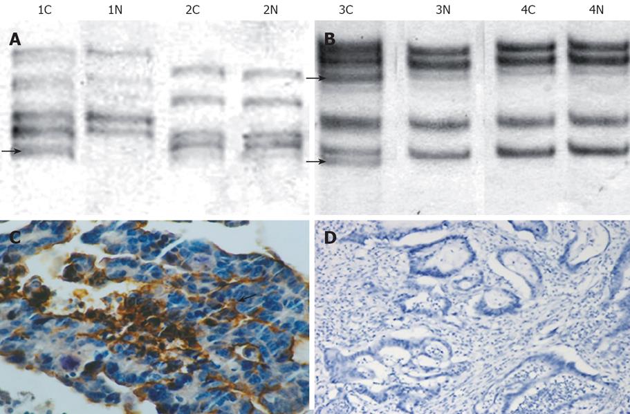
Microsatellite fragments of D17S396 were amplified. The positive rate of D17S396 MSI (Figure 2A), LOH (Figure 2B) and nm23H1 protein (Figure 2C) was 26.67%, 20.00% and 53.33%, respectively, in 30 cases of colon cancer (Table 4).
MSI and LOH were independent of the histological type, the degree of differentiation and serosa infiltration of colon cancer. MSI was related to the clinical TNM stage. In TNM staging, the frequency of MSI was higher in stageI + II than in stages III + IV of colon cancer (43.75% vs 7.14%, P = 0.023). In contrast, LOH was higher in stage III + IV than in stageI + II of colon cancer (35.71% vs 11.76%, P = 0.046). In addition, LOH tended to decrease with lymph node metastasis (5.26% vs 45.45%, P = 0.003).
The positive rate of nm23H1 protein was closely correlated with the biological behaviors, differentiation degree and clinical stage of colon cancer. The expression of nm23H1 protein trended to increase with the differentiation degree of tubular adenocarcinoma (P = 0.004). The positive rate of nm23H1 was higher in stageI + II than in stage III + IV of colon cancer (81.25% vs 21.43%, P < 0.0001), and higher in negative than in positive lymph node metastasis patients (68.42% vs 27.27%, P = 0.074). Computer imaging analysis showed that there was no difference in nm23H1 protein expression level between the two groups of patients (Table 3).
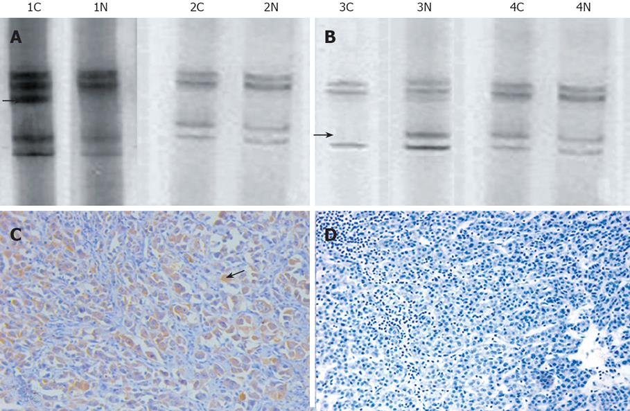
The positive rate of D17S396 MSI (Figure 3A), LOH (Figure 3B) and nm23H1 protein (Figure 3C) was 12.50%, 22.92% and 56.25%, respectively, in 48 cases of HCC (Table 5).
MSI and LOH were independent of the differentiation degree, liver infiltration and lymph node metastasis of HCC. The frequency of MSI showed no correlation to the differentiation degree, liver infiltration and lymph node metastasis of HCC. However, LOH was higher in stage III + IV than in stageI + II of HCC (52.63% vs 3.45%, P < 0.0001).
The positive rate of nm23H1 protein was related with lymph node metastasis and clinical stage of HCC. The expression of nm23H1 protein was higher in stageI + II than in stage III + IV of HCC (75.86% vs 26.32%, P < 0.0001), and in negative lymph node metastasis patients than in positive lymph node metastasis patients (82.14% vs 20.00%, P < 0.0001). The same phenomenon occurred in negative and positive LOH patients (64.86% vs 27.27%, P = 0.027, Table 2). However, MSI had no effect on the expression of nm23H1 protein (Table 2). Computer imaging analysis showed that there was no difference in the nm23H1 protein expression level between the two groups of patients (Table 3).
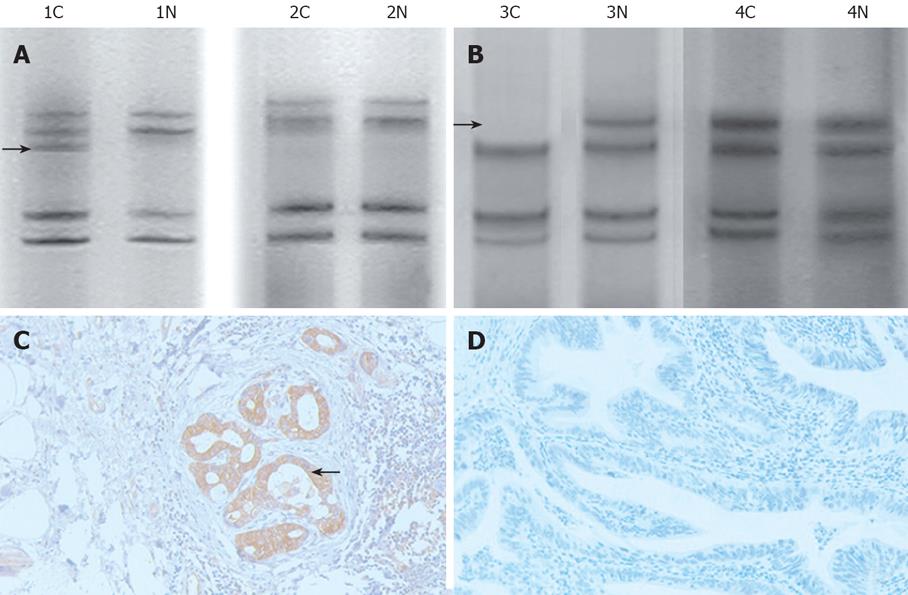
In gallbladder carcinoma, the positive rate of D17S396 MSI (Figure 4A), LOH (Figure 4B) and nm23H1 protein (Figure 4C) was 22.40%, 19.14% and 46.81%, respectively, in 47 cases of gallbladder carcinoma (Table 6).
MSI and LOH were correlated to the differentiation degree, liver infiltration, lymph node metastasis and TNM staging of gallbladder carcinoma. MSI trended to increase with the differentiation degree of gallbladder carcinoma (P < 0.0001). Furthermore, MSI was higher in negative than in positive liver infiltration patients (34.48% vs 5.56%, P = 0.023), and in negative than in positive lymph node metastasis patients (37.04% vs 5.00%, P < 0.0001). The frequency of MSI was higher in stageI + II than in stage III + IV of gallbladder carcinoma (43.48% vs 4.17%, P = 0.001). On the other hand, the frequency LOH was lower than that of MSI (6.90% vs 38.89%, P = 0.006).
The positive rate of nm23H1 protein was higher in stageI + II than in stage III + IV gallbladder carcinoma (65.22% vs 29.17%, P = 0.013), and in negative than in positive lymph node metastasis patients (62.96% vs 25.00%, P = 0.009). The same phenomenon occurred in the negative and positive LOH patients (55.26% vs 11.11%, P = 0.016, Table 2). However, MSI had no effect on the expression of nm23H1 protein. Computer imaging analysis showed that there was no difference in nm23H1 protein expression level between the two groups of patients (Table 3).
Microsatellites are short tandem repeat sequences with unknown functions scattered throughout the human genome. LOH and MSI are the phenotypes with genetic instability caused by abnormalities of tumor suppressor and DNA mismatch repair (MMR) genes. MSI is associated with slippage of DNA polymerase during DNA synthesis resulting in changing units of repetitive sequences, while LOH is allelic loss in a certain region of chromosome. Genetic instability of oncogenes, such as MSI and LOH, is probably associated with mutations of genes responsible for tumor-genesis, and plays an important role in tumor pathology[17].
MSI was first found in hereditary non-polyposis colorectal cancer (HNPCC), and then in some kinds of sporadic tumors, such as colon cancer[18], gastric cancer[19], uterus cancer[20], breast cancer[21], prostate cancer[22] and pancreatic cancer[23]. Alexander[24] examined the utility of histopathology for the identification of MSI-H cancers by evaluating the features of 323 sporadic carcinomas using specified criteria and comparing the results to MSI-H status, showing that MSI is more often found in patients with early stage cancers. In our experiment, the MSI frequency was significantly correlated with the clinical TNM staging and lymph node metastasis of gastric, colonic and gallbladder cancers. The frequency of MSI was higher in stagesIand II than in stages III and IV of these cancers. Furthermore, the frequency tended to decrease with lymph node metastasis. Our data indicate that MSI of nm23H1 may be a molecule marker of early digestive system cancer.
Berney[25] investigated the relationship between LOH and MSI in liver metastasis and nm23 protein expression, showing that the increasing proportion of LOH in nm23H1 is positively associated with liver metastasis. Candusso[26] found that LOH often occurs in later stage cancer with lymph metastasis. The frequency of LOH in 4 kinds of digestive system cancer in our study was significantly higher in stage TNM III + IV than in stage TNM I + II. Moreover, the frequency of LOH was significantly higher in patients with lymph node metastasis than in those without lymph node metastasis, suggesting that LOH of nm23H1 gene may occur in later stage digestive system cancer and accelerate lymphatic metastasis, and thus can be used as an estimate marker for malignant degree, lymphatic metastasis and prognosis. In the present study, the difference in pathology of MSI and LOH had a strong influence on the four digestive system cancers, indicating that MSI and LOH can control the carcinogenesis and metastasis through different pathways.
Lower expression of nm23H1 is closely associated with high metastasis potential and poor prognosis of human mammary cancer, gastric cancer, lung cancer, melanoma, and ovary cancer. The lower the nm23H1 expression is, the poorer the prognosis is. Steeg[27] found that nm23H1 can prevent tumor metastasis by inhibiting the ability of cancer cells to clone. In our study, TNM stage, lymphatic metastasis, and local expression of nm23H1 protein were significantly reduced in gastric cancer, colonic cancer, HCC and gallbladder cancer. Also, the expression of m23H1 protein was higher in gastric and colon cancer, which was associated with the degree of differentiation. These findings suggest that nm23H1 is actively involved in cancer metastasis as a prohibitive gene, and restrains the digestive system cancer, and can serve as a valuable biological marker in the evaluation of tumor development and prognosis.
In our study, the frequency of nm23H1 protein was higher in negative than in positive LOH patients, suggesting that LOH on D17S396 can induce loss or aberrance of anti-oncogenes, and decrease the expression of nm23H1 protein, which endows cancer with a high invasiveness and a poor prognosis. In contrast, the frequency of nm23H1 protein was higher in positive than in negative MSI patients, suggesting that MSI can accelerate the expression of nm23H1 protein. However, its molecular mechanism is not clear, and remains to be elucidated.
nm23H1 has been regarded as a metastasis-associated gene in various tumors. Genetic instabilities of oncogenes, such as microsatellite instability (MSI) and loss of heterozygosity (LOH), are probably associated with mutations of genes responsible for tumor-genesis, and play an important role in tumor pathology. Studies on MSI and LOH of digestive system cancer have been focused on the genetic instability of P53, P16 and FHIT, but few studies are available on gene nm23H1.
The present study investigated the MSI and LOH of gene nm23H1 at locus D17S396 in Chinese patients with digestive system cancers and their influence on the nm23H1 protein expression, to reveal their clinical pathological characteristics.
The results of this study indicate that MSI and LOH may independently control the biological behaviors of digestive system cancers. Enhanced expression of nm23H1 protein could efficiently inhibit cancer metastasis and improve the prognosis.
MSI could serve as an early biological marker of digestive system cancers. The data strongly suggest that LOH usually appears in the late digestive system cancer, indicating a higher malignancy and a poor prognosis.
This is an interesting study, which showed a certain significant correlation between nm23H1 genetic instability and clinical pathological characteristics of different kinds of digestive system cancer, which may serve as a marker for the diagnosis of such cancers.
Peer reviewer: Marc Basson, MD, PhD, MBA, Chief of Surgery, John D. Dingell VA Medical Center, 4646 John R. Street, Detroit, MI 48301, United States
S- Editor Zhong XY L- Editor Wang XL E- Editor Ma WH
| 1. | Cai YC, So CK, Nie AY, Song Y, Yang GY, Wang LD, Zhao X, Kinzy TG, Yang CS. Characterization of genetic alteration patterns in human esophageal squamous cell carcinoma using selected microsatellite markers spanning multiple loci. Int J Oncol. 2007;30:1059-1067. |
| 2. | Chakrabarti S, Sengupta S, Sengupta A, Basak SN, Roy A, Panda C, Roychoudhury S. Genomic instabilities in squamous cell carcinoma of head and neck from the Indian population. Mol Carcinog. 2006;45:270-277. |
| 3. | Inoue Y, Miki C, Watanabe H, Ojima E, Kusunoki M. Genomic instability and tissue expression of angiogenic growth factors in sporadic colorectal cancer. Surgery. 2006;139:305-311. |
| 4. | Nowacka-Zawisza M, Brys M, Hanna RM, Zadrozny M, Kulig A, Krajewska WM. Loss of heterozygosity and microsatellite instability at RAD52 and RAD54 loci in breast cancer. Pol J Pathol. 2006;57:83-89. |
| 5. | Yoshida K, Miki Y. Role of BRCA1 and BRCA2 as regulators of DNA repair, transcription, and cell cycle in response to DNA damage. Cancer Sci. 2004;95:866-871. |
| 6. | Juvan R, Hudler P, Gazvoda B, Repse S, Bracko M, Komel R. Significance of genetic abnormalities of p53 protein in Slovenian patients with gastric carcinoma. Croat Med J. 2007;48:207-217. |
| 7. | Xiao YP, Wu DY, Xu L, Xin Y. Loss of heterozygosity and microsatellite instabilities of fragile histidine triad gene in gastric carcinoma. World J Gastroenterol. 2006;12:3766-3769. |
| 8. | Horak CE, Lee JH, Elkahloun AG, Boissan M, Dumont S, Maga TK, Arnaud-Dabernat S, Palmieri D, Stetler-Stevenson WG, Lacombe ML. Nm23-H1 suppresses tumor cell motility by down-regulating the lysophosphatidic acid receptor EDG2. Cancer Res. 2007;67:7238-7246. |
| 9. | Hsu NY, Chen CY, Hsu CP, Lin TY, Chou MC, Chiou SH, Chow KC. Prognostic significance of expression of nm23-H1 and focal adhesion kinase in non-small cell lung cancer. Oncol Rep. 2007;18:81-85. |
| 10. | Korabiowska M, Honig JF, Jawien J, Knapik J, Stachura J, Cordon-Cardo C, Fischer G. Relationship of nm23 expression to proliferation and prognosis in malignant melanomas of the oral cavity. In Vivo. 2005;19:1093-1096. |
| 11. | Ouatas T, Salerno M, Palmieri D, Steeg PS. Basic and translational advances in cancer metastasis: Nm23. J Bioenerg Biomembr. 2003;35:73-79. |
| 12. | Ferrari D, Lombardi M, Ricci R, Michiara M, Santini M, De Panfilis G. Dermatopathological indicators of poor melanoma prognosis are significantly inversely correlated with the expression of NM23 protein in primary cutaneous melanoma. J Cutan Pathol. 2007;34:705-712. |
| 13. | Guan-Zhen Y, Ying C, Can-Rong N, Guo-Dong W, Jian-Xin Q, Jie-Jun W. Reduced protein expression of metastasis-related genes (nm23, KISS1, KAI1 and p53) in lymph node and liver metastases of gastric cancer. Int J Exp Pathol. 2007;88:175-183. |
| 14. | Kapitanovic S, Cacev T, Berkovic M, Popovic-Hadzija M, Radosevic S, Seiwerth S, Spaventi S, Pavelic K, Spaventi R. nm23-H1 expression and loss of heterozygosity in colon adenocarcinoma. J Clin Pathol. 2004;57:1312-1318. |
| 15. | Sgouros J, Galani E, Gonos E, Moutsatsou P, Belechri M, Skarlos D, Dionyssiou-Asteriou A. Correlation of nm23-H1 gene expression with clinical outcome in patients with advanced breast cancer. In Vivo. 2007;21:519-522. |
| 16. | Jackson AL, Loeb LA. Microsatellite instability induced by hydrogen peroxide in Escherichia coli. Mutat Res. 2000;447:187-198. |
| 17. | Storchova Z, Pellman D. From polyploidy to aneuploidy, genome instability and cancer. Nat Rev Mol Cell Biol. 2004;5:45-54. |
| 18. | Svrcek M, El-Bchiri J, Chalastanis A, Capel E, Dumont S, Buhard O, Oliveira C, Seruca R, Bossard C, Mosnier JF. Specific clinical and biological features characterize inflammatory bowel disease associated colorectal cancers showing microsatellite instability. J Clin Oncol. 2007;25:4231-4238. |
| 19. | Sakurai M, Zhao Y, Oki E, Kakeji Y, Oda S, Maehara Y. High-resolution fluorescent analysis of microsatellite instability in gastric cancer. Eur J Gastroenterol Hepatol. 2007;19:701-709. |
| 20. | An HJ, Kim KI, Kim JY, Shim JY, Kang H, Kim TH, Kim JK, Jeong JK, Lee SY, Kim SJ. Microsatellite instability in endometrioid type endometrial adenocarcinoma is associated with poor prognostic indicators. Am J Surg Pathol. 2007;31:846-853. |
| 21. | Pizzi C, Di Maio M, Daniele S, Mastranzo P, Spagnoletti I, Limite G, Pettinato G, Monticelli A, Cocozza S, Contegiacomo A. Triplet repeat instability correlates with dinucleotide instability in primary breast cancer. Oncol Rep. 2007;17:193-199. |
| 22. | Burger M, Denzinger S, Hammerschmied CG, Tannapfel A, Obermann EC, Wieland WF, Hartmann A, Stoehr R. Elevated microsatellite alterations at selected tetranucleotides (EMAST) and mismatch repair gene expression in prostate cancer. J Mol Med. 2006;84:833-841. |
| 23. | House MG, Herman JG, Guo MZ, Hooker CM, Schulick RD, Cameron JL, Hruban RH, Maitra A, Yeo CJ. Prognostic value of hMLH1 methylation and microsatellite instability in pancreatic endocrine neoplasms. Surgery. 2003;134:902-908; discussion 909. |
| 24. | Alexander J, Watanabe T, Wu TT, Rashid A, Li S, Hamilton SR. Histopathological identification of colon cancer with microsatellite instability. Am J Pathol. 2001;158:527-535. |
| 25. | Berney CR, Fisher RJ, Yang J, Russell PJ, Crowe PJ. Genomic alterations (LOH, MI) on chromosome 17q21-23 and prognosis of sporadic colorectal cancer. Int J Cancer. 2000;89:1-7. |
| 26. | Candusso ME, Luinetti O, Villani L, Alberizzi P, Klersy C, Fiocca R, Ranzani GN, Solcia E. Loss of heterozygosity at 18q21 region in gastric cancer involves a number of cancer-related genes and correlates with stage and histology, but lacks independent prognostic value. J Pathol. 2002;197:44-50. |
| 27. | Palmieri D, Horak CE, Lee JH, Halverson DO, Steeg PS. Translational approaches using metastasis suppressor genes. J Bioenerg Biomembr. 2006;38:151-161. |


