Published online Aug 14, 2008. doi: 10.3748/wjg.14.4810
Revised: July 19, 2008
Accepted: July 26, 2008
Published online: August 14, 2008
AIM: To explore the effect of trichostatin A (TSA) on apoptosis and acetylated histone H3 levels in gastric cancer cell lines BGC-823 and SGC-7901.
METHODS: The effect of TSA on growth inhibition and apoptosis was examined by MTT, fluorescence microscopy and PI single-labeled flow cytometry. The acetylated histone H3 level was detected by Western blot.
RESULTS: TSA induced apoptosis in gastric cancer cell lines BGC-823 and SGC-7901 was in a dose and time-dependent manner. Apoptotic cells varied significantly between TSA treated groups (37.5 ng/mL 72 h for BGC-823 cell line and 75 ng/mL 72 h for SGC-7901 cell line) and control group (0.85 ± 0.14 vs 1.14 ± 0.07, P = 0.02; 0.94 ± 0.07 vs 1.15 ± 0.06, P = 0.02). Morphologic changes of apoptosis, including nuclear chromatin condensation and fluorescence strength, were observed under fluorescence microscopy. TSA treatment in BGC-823 and SGC-7901 cell lines obviously induced cell apoptosis, which was demonstrated by the increased percentage of sub-G1 phase cells, the reduction of G1-phase cells and the increase of apoptosis rates in flow cytometric analysis. The result of Western blot showed that the expression of acetylated histone H3 increased in BGC-823 and SGC-7901 TSA treatment groups as compared with the control group.
CONCLUSION: TSA can induce cell apoptosis in BGC-823 and SGC-7901 cell lines. The expression of acetylated histone H3 might be correlated with apoptosis.
- Citation: Zou XM, Li YL, Wang H, Cui W, Li XL, Fu SB, Jiang HC. Gastric cancer cell lines induced by trichostatin A. World J Gastroenterol 2008; 14(30): 4810-4815
- URL: https://www.wjgnet.com/1007-9327/full/v14/i30/4810.htm
- DOI: https://dx.doi.org/10.3748/wjg.14.4810
Gastric cancer is the second most common cancer worldwide[1]. It is often not detected until an advanced stage; consequently, the 5-year survival rates are low (10%-20%). Owing to local invasion and metastasis, radiation therapy or chemotherapy does not significantly increase the length or improve the quality of life of patients with advanced gastric cancer. Therefore, there is growing interest in the development of novel neoadjuvant and adjuvant treatment modalities.
It is widely accepted that histone acetylation is essential to establish a transcriptional competent state of chromatin[2-4]. The reversible (de)acetylation of the N-terminal histone tails by specific histone acetylases and deacetylases (HDAC) is involved in the regulation of gene expression. Dysfunction of histone acetylases and HDACs are associated with different types of cancer[5-8]. Various HDAC inhibitors (HDACIs) have been described to induce cell cycle arrest, differentiation, and apoptosis in cell lines[9-12]. Many of these have potent antitumor activities in vivo[13]. One of the most effective and best studied HDAC inhibitors is trichostatin A (TSA). The crystallographic analysis of TSA and a histone deacetylase homologue indicates that TSA interacts reversibly with the HDAC catalytic site preventing binding of the substrate[13]. Considering that HDAC inhibitors are able to induce apoptosis in different cell types[14-16], we intend to know their potential to induce apoptosis in gastric cancer cell lines BGC-823 and SGC-7901. We also studied the effect of TSA-induced acetylated histone H3 level on these cell lines.
Stock solutions of TSA (Sigma-Aldrich, USA) in ethanol were stored at -20°C. Dimethylthiazole diphenyl tetrazolium bromide (MTT), propidium iodide (PI, Beijing Zhongshan Golden Bridge Biotechnology, China) and Hoechst 33342 (Sigma-Aldrich) were used. Antibody against GAPDH was purchased from Santa Cruz Biotechnology (Santa Cruz Biotechnology, USA). Anti-acetyl-histone H3 (rabbit polyclonal IgG) and Goat Anti-Rabbit IgG, HRP-conjugate were purchased from Upstate Biotechnology (Upstate Biotechnology, USA).
Human gastric epithelial cell lines BGC-823 and SGC-7901 were obtained from Institute of Tumor Research of Heilongjiang. BGC-823 cells and SGC-7901 cells were cultured and maintained in RPMI 1640, supplemented with fetal bovine serum 10% (v/v), penicillin 100 IU/mL, and streptomycin 100 μg/mL in a humidified atmosphere of 5% CO2 in air at 37°C.
MTT assay was used to obtain the number of living cells in the sample. Cells were seeded on 96-well plates at a predetermined optimal cell density to ensure exponential growth in the duration of the assay. After a 24 h preincubation, growth medium was replaced with experimental medium containing the appropriate drug or control. Six duplicate wells were set up for each sample, and cells untreated with drug served as control. Treatment was conducted at 12, 24, 48 and 72 h with final TSA concentrations of 37.5, 75, 150, 300 and 600 ng/mL, respectively. After incubation, 10 μL MTT (6 g/L, Sigma) was added to each well and the incubation was continued for 4 h at 37°C. After removal of the medium, MTT stabilization solution (dimethylsulphoxide: ethanol = 1:1) was added to each well, and shaken for 10 min until all crystals were dissolved. Then, optical density was detected in a microplate reader at 550 nm wavelength using an ELISA reader. The negative control well without cells was used as zero point of absorbance. Each assay was performed in triplicate.
Chromatin condensation was detected by nuclear staining with Hoechst 33342. BGC-823 and SGC-7901 cells were collected by centrifugation (500 ×g for 5 min at 4°C) and washed twice with PBS. Cells were fixed in 10% formaldehyde and stored at 4°C. For analysis, cells were washed in PBS, then Hoechst 33 342 (5 mg/L) was directly added to the medium by gently shaking at 4°C for 5 min. Stained nuclei were visualized under a Zeiss Axiophot fluorescence microscope at 400 × magnification with an excitation wavelength of 355-366 nm and an emission wavelength of 465-480 nm. Four independent replicates were used. In this way, apoptotic BGC-823 and SGC-7901 cells were stained brightly blue because of their chromatin condensation, while normal BGC-823 and SGC-7901 cells were evenly stained slightly blue.
BGC-823 and SGC-7901 cells were treated as indicated. Floating and adherent cells were collected by centrifugation (500 ×g for 5 min at 4°C) and washed twice with PBS. Cells were fixed in 90% ethanol and stored at -20°C. For analysis, cells were washed in PBS and stained by suspension in PI (50 mg/L) containing RNase A (2 mg/L) for 30 min at 4°C. Stained cells were analyzed on a FACScan (Becton- Dickinson, Heidelberg, Germany).
Cells treated as indicated were harvested in 5 mL of medium, pelleted by centrifugation (1000 ×g for 5 min at 4°C), then washed twice with ice-cold PBS and lysed in ice-cold HEPES buffer [HEPES (pH 7.5) 50 mmol/L, NaCl 10 mmol/L, MgCl2 5 mmol/L, EDTA 1 mmol/L, glycerol 110% (v/v), Triton X-100 1% (v/v), a cocktail of protease inhibitors, and 1 mg/L TSA on ice for 30 min. The lysates were clarified by centrifugation (15 000 ×g for 10 min at 4°C) and the supernatants then either analyzed immediately or stored at -80°C. Equivalent amounts of protein (50 μg) from total cell lysates were resolved by SDS-PAGE using precast 12% Bis-Tris gradient gels and transferred onto polyvinylidene difluoride (PVDF) membranes. Membranes were blocked overnight at 4°C in blocking buffer [nonfat dried milk 5% (v/v), NaCl 150 mmol/L, Tris (pH 8.0) 10 mmol/L and 0.05% Tween 20 (v/v)]. Proteins were detected by incubation with primary antibodies at appropriate dilutions in blocking buffer overnight at 4°C. Unbound antibody was removed by washing with Tris-buffered saline (pH 7.2) containing 0.5% Tween 20 (TBS-T). The membrane was then incubated at room temperature with horseradish peroxidase-conjugated secondary antibody. After extensive washing with TBS-T, bands were visualized by enhanced chemiluminescence followed by exposure to autoradiography.
TSA inhibited cellular proliferation and survival in BGC-823 and SGC-7901 cell lines. It resulted in a significant decrease in the cell population of BGC-823 and SGC-7901 compared with control, following treatment with TSA. Inhibition of TSA was dependent on the dose and incubation time (Tables 1 and 2).
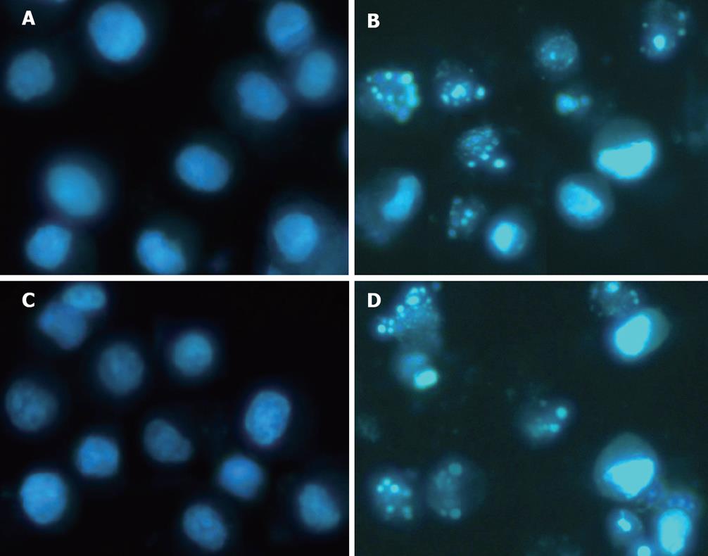
To investigate the effects of TSA induced cytotoxicity, morphologic changes of apoptosis were observed under fluorescence microscope. At 72 h, cells treated with or without TSA (37.5 ng/mL in BGC-823 and 75 ng/mL in SGC-7901) were stained by Hoechst 33 342, a classical way of identifying apoptotic cells, to observe nuclei morphology. The result indicated that nuclei of most BGC-823 and SGC-7901 cells treated with TSA were stained highly condensed, bright nucleus; while the cells in control group were stained average slightly blue (Figure 1).
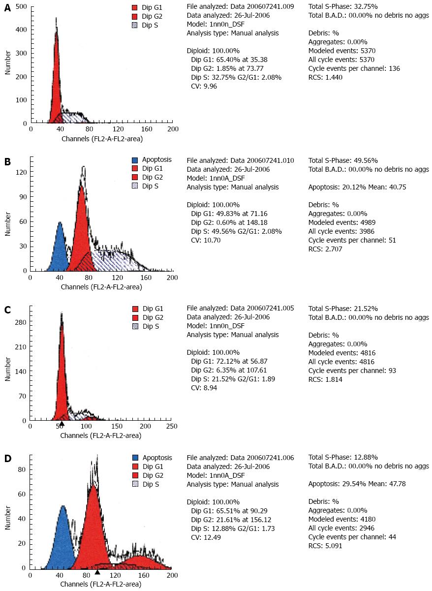
TSA treatment (37.5 ng/mL per 72 h) sensitively induced apoptosis of BGC-823 cells, which was demonstrated by the raised percentage of sub-G1 phase cells, the increase of apoptosis rates (20.12%) in flow cytometry (Figure 2). Cell cycle effects were examined by FACS analysis. The reduction of G1-phase cells (65.40%-49.83%) and the increment of S-phase cells (32.75%-49.56%) were observed in BGC-823 cells (Figure 2A, B). TSA induced apoptosis in SGC-7901 cells. Upon treatment with TSA (75 ng/mL) for 72 h in SGC-7901 cells, the reduction of G1-phase cells (72.12%-65.51%), S-phase cells (21.52%-12.88%) and the increment of G2-phase cells (6.35%-21.61%), and apoptosis rates (29.54%) were observed by FACS analysis (Figure 2C, D).
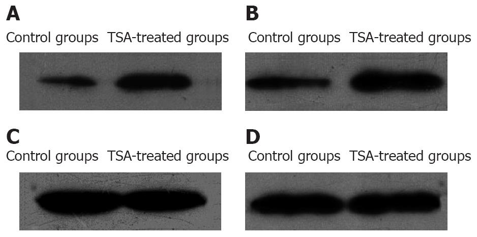
Western blot analysis was used to detect the level of acetylated histone H3 of BGC-823 cells and SGC-7901 cells treated with TSA (37.5 ng/mL per 48 h in BGC-823 cells and 75 ng/mL per 48 h in SGC-7901 cells). It was shown that there was an increase of acetylated histone H3 level in TSA treated cells (Figure 3).
Histones are small-sized and basic-charged proteins essential for chromatin folding. Posttranslational modifications such as acetylation, methylation, and phosphorylation have been suggested to be involved in the regulation of gene expression, cell division, nucleosome assembly, and DNA repair processes via alterations in the nucleosome architecture[17]. To date, acetylation is the best understood among these modifications. Chromatin structure and, thereby, transcription is controlled by the level of acetylation of histones, which is determined by the balance between histone acetyl transferase (HAT) activity and histone deacetylase (HDAC) activity. While proteins with HAT activity have been demonstrated to function as transcriptional coactivators, proteins with HDAC activity induce transcriptional repression[18]. Therefore, acetylation of histones seems to be predominantly implicated in the regulation of gene transcription due to nucleosome remodeling. The aberrant utilization of HDACs is believed to be a contributing factor in carcinogenesis.
HDAC inhibitors are members of a new class of agents able to regulate gene expression by modulating chromatin structure. HDAC inhibitors have been known to induce differentiation, growth arrest, and apoptosis in cancer cells[15,19,20]. TSA, an antifungal antibiotic with cytostatic and differentiating properties in mammalian cell culture, is a potent and specific inhibitor of HDAC activity. X-ray crystallographic studies showed that TSA has a tubular structure with a zinc atom at its base and TSA fits into this structure with the hydroxamic moiety of the inhibitor binding to the zinc. Numerous studies have reported that TSA can induce apoptosis in cancer cells[21-23].
Moderately and poorly differentiated gastric adenocarcinomas are the main types of gastric cancer in China. The rate of each was 71.0% and 58.7% in early gastric cancer and advanced gastric cancer, respectively. The cell lines BGC-823 and SGC-7901 were derived from human gastric adenocarcinoma. The stem cells in tumor tissue have infinite proliferating ability, and can form colonies in vitro. The BGC-823 cell line is a poorly-differentiated human gastric adenocarcinoma cell line. SGC-7901 cell was a moderately-differentiated human stomach adenocarcinoma cell line with mutant p53. Here, we examined the effect of TSA on BGC-823 and SGC-7901 cells.
We found that low concentrations of TSA can significantly reduce the growth of BGC-823 and SGC-7901 cells, and the effects of TSA on BGC-823 and SGC-7901 cells are dose and time-dependent. The mechanism leading to these effects remained unknown. Accumulating evidence suggests that inhibition of HDAC activity leads to relaxation of the structure of chromatin associated with a specific set of programmed genes. The relaxed chromatin structure allows these genes to be expressed, which, in turn, arrests tumor cell growth[13,24]. This suggests that induction of histone hyperacetylation by HDAC inhibitors is responsible for the antiproliferative activity through selective induction of genes that play important roles in the cell cycle and cell morphology[25]. It has been shown that reversible acetylation of lysine on histone H3 plays an important role in regulating gene transcription[26]. Our findings indicate that acetylated histone H3 expression levels increased in BGC-823 and SGC-7901 cells following TSA treatment. Increased acetylated histone H3 expression levels in BGC-823 and SGC-7901 cells may be an important event in mediating the apoptosis of BGC-823 and SGC-7901 cells induced by TSA. These findings generate the necessity to investigate the mechanism of TSA in the treatment of BGC-823 and SGC-7901 cells. Therefore, we suppose that HDAC inhibitors cause acetylated histones to accumulate in tumor tissues, and this accumulation can be used as a trigger of the biologic activity of the HDAC inhibitors.
In summary, in this study we showed that TSA can induce apoptosis in gastric cancer cell lines BGC-823 and SGC-7901. We demonstrated that the expression of acetylated histone H3 might be correlated with apoptosis. Further work will be necessary to explore additional mechanisms that lead to induction of apoptosis.
Histones are small-sized and basic-charged proteins essential for chromatin folding. The acetylation state of histones is reversibly regulated by histone acetyltransferase (HAT) and deacetylase (HDAC). An imbalance of this reaction leads to an aberrant behavior of the cells in morphology, cell cycle, differentiation, and carcinogenesis. HDAC is especially known to play an important role in carcinogenesis.
There is increasing evidence that HDAC inhibitors are effective therapeutic agents in the treatment of a variety of cancers refractory to conventional anticancer agents. Several structurally diverse HDAC inhibitors, such as trichostatin A (TSA), amicrobial metabolite, or butyrates, have been identified and their in vitro activity in transformed cells makes them promising agents for cancer therapy. Although extensive studies have been done, roles of HDAC inhibitor in gastric cancer are still unclear.
This study investigates the effects of the HDAC inhibitor TSA on gastric cancer cell lines BGC-823 and SGC-7901. The results demonstrate that low concentrations of TSA can significantly reduce the growth of BGC-823 and SGC-7901 cells and the effects of TSA on BGC-823 and SGC-7901 cells are dose and time-dependent. These findings indicate that acetylated histone H3 expression levels increased in BGC-823 and SGC-7901 cells following TSA treatment.
It can be seen from this paper that the HDAC inhibitor, TSA, can induce apoptosis of BGC-823 and SGC-7901 cells. It suggests that HDAC is a promising target for the development of anticancer drugs for gastric cancer.
This paper examined the role of TSA in gastric cancer cell lines BGC-823 and SGC-7901. The results show that TSA can induce apoptosis of BGC-823 and SGC-7901 cells. And the expression of acetylated histone H3 might be correlated with apoptosis. It suggests that TSA has a potential against gastric cancer.
Peer reviewer: Dr. Jordi Camps, Centre de Recerca Biomèdica, Hospital Universitari de Sant Joan, C. Sant Joan s/n, Reus 43201, Spain
S- Editor Zhong XY L- Editor Ma JY E- Editor Yin DH
| 1. | Dicken BJ, Bigam DL, Cass C, Mackey JR, Joy AA, Hamilton SM. Gastric adenocarcinoma: review and considerations for future directions. Ann Surg. 2005;241:27-39. |
| 2. | Kadonaga JT. Eukaryotic transcription: an interlaced network of transcription factors and chromatin-modifying machines. Cell. 1998;92:307-313. |
| 3. | Zhang W, Bone JR, Edmondson DG, Turner BM, Roth SY. Essential and redundant functions of histone acetylation revealed by mutation of target lysines and loss of the Gcn5p acetyltransferase. EMBO J. 1998;17:3155-3167. |
| 5. | Pruitt K, Zinn RL, Ohm JE, McGarvey KM, Kang SH, Watkins DN, Herman JG, Baylin SB. Inhibition of SIRT1 reactivates silenced cancer genes without loss of promoter DNA hypermethylation. PLoS Genet. 2006;2:e40. |
| 6. | Shukla SD, Aroor AR. Epigenetic effects of ethanol on liver and gastrointestinal injury. World J Gastroenterol. 2006;12:5265-5271. |
| 7. | De Schepper S, Bruwiere H, Verhulst T, Steller U, Andries L, Wouters W, Janicot M, Arts J, Van Heusden J. Inhibition of histone deacetylases by chlamydocin induces apoptosis and proteasome-mediated degradation of survivin. J Pharmacol Exp Ther. 2003;304:881-888. |
| 8. | Chen YX, Fang JY, Zhu HY, Lu R, Cheng ZH, Qiu DK. Histone acetylation regulates p21WAF1 expression in human colon cancer cell lines. World J Gastroenterol. 2004;10:2643-2646. |
| 9. | Subramanian C, Opipari AW Jr, Bian X, Castle VP, Kwok RP. Ku70 acetylation mediates neuroblastoma cell death induced by histone deacetylase inhibitors. Proc Natl Acad Sci USA. 2005;102:4842-4847. |
| 10. | Fenic I, Sonnack V, Failing K, Bergmann M, Steger K. In vivo effects of histone-deacetylase inhibitor trichostatin-A on murine spermatogenesis. J Androl. 2004;25:811-818. |
| 11. | Toth KF, Knoch TA, Wachsmuth M, Frank-Stohr M, Stohr M, Bacher CP, Muller G, Rippe K. Trichostatin A-induced histone acetylation causes decondensation of interphase chromatin. J Cell Sci. 2004;117:4277-4287. |
| 12. | Romanski A, Bacic B, Bug G, Pfeifer H, Gul H, Remiszewski S, Hoelzer D, Atadja P, Ruthardt M, Ottmann OG. Use of a novel histone deacetylase inhibitor to induce apoptosis in cell lines of acute lymphoblastic leukemia. Haematologica. 2004;89:419-426. |
| 13. | Marks PA, Richon VM, Rifkind RA. Histone deacetylase inhibitors: inducers of differentiation or apoptosis of transformed cells. J Natl Cancer Inst. 2000;92:1210-1216. |
| 14. | Batova A, Shao LE, Diccianni MB, Yu AL, Tanaka T, Rephaeli A, Nudelman A, Yu J. The histone deacetylase inhibitor AN-9 has selective toxicity to acute leukemia and drug-resistant primary leukemia and cancer cell lines. Blood. 2002;100:3319-3324. |
| 15. | Glick RD, Swendeman SL, Coffey DC, Rifkind RA, Marks PA, Richon VM, La Quaglia MP. Hybrid polar histone deacetylase inhibitor induces apoptosis and CD95/CD95 ligand expression in human neuroblastoma. Cancer Res. 1999;59:4392-4399. |
| 16. | Kim DH, Kim M, Kwon HJ. Histone deacetylase in carcinogenesis and its inhibitors as anti-cancer agents. J Biochem Mol Biol. 2003;36:110-119. |
| 17. | Grunstein M. Histone acetylation in chromatin structure and transcription. Nature. 1997;389:349-352. |
| 18. | de Ruijter AJ, van Gennip AH, Caron HN, Kemp S, van Kuilenburg AB. Histone deacetylases (HDACs): characterization of the classical HDAC family. Biochem J. 2003;370:737-749. |
| 19. | Saito A, Yamashita T, Mariko Y, Nosaka Y, Tsuchiya K, Ando T, Suzuki T, Tsuruo T, Nakanishi O. A synthetic inhibitor of histone deacetylase, MS-27-275, with marked in vivo antitumor activity against human tumors. Proc Natl Acad Sci USA. 1999;96:4592-4597. |
| 20. | Butler LM, Agus DB, Scher HI, Higgins B, Rose A, Cordon-Cardo C, Thaler HT, Rifkind RA, Marks PA, Richon VM. Suberoylanilide hydroxamic acid, an inhibitor of histone deacetylase, suppresses the growth of prostate cancer cells in vitro and in vivo. Cancer Res. 2000;60:5165-5170. |
| 21. | Chen WK, Chen Y, Gu JX, Cui GH. [Effect of trichostatin A on histone acetylation level and apoptosis in HL-60 cells]. Zhongguo Shiyan Xueyexue Zazhi. 2004;12:324-328. |
| 22. | Duan H, Heckman CA, Boxer LM. Histone deacetylase inhibitors down-regulate bcl-2 expression and induce apoptosis in t(14;18) lymphomas. Mol Cell Biol. 2005;25:1608-1619. |
| 23. | Hong ZY, Yi LS, Miao XY, Lu YP, Zhou JF, Liu WL. [Mechanism of apoptosis induced by trichostatin a in leukemia Molt-4 cells analyzed by microarray]. Ai Zheng. 2006;25:946-953. |
| 24. | Wang J, Saunthararajah Y, Redner RL, Liu JM. Inhibitors of histone deacetylase relieve ETO-mediated repression and induce differentiation of AML1-ETO leukemia cells. Cancer Res. 1999;59:2766-2769. |
| 25. | Han JW, Ahn SH, Park SH, Wang SY, Bae GU, Seo DW, Kwon HK, Hong S, Lee HY, Lee YW. Apicidin, a histone deacetylase inhibitor, inhibits proliferation of tumor cells via induction of p21WAF1/Cip1 and gelsolin. Cancer Res. 2000;60:6068-6074. |
| 26. | Davie JR. Covalent modifications of histones: expression from chromatin templates. Curr Opin Genet Dev. 1998;8:173-178. |


