Published online Feb 14, 2007. doi: 10.3748/wjg.v13.i6.973
Revised: December 1, 2006
Accepted: January 17, 2007
Published online: February 14, 2007
Jejunostomy feeding tubes provide surgeons with an excellent method for providing nutritional support, but there are several complications associated with a tube jejunostomy, including complications resulting from placement of the tube, mechanical problems related to the location or function and development of focally thickened small-bowel folds. A 76-year old man who presented with multiple medical diseases was admitted to our hospital due to aspiration pneumonia with acute respiratory failure and septic shock. He underwent exploratory laparotomy with feeding jejunostomy using a 14-French nasogastric tube for nutritional support. However, occlusion of the feeding tube was found 30 d after operation, and a rare complication of knot formation in the tube occurred after a new tube was replaced. On the following day, the tube was removed and replaced with a similar tube, which was placed into the jejunum for only 15 cm. The patient’s feedings were maintained smoothly for two months. Knot formation in the feeding tube seems to be very rare. To our knowledge, this is the third case in the literature review. Its incidence is probably related to the length of the tube inserted into the lumen.
- Citation: Liao GS, Hsieh HF, Wu MH, Chen TW, Yu JC, Liu YC. Knot formation in the feeding jejunostomy tube: A case report and review of the literature. World J Gastroenterol 2007; 13(6): 973-974
- URL: https://www.wjgnet.com/1007-9327/full/v13/i6/973.htm
- DOI: https://dx.doi.org/10.3748/wjg.v13.i6.973
During the past 3 decades, enteral nutrition has become increasingly popular because of improved nutritional formulas, advances in catheter technology, and development of less invasive techniques (including endoscopic, fluoroscopic, and laparoscopic techniques) for placement of feeding tubes for this purpose[1]. As a result, jejunostomy tubes are used more frequently than before. Despite the benefit of the enteral route for maintaining nutrition, complications have been reported in 2%-12% of patients in whom jejunostomy tubes were placed[2].
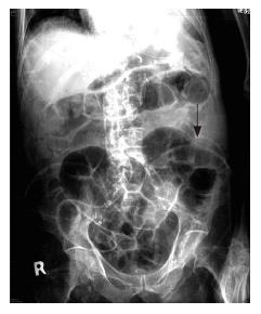
Here we present a case of 76-year old man who had a history of bilateral middle cerebral artery stenosis with multiple cerebral infarctions and was bedridden for five years. He was living in a nursing home when he was admitted to our hospital due to the impression of aspiration pneumonia with acute respiratory failure and septic shock. He underwent exploratory laparotomy with feeding jejunostomy using a 14-French nasogastric tube for enteral feedings. However, occlusion of the feeding tube was found 30 d after operation. We tried flushing the tube with warm water through a large piston syringe by gentle push-and-pull motions, but in vain. So we replaced it with a new tube. When we inserted the 14-French nasogastric tube into the upper jejunum, we advanced it at least one third of a meter distally beyond its entrance site. When the patency of the tube was checked, we were unable to put any feedings through the tube. An abdominal roentgenographic study demonstrated that a knot was formed in the tube (Figure 1, arrow).
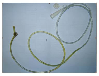
On the following day, with the patient under intravascular general anesthesia and local anesthesia, the tube was removed (Figure 2), and replaced with a similar tube which was inserted into the jejunum only for 15 cm. The patient’s feedings were maintained smoothly for two months.
An enterostomal device, which is inserted percutaneously through a stoma created on the abdomen (gastrostomy or jejunostomy), can be placed by surgical, endoscopic, laparoscopic, or radiologic techniques[3]. It is indicated when the nasal route is contraindicated or when the patient is expected to need long-term enteral feedings. Jejunostomy tube may be placed for long-term postpyloric enteral feedings when bypassing the stomach is desirable. Patients who will benefit from jejunostomy feedings include those with gastric disease, abnormal gastric and duodenal emptying, upper gastrointestinal obstruction or fistula, absent gag reflex, or significant risk of esophageal reflux and aspiration[4,5].
Abnormalities related to the placement of the tube include complications resulting from placement of the tube (i.e., small-bowel obstruction, nonobstructive small bowel narrowing, sealed-off extraluminal tracks or collections, extravasation of contrast material to the skin, jejunal hematomas, small bowel intussusception, and pneumatosis cystoides intestinalis), mechanical problems related to the location or function (i.e., coiling, kinking, occlusion, malpositioning, disruption, or knotting of the jejunostomy tube and retrograde flow of contrast material) and development of focally thickened small bowel folds as an isolated finding at or just distal to the intraluminal end of the tube[1,6-8].
To avoid a clogged feeding tube, enteral feeding devices should be thoroughly flushed every 4 to 6 h during continuous feedings and whenever feedings are on hold, before and after administration of feedings and medications or after the residuals are checked. A large syringe (30 to 60 mL) should always be used for flushing to prevent rupture of the tube. The tube should be irrigated with 20 to 30 mL of tepid water. No fluid has been found to be superior to water for maintaining patency. A stylet should never be used to unclog a tube because it can puncture the tube or part of the gastrointestinal tract. We may try one of the newer plastic declogging devices to mechanically unclog a feeding tube[9-11].
Coiling, kinking, malpositioning, occlusion, or disruption of the jejunostomy tube have been frequently found in patients, but knot formation in the feeding tube seems to be rare[12,13]. To our knowledge, this seems to be the third case in the literature review. Its occurrence is probably greater when the tube is inserted farther into the lumen, suggesting that the mechanism of knot formation is similar to that of supercoiling and concatenate formation. To avoid knot formation the feeding tube should not be inserted too long into the lumen. No more than 15 cm seems appropriate in our experience.
S- Editor Liu Y L- Editor Wang XL E- Editor Lu W
| 1. | Carucci LR, Levine MS, Rubesin SE, Laufer I, Assad S, Herlinger H. Evaluation of patients with jejunostomy tubes: imaging findings. Radiology. 2002;223:241-247. [RCA] [PubMed] [DOI] [Full Text] [Cited by in Crossref: 25] [Cited by in RCA: 28] [Article Influence: 1.2] [Reference Citation Analysis (0)] |
| 2. | Pagana KD. Preventing complications in jejunostomy tube feedings. Dimens Crit Care Nurs. 1987;6:28-38. [RCA] [PubMed] [DOI] [Full Text] [Cited by in Crossref: 1] [Cited by in RCA: 2] [Article Influence: 0.1] [Reference Citation Analysis (0)] |
| 3. | Keymling M. Technical aspects of enteral nutrition. Gut. 1994;35:S77-S80. [RCA] [PubMed] [DOI] [Full Text] [Cited by in Crossref: 14] [Cited by in RCA: 14] [Article Influence: 0.5] [Reference Citation Analysis (0)] |
| 4. | Abdel-Lah Mohamed A, Abdel-Lah Fernández O, Sánchez Fernández J, Pina Arroyo J, Gómez Alonso A. Surgical access routes in enteral nutrition. Cir Esp. 2006;79:331-341. [RCA] [PubMed] [DOI] [Full Text] [Cited by in Crossref: 3] [Cited by in RCA: 5] [Article Influence: 0.3] [Reference Citation Analysis (0)] |
| 5. | Kaur N, Gupta MK, Minocha VR. Early enteral feeding by nasoenteric tubes in patients with perforation peritonitis. World J Surg. 2005;29:1023-1027; discussion 1027-1028;. [PubMed] |
| 6. | Tapia J, Murguia R, Garcia G, de los Monteros PE, Oñate E. Jejunostomy: techniques, indications, and complications. World J Surg. 1999;23:596-602. [RCA] [PubMed] [DOI] [Full Text] [Cited by in Crossref: 86] [Cited by in RCA: 88] [Article Influence: 3.4] [Reference Citation Analysis (0)] |
| 7. | Prahlow JA, Barnard JJ. Jejunostomy tube failure: malnutrition caused by intraluminal antegrade jejunostomy tube migration. Arch Phys Med Rehabil. 1998;79:453-455. [RCA] [PubMed] [DOI] [Full Text] [Cited by in Crossref: 12] [Cited by in RCA: 14] [Article Influence: 0.5] [Reference Citation Analysis (0)] |
| 8. | Cataldi-Betcher EL, Seltzer MH, Slocum BA, Jones KW. Complications occurring during enteral nutrition support: a prospective study. JPEN J Parenter Enteral Nutr. 1983;7:546-552. [RCA] [PubMed] [DOI] [Full Text] [Cited by in Crossref: 225] [Cited by in RCA: 196] [Article Influence: 4.7] [Reference Citation Analysis (0)] |
| 9. | Nicholas JM, Cornelius MW, Tchorz KM, Tremblay LN, Spiegelman ER, Easley KA, Small W, Feliciano DV, Powell MA, Poklepovic J. A two institution experience with 226 endoscopically placed jejunal feeding tubes in critically ill surgical patients. Am J Surg. 2003;186:583-590. [RCA] [PubMed] [DOI] [Full Text] [Cited by in Crossref: 16] [Cited by in RCA: 17] [Article Influence: 0.8] [Reference Citation Analysis (0)] |
| 10. | Myers JG, Page CP, Stewart RM, Schwesinger WH, Sirinek KR, Aust JB. Complications of needle catheter jejunostomy in 2,022 consecutive applications. Am J Surg. 1995;170:547-550; discussion 550-551. [PubMed] |
| 11. | Holmes JH, Brundage SI, Yuen P, Hall RA, Maier RV, Jurkovich GJ. Complications of surgical feeding jejunostomy in trauma patients. J Trauma. 1999;47:1009-1012. [RCA] [PubMed] [DOI] [Full Text] [Cited by in Crossref: 55] [Cited by in RCA: 44] [Article Influence: 1.7] [Reference Citation Analysis (0)] |
| 12. | Myers A, Thurston W, Ho CS. Spontaneous knotting of a transgastric jejunostomy tube: case report. Can Assoc Radiol J. 1997;48:22-24. [PubMed] |
| 13. | Cappell MS, Scarpa PJ, Nadler S, Miller SH. Complications of nasoenteral tubes. Intragastric tube knotting and intragastric tube breakage. J Clin Gastroenterol. 1992;14:144-147. [RCA] [PubMed] [DOI] [Full Text] [Cited by in Crossref: 20] [Cited by in RCA: 18] [Article Influence: 0.5] [Reference Citation Analysis (0)] |


