Published online Jun 14, 2007. doi: 10.3748/wjg.v13.i22.3141
Revised: March 3, 2007
Accepted: March 15, 2007
Published online: June 14, 2007
Intrahepatic cholangiocarcinoma is a malignant neoplasm arising from the biliary epithelium, which frequently invades adjacent organs or metastasizes to other visceral organs such as the lungs, bones, adrenals, and brain. However, distant skeletal muscle metastasis of cholangiocarcinoma has never been described before to the best of our knowledge and, furthermore, Budd-Chiari syndrome secondary to intrahepatic cholangiocarcinoma is also extremely rare. Here we present the first case overall of distant muscle metastasis from intrahepatic cholangiocarcinoma presenting as Budd-Chiari syndrome. A 44-year-old man admitted to the hospital with complaints of abdominal distension, edema of both legs, back pain and anorexia of 30 d' duration. Computed tomography and ultrasonography-guided percutaneous muscle biopsy established intrahepatic cholangiocarcinoma with disseminated thrombosis from inferior vena cava to bilateral iliac and femoral veins, and multiple skeletal muscle metastases in bilateral buttock and erector spinal muscle.
- Citation: Kwon OS, Jun DW, Kim SH, Chung MY, Kim NI, Song MH, Lee HH, Kim SH, Jo YJ, Park YS, Joo JE. Distant skeletal muscle metastasis from intrahepatic cholangiocarcinoma presenting as Budd-Chiari syndrome. World J Gastroenterol 2007; 13(22): 3141-3143
- URL: https://www.wjgnet.com/1007-9327/full/v13/i22/3141.htm
- DOI: https://dx.doi.org/10.3748/wjg.v13.i22.3141
Intrahepatic cholangiocarcinoma is a malignant neoplasm arising from the biliary epithelium and a devastating malignancy that presents late, is notoriously difficult to diagnose, and often invades adjacent organs or metastasizes to other visceral organs such as lungs, bones, adrenals, and brain. Skeletal muscle is one of the most uncommon sites of metastasis from any malignancy. Although direct muscle invasion by primary malignancy is well recognized, few cases of metastasis to skeletal muscle distant from the primary carcinoma have been published[1]. Primary carcinoma sites to distant skeletal muscle metastasis included the stomach, esophagus, lung, colon, and pancreas[2]. However intrahepatic cholangiocarcinoma has never been mentioned as the primary carcinoma site for skeletal muscle metastases to the best of our knowledge. Budd-Chiari syndrome which is defined as any pathophysiologic process that results in interruption of the normal flow of blood out of the liver, and is commonly associated with a hypercoagulable state which is often secondary to malignancy. But the Budd-Chiari syndrome secondary to intrahepatic cholangiocarcinoma is so rare that only three cases have been reported in the literature so far.
We report the first case of distant skeletal muscle metastasis of intrahepatic cholangiocarcinoma presenting as Budd-Chiari syndrome and acute thrombus extended down into the bilateral iliac veins and femoral veins.
A 44-year-old man visited the gastroenterology department with complaints of abdominal distension, dyspnea, low extremity edema, back pain and anorexia of one month’s duration. He was previously healthy and his past medical and family histories were not remarkable. He consumed alcohol (3 bottles of Soju distilled liquor per week) until one month before admission.
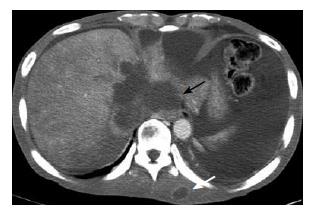
Physical examination on admission revealed a height of 174 cm, body weight of 78 kg, temperature of 36.4°C, blood pressure of 110/60 mmHg, and pulse rate of 78/min. Upon examination, the abdomen was not tender, distended with a shifting dullness. The liver was not palpable below the costal margin. Ascites was severe and both low limb edema was moderate. Patient laboratory tests included a red blood cell count (RBC) of 3.93 × 10¹²/L, hemoglobin concentration of 121 g/L, hematocrit of 35.9%, white blood cell count of 5.8 × 10⁹/L, platelet count of 1.18 × 10¹¹/L, prothrombin activity of 75.9%, activated partial thromboplastin time of 30.1 s, aspartate aminotransferase (AST) level of 50 IU/L, alanine aminotransferase (ALT) level of 26 IU/L, alkaline phosphatase level of 813 IU/L, γ-glutamyl transferase level of 144 IU/L, lactate dehydrogenase level of 494 IU/L, total bilirubin level of 0.8 mg/dL, and albumin level of 37 g/L. Renal function tests showed a blood urea nitrogen level of 39.5 mg/dL and a creatinine level of 1.6 mg/dL. HBsAg and anti-HBeAb were positive but anti-HBs antibodies, anti-HBc antibodies, HBeAg, HBV DNA, and anti-hepatitis C virus antibodies were all negative. Peritoneal fluid analysis revealed WBC of 6.90 × 10⁸/L, RBC of 1.05 × 10⁹/L, polymorphonuclear cell of 19%, lymphocytes of 81%, albumin level of 13 g/L, and the serum ascites albumin gradient (SAAG) was 24 g/L. Levels of carcinoembryonic antigen and alpha-fetoprotein were normal. The levels of thyroxin stimulating hormone and thyroid hormones were within normal limits. An abdominal ultrasonography showed a large amount of ascites, splenomegaly and hydronephrosis of the left kidney. The ascites was intractable and the edema and pain of both lower extremities were aggravated. Abdominal computed tomography (CT) without contrast enhancement was performed and it showed a large lobulated area of low density in the left lobe of liver (7 cm × 4 cm), thrombosis in inferior vena cava (IVC), two round, low density areas in the right psoas muscle (2cm) and an oval low density area at the left paravertebral area between left psoas muscle and quadratus lumbrom muscle (3.5 cm × 2 cm), mild left hydronephrosis and a large amount of ascites. For further evaluation, CT scan with contrast enhancement from chest to lower extremity was performed. The constrasted CT scan revealed a large liver mass (8 cm × 8 cm) with peripheral rim enhancement around intrahepatic portion of IVC, extensive thrombosis in IVC to bilateral iliac and femoral veins, multiple muscle metastasis in bilateral buttock, left erector spinae, right psoas muscles, multiple bony metastasis in the 10th and 11th thorasic spine and the 1st lumbar spine, and multiple patches or nodules of increased density in the lungs suggesting metastasis (Figure 1). Tests for coagulation activity showed D-dimer level of 6200 ng/mL (normal less than 300 ng/mL), antithrombin III level of 20 mg/dL (normal 21.0-34.0 mg/dL), protein-C activity level of 55% (normal 70%-130%), and protein-S activity level of 61% (normal 77%-143%).
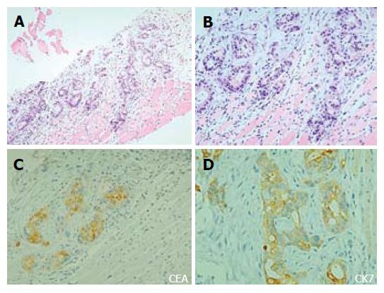
Ultrasonography-guided percutaneous needle biopsy was performed at the erector spinae muscle which was suspected to have metastatic lesions. Microscopic examinations showed that the skeletal muscle was infiltrated by neoplastic glands accompanying moderate desmoplasia (Figure 2). Immunohistochemical staining showed that the neoplastic cells were strongly positive for cytokeratin 7 and CEA, weak and focally positive for cytokeratin 20, and negative for TTF-1. The findings of CT scan and the histopathologic feature would be compatible with intraductal cholangiocarcinoma and its distant skeletal muscle metastasis.
The incidence of skeletal muscle metastases is reported to be less than 1% of metastases of hematogeneous origin, despite of the fact that skeletal muscle accounts for nearly 50% of the total body weight and is characterized by rich blood supply[3]. The cause for the low incidence of skeletal muscular metastases of primary cancer is still unclear, but may be related to various factors as follows: tumor suppressors in skeletal muscles[4], the constant movement of skeletal muscles which may represent a difficult condition for the implantation and growth of metastatic cells under the high tissue pressure related to the exercise- associated increase of blood flow, the local production of lactic acid which would create an unfavorable environment for metastatic cell growth, the inhibition of cell invasion by protease inhibitors located in the basement membrane, and the antitumor activity of lymphocytes or natural killer cells within the skeletal muscle[3].
The reason why distant skeletal muscle metastasis from intrahepatic cholangiocarcinoma has been not been reported may be because of the relative rarity and poor prognosis of intrahepatic cholangiocarcinoma, and another possibility is that physicians and patients tend to overlook the distant skeletal muscle metastasis, which is frequentely asymptomatic, nonspecific, and in hidden locations[5]. Under-diagnosis of skeletal muscle metastases may contribute to their apparent low incidence. In 194 autopsies involving tumor metastasis to skeletal muscles, which were performed at the Marque de Valdecilla National Medical Center from 1980 to 1982, metastases to skeletal muscle were noted in 11%, and 20% of the patients with carcinoma had muscle metastasis[6].
Budd-Chiari syndrome secondary to intrahepatic cholangiocarcinoma is very rare, only three cases have been reported to our knowledge. The first was from a case series that attempted to etiopathophysiologically classify Budd-Chiari syndrome[7]. The second was in a woman who had presented with recurrent venous thrombosis during the third episode endoscopic cholangiopancreatography, and guided biopsy established a diagnosis of cholangiocarcinoma at the mid portion of common bile duct[8]. Law et al[9] reported the third case of metastatic intrahepatic cholangiocarcinoma presenting as acute Budd-Chiari syndrome with a large thrombus in the inferior vena cava and plain thrombus extending from the right atrium down into the iliac veins by postmortem examination.
In summary, intrahepatic cholangiocarcinoma presenting Budd-Chiari syndrome is extremely rare, and distant skeletal muscle metastasis from intrahepatic cholangiocarcinoma presenting with Budd-Chiari syndrome has not been reported previously to our knowledge. In the present report, we described the first case of metastasis to the distant erector spinae muscle from intrahepatic cholangiocarcinoma presenting as Budd-Chiari syndrome. Intrahepatic cholangiocarcinoma tends to have a poor prognosis because of its typically late presentation. In our case, the presentation of distant skeletal muscle metastasis and Budd-Chiari syndrome is a reflection of the severe malignant potential of this carcinoma and our limited options in the management of this disease.
S- Editor Wang J L- Editor Ma JY E- Editor Lu W
| 1. | Beşe NS, Ozgüroğlu M, Dervişoğlu S, Kanberoğlu K, Ober A. Skeletal muscle: an unusual site of distant metastasis in gastric carcinoma. Radiat Med. 2006;24:150-153. [RCA] [PubMed] [DOI] [Full Text] [Cited by in Crossref: 20] [Cited by in RCA: 19] [Article Influence: 1.0] [Reference Citation Analysis (0)] |
| 2. | Heyer CM, Rduch GJ, Zgoura P, Stachetzki U, Voigt E, Nicolas V. Metastasis to skeletal muscle from esophageal adenocarcinoma. Scand J Gastroenterol. 2005;40:1000-1004. [RCA] [PubMed] [DOI] [Full Text] [Cited by in Crossref: 17] [Cited by in RCA: 21] [Article Influence: 1.1] [Reference Citation Analysis (0)] |
| 3. | Sudo A, Ogihara Y, Shiokawa Y, Fujinami S, Sekiguchi S. Intramuscular metastasis of carcinoma. Clin Orthop Relat Res. 1993;296:213-217. [PubMed] |
| 4. | Bar-Yehuda S, Barer F, Volfsson L, Fishman P. Resistance of muscle to tumor metastases: a role for a3 adenosine receptor agonists. Neoplasia. 2001;3:125-131. [RCA] [PubMed] [DOI] [Full Text] [Cited by in Crossref: 65] [Cited by in RCA: 70] [Article Influence: 2.9] [Reference Citation Analysis (0)] |
| 5. | Cione GP, Arciero G, De Angelis CP, Marano A, Farella N, Cerrone C, Cerbone D, Parmeggiani D, Cimmino G, Perrotta M. Intrahepatic cholangiocarcinoma: case report. Suppl Tumori. 2005;4:s46-s47. [PubMed] |
| 6. | Acinas García O, Fernández FA, Satué EG, Buelta L, Val-Bernal JF. Metastasis of malignant neoplasms to skeletal muscle. Rev Esp Oncol. 1984;31:57-67. [PubMed] |
| 7. | De BK, De KK, Sen S, Biswas PK, Das TK, Das S, Hazra B. Etiology based prevalence of Budd-Chiari syndrome in eastern India. J Assoc Physicians India. 2000;48:800-803. [PubMed] |
| 8. | Bandyopadhyay SK, Sarkar N, Ghosh S, Dasgupta S. Cholangiocarcinoma presenting with recurrent venous thrombosis. J Assoc Physicians India. 2003;51:824-825. [PubMed] |
| 9. | Law JK, Davis J, Buckley A, Salh B. Intrahepatic cholangiocarcinoma presenting as the Budd-Chiari syndrome: a case report and literature review. Can J Gastroenterol. 2005;19:723-728. [PubMed] |


