Published online Apr 7, 2007. doi: 10.3748/wjg.v13.i13.1980
Revised: February 18, 2007
Accepted: March 14, 2007
Published online: April 7, 2007
AIM: To explore the pathogenesis of the rectovestibular disruption (RVD) defect and to recommend a successful repair, and prevention of it.
METHODS: Clinical records of 15 girls, age ranged from 3 to 15 (median, 7.5) years, with acquired rectovestibular fistula (RVF) mistreated before were retrospectively reviewed. All of them presented an abnormal appearance of perineum and were suffering from some degree of fecal incontinence, and those were graded III to IV by Li Zheng’s Score. Repair of anal sphincters and reconstruction of perineum body and skin by anterior perineal rectoanoplasty were performed in all cases.
RESULTS: Operation in all cases was successful. The perineum looked practically normal and fecal continence score rose up to VI by Li Zheng’s Score.
CONCLUSION: The conventional treatment for anal fistula, lay-open or string-treatment, should be considered as malpractice of RVF, and certainly leads to the RVD defect, and the anterior perineal rectoanoplasty could cure it satisfactorily.
- Citation: Zhang TC, Pang WB, Chen YJ, Zhang JZ. Recto-vestibular disruption defect resulted from the malpractice in the treatment of the acquired recto-vestibular fistula in infants. World J Gastroenterol 2007; 13(13): 1980-1982
- URL: https://www.wjgnet.com/1007-9327/full/v13/i13/1980.htm
- DOI: https://dx.doi.org/10.3748/wjg.v13.i13.1980
Acquired rectovestibular fistula (RVF) is always resulted from anal infection in newborn period. Following the healing of the acute infection, a well-epithelialized tract between the vestibular and anal canal develops, passing feces from the vestibular perforation. As the stool becomes solid, the leakage stops, and ARF does not give any remarkable inconvenience on children’s quality of life, growth and development[1]. The indication of operation is mainly about psychological concern of parents and children. However, malpractice of the surgical treatment of rectovestibular fistula, including lay-open or string treatment of fistula, will certainly cause total perineal disruption resulting to vestibular cosmetic defect and remarkable fecal incontinence. From June 1996 to April 2006, 15 girls with such defects were admitted to Beijing Children’s Hospital, and treated successfully by anterior perineal anorectoplasty.
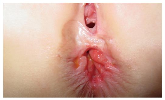
Records of fifteen girls, age ranged from 3 to 15 (average 7.5) years, were enrolled in this study. All of them came with a perineal defect and fecal incontinence due to malpractice in the treatment of RVF by lay-open or string treatment performed in other hospitals half to five years ago. These children had no normal perineal skin between the vestibular fossa and anus or underneath muscles, but had a mucosal patch between the vaginal orifice and anterior wall of the rectum. Sphincters ani was divided and its broken ends shrank backward to both sides and behind the rectum; consequently, it lost the normal appearance of perineum (Figure 1). All children were suffering from fecal soiling and some degree of anal incontinence partly with loose stool, which was graded III-IV by Li Zheng’s score[2].
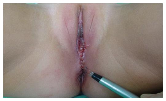
Metronidazole tablet (10 mg/kg) and oral garamycine (4 mg/kg) were given pre-operatively for three days. Food but water was restricted one day prior to operation. Saline enemas were given in the night before and on the morning of operation. The children were laid in lithotomy position. Almost half roll of sterile bandage lubricated with liquid paraffin, with the tail soaked with iodophor, was inserted and stacked in the rectum to prevent rectal discharge soiling out. The bandage tail was tied by a long heavy thread, which was left outside the anus for easier removal after operation. The embedded broken ends of the sphincter ani externus were detected by electric nerve stimulator. Four to six stay sutures were applied to the mucosal patch at mucocutaneous junction around vagina and anus, respectively. The stay sutures were stretched, and a half-circular incision anterior to the anus up to the level of two broken underneath ends of the external sphincter was made by sharp point of Bovie. Inward dissection along the bilateral wings of disrupted perineum was carried to separate vagina from rectum completely for about 5 cm in length. And the two broken ends of the sphincter ani externus were found by means of electric nerve stimulator. The transversus perinei, levator ani, and broken sphincter ani externus were approximately sutured layer by layer, the skin cut edge was approximately sutured around the rectal opening and behind the vagina orifice, and the skin cut edge between the anus and vagina was sutured vertically (anterior-posteriorly) interruptedly with 5-0 Dexon. Thus, the perineal body sphincter as well as skin bridge were all re-established (Figure 2). Routinely, food but water was restricted for three days. Antibiotics were given intravenously for 3 or 5 d. Perianal area was kept clean and dry by warm ventilation three to five times a day for some five days. The daily anal dilation began two weeks after the operation, and continued for 6 mo.
During operation, vagina was injured and repaired immediately in two of the cases, but incision healed by first intention in all 15 cases. All children were followed up for 3 mo to 9.5 years (average 7.5 years). In two children, anal dilation was difficult at beginning, because their sphincter ani externus was too tight and the anus was too small after operation. Perineal appearance of all children resumed normal, and the bowel control was good, with bowel movement one or two times a day, without soiling, incontinence or constipation, with grade VI on Li Zheng’s score.
RVF with normal anus is considered acquired, in spite of a lot of disputes[3-6], as secondary to perineal infection in neonatal period. In recent 10 years, 319 cases, averaged to 30 per year, were admitted to the Surgical Department of Beijing Chidren’s Hospital. Clinically, the fistula had perfect mucosal lining, but none of the patients had a definite history of fistula at birth. Pathological section never showed normal histology of mucosa, submucosa or continuation of smooth muscle in the fistula tract[1,7]. Anyway, the opening was not big enough to allow solid stool leakage, and these patients could have normal marriage and childbirth, so the treatment should be safe and simple.
The conventional treatment of anal fistula is lay-open by perpendicular cutting of sphincters and the tract at one site, or string-treatment by gradual cutting through by a rubber band. In rectovestibular fistula, the external opening is in the vestibule and inner rectum opening lies in the anterior wall of rectum at the dentate line, with the tract crossing beneath the sphincter ani. Dividing the wall of fistula tract and the sphincter by lay-open or by string-treatment, the cut ends of sphincter will retract apart, leaving a big mucosal patch between the rectum and vestibule. The defect will not heal. Loss of normal perineal appearance and normal function undoubtedly makes the mother and child eager to have it repaired. Among our 15 cases, five resulted from lay-open, and ten from string-treatment. Nine were suffering from fecal soiling or fecal incontinence graded III-IV by Li Zheng’s score. Anorectal surgeons should be alert to avoid the malpractices of lay-open and string treatment in acquired rectovestibular fistula.
In our series of 319 cases in the last 10 years, one stage repairing the fistula through either transrectal approach or through vestibular opening had obtained successful rate above 90%[8,9].
With anterior perineal anorectoplasty conventionally used for imperforated anus, and rectovestibular fistula with normal anus[10-12], we recommand it here for rectovestibular disruption (RVD). It provided a good exposure of operation field to separate the rectovaginal septum, and to find the broken ends of sphincter ani externus for reunion. It has been successfully proved in all our 15 cases. Key points for successful operation include: (1) bandage packing in the rectum to prevent fecal contamination of operation field; (2) sharp dissection of natural clearance by Bovie to prevent bleed and creat a clean operation field; (3) bipolar coagulation of the two main arterioles 1 cm inward at 1 and 11 o’clock to lessen active oozing; (4) no dead space left between the vagina and rectum; (5) repair of the vagina wall or rectum wall immediately if injured during operation, as much as possible not injuring the rectum wall. In our group, vaginal wall was injured in two cases which were immediately sutured interruptedly with 5-0 Dexon. The wound healed well without any complication.
All children in this group were older than three year of age. All of them had satisfactory result. It proved, at least, that anterior perineal anorectoplasty is preferable for children older than 3 years. Besides, the anorectoplasty ought to be performed 6 mo to three years after the injured wound of perineum healed up. If delayed, the broken sphincter ani might be atrophic and contracted back the anus canal. Two girls in this series had anorectoplasty three and half years and four years, respectively, after the string-treatment. Both of them had a small anal orifice after the operation, and had difficult dilation for certain time.
Diverting colostomy has been a routine for better perineum hygiene in Western World[13], but for the suffering in RVD is minor, it seems not worthwhile to make two additional abdominal operations, which will increase the operation risk as well as the economic burden. In our 15 cases of anterior perineal anorectoplasty for RVD, diverting colostomy seems unnecessary, provided we pay more attention to lessen the operative trauma and contamination, and to avoid early bowel movement.
S- Editor Liu Y L- Editor Kumar M E- Editor Ma WH
| 1. | Zhang JZ. Anorectal Diseases Among Children. First edition. Beijing: International Academic Publishers 1993; 239-242. |
| 2. | Wang HZ, Li Z. Primary advice on standard of anal function measuring after anoplasty. Zhonghua Xiao'er Waike Zazhi. 1985;6:116-117. |
| 3. | Brem H, Guttman FM, Laberge JM, Doody D. Congenital anal fistula with normal anus. J Pediatr Surg. 1989;24:183-185. [RCA] [PubMed] [DOI] [Full Text] [Cited by in Crossref: 19] [Cited by in RCA: 19] [Article Influence: 0.5] [Reference Citation Analysis (0)] |
| 4. | Bianchini MA, Fava G, Cortese MG, Vinardi S, Costantino S, Canavese F. A rare anorectal malformation: a very large H-type fistula. Pediatr Surg Int. 2001;17:649-651. [RCA] [PubMed] [DOI] [Full Text] [Cited by in Crossref: 6] [Cited by in RCA: 6] [Article Influence: 0.3] [Reference Citation Analysis (0)] |
| 5. | Yazlcl M, Etensel B, Gürsoy H, Ozklsaclk S. Congenital H-type anovestibuler fistula. World J Gastroenterol. 2003;9:881-882. [PubMed] |
| 6. | Zhang JZ. Etiological and pathological study of fistula-in-ano in children. Zhonghua Xiao'er Waike Zazhi. 1988;9:111-112. |
| 7. | Sun L, Wang YX, Liu Y. Histopathological study of fistula-in-ano in female. Zhonghua Xiao'er Waike Zazhi. 1995;16:136-137. |
| 8. | Chen YJ, Niu ZY, Zhang JZ, Liu YT, Li L, Wang DY. Transperineal approach to repair acquired rectovestibular fistula in female. Shiyong Erke Linchuang Zazhi. 2001;16:242-243. |
| 9. | Chen YJ, Zhang TC, Zhang JZ. Transanal approach in repairing acquired rectovestibular fistula in females. World J Gastroenterol. 2004;10:2299-2300. [PubMed] |
| 10. | Okada A, Kamata S, Imura K, Fukuzawa M, Kubota A, Yagi M, Azuma T, Tsuji H. Anterior sagittal anorectoplasty for rectovestibular and anovestibular fistula. J Pediatr Surg. 1992;27:85-88. [RCA] [PubMed] [DOI] [Full Text] [Cited by in Crossref: 46] [Cited by in RCA: 59] [Article Influence: 1.8] [Reference Citation Analysis (0)] |
| 11. | Wakhlu A, Pandey A, Prasad A, Kureel SN, Tandon RK, Wakhlu AK. Anterior sagittal anorectoplasty for anorectal malformations and perineal trauma in the female child. J Pediatr Surg. 1996;31:1236-1240. [RCA] [PubMed] [DOI] [Full Text] [Cited by in Crossref: 24] [Cited by in RCA: 29] [Article Influence: 1.0] [Reference Citation Analysis (0)] |
| 12. | Aziz MA, Banu T, Prasad R, Khan AR. Primary anterior sagittal anorectoplasty for rectovestibular fistula. Asian J Surg. 2006;29:22-24. [RCA] [PubMed] [DOI] [Full Text] [Cited by in Crossref: 18] [Cited by in RCA: 22] [Article Influence: 1.2] [Reference Citation Analysis (0)] |
| 13. | Mirza I, Zia-ul-Miraj M. Management of perineal canal anomaly. Pediatr Surg Int. 1997;12:611-612. [RCA] [PubMed] [DOI] [Full Text] [Cited by in Crossref: 6] [Cited by in RCA: 8] [Article Influence: 0.3] [Reference Citation Analysis (0)] |


